https://doi.org/10.1007/s00167-019-05590-4 KNEE
Steep posterior slope and shallow concave shape of the medial tibial plateau are risk factors for medial meniscus posterior root tears
Yuki Okazaki1 · Takayuki Furumatsu1 · Yuya Kodama1 · Yusuke Kamatsuki1 · Yoshiki Okazaki1 · Takaaki Hiranaka1 · Shota Takihira1 · Tomonori Tetsunaga1 · Kenta Saiga1 · Toshifumi Ozaki1
Received: 21 January 2019 / Accepted: 19 June 2019
© European Society of Sports Traumatology, Knee Surgery, Arthroscopy (ESSKA) 2019
Abstract
Purpose Bone morphological factors are important for menisci. Their association with medial meniscus posterior root tears, however, has not yet been studied. This study aimed to compare sagittal medial tibial slope and medial tibial plateau depth between knees with and without medial meniscus posterior root tears.
Methods Nine healthy volunteers, 24 patients who underwent anterior cruciate ligament reconstruction, and 36 patients who underwent medial meniscus posterior root pullout repair were included. Magnetic resonance imaging examinations were performed in the 10°-knee-flexed position. The medial tibial slope and medial tibial plateau depth were compared among the groups.
Results In healthy volunteers, the anterior cruciate ligament reconstruction group, and the medial meniscus posterior root tear group, the medial tibial slopes were 3.5° ± 1.4°, 4.0° ± 1.9°, and 7.2° ± 1.9°, respectively, and the medial tibial plateau depths were 2.1 ± 0.7 mm, 2.2 ± 0.6 mm, and 1.2 ± 0.5 mm, respectively. Patients with medial meniscus posterior root tears had a significantly steep medial tibial slope and shallow medial tibial plateau concavity compared to those of healthy volunteers (P < 0.01) and the anterior cruciate ligament group (P < 0.01). In the multivariate logistic regression analysis, body mass index, medial tibial slope, and medial tibial plateau depth were significantly associated with medial meniscus posterior root tears.
Conclusions A steep posterior slope and a shallow concave shape of the medial tibial plateau are risk factors for medial meniscus posterior root tear.
Level of evidence Level III: Case–control study.
Keywords Medial meniscus · Posterior root tear · Magnetic resonance imaging · Medial tibial slope · Medial tibial plateau depth
Abbreviations
ACL Anterior cruciate ligament BMI Body mass index
ICC Intra-class correlation coefficient LTS Lateral tibial slope
MMPRT Medial meniscus posterior root tear MRI Magnetic resonance imaging MTPD Medial tibial plateau depth
MTS Medial tibial slope PRT Posterior root tear
Introduction
The interest in medial meniscus posterior root tears (MMPRT) has been increasing, and they have been widely studied. Knee motions associated with descending stairs and downhill slopes are the most common injury patterns of MMPRTs [9]. MMPRT pullout repair is recommended to prevent subsequent cartilage degeneration or meniscus extrusion [4], and favourable clinical outcomes have been reported after transtibial pullout repair of MMPRT [16, 22].
Severe chondral lesions, varus alignment, and older age have been reported to predict a poor prognosis after MMPRT
* Takayuki Furumatsu matino@md.okayama-u.ac.jp
1 Department of Orthopaedic Surgery, Okayama University Graduate School of Medicine, Dentistry, and Pharmaceutical Sciences, 2-5-1 Shikatacho, Kitaku, Okayama 700-8558, Japan
fixation, and MMPRT pullout repair is recommended in patients with a femorotibial angle < 180° and mild cartilage lesions [5, 24].
To date, many studies about the association between tibial geometry or biomechanics and the anterior cruciate ligament (ACL) or meniscus have been reported [6, 12, 19, 26, 27, 30]. The geometry of the tibial plateau has a direct influence on the biomechanics of the tibiofemoral joint [3, 7]. An important characteristic of the tibial plateau is its posterior slope (with the anterior elevation being higher than the posterior elevation) [7, 10]. Biomechanical stud- ies have shown that a steep medial tibial slope (MTS) leads to increased anterior tibial translation under load, which may increase the forces acting on the menisci [1, 7, 10, 20, 21, 25]. Furthermore, a biconcave medial tibial plateau is reported to be more frequently associated with a complex medial meniscus tear [2] and a shallower MTS and lateral tibial slope (LTS) may result in impingement of the poste- rior horn of the medial meniscus and the anterior horn of the lateral meniscus, respectively. However, no significant association was found between medial tibial plateau depth (MTPD) and meniscal tears [15].
These associations in relation to MMPRT, however, have not been studied thus far and were, therefore, the subject of the present study. The purpose of this study was to inves- tigate the unique bone geometry in patients with MMPRT.
This study hypothesized that patients with MMPRT would have a steeper MTS and shallower MTPD compared to sub- jects without MMPRT.
Materials and methods
A retrospective design was used to compare sagittal MTS and MTPD among healthy volunteers (group V), patients who underwent ACL reconstruction (group A), and patients
who underwent MMPRT pullout repair (group M). A chart review was conducted using our electronic medical record system to identify healthy volunteers and all patients under- going ACL reconstruction or MMPRT pullout repair at our institution between January 2013 and November 2018.
Exclusion criteria were concomitant injuries such as ACL rupture and MMPRT and a history of previous surgery at the index knee.
A total of 26 healthy volunteers underwent open MRI examinations, 49 patients underwent ACL reconstruction, and 44 patients underwent MMPRT pullout repair. Of those, 9 (35%), 24 (49%), and 36 (82%) were women. For the pur- pose of this study, only women were included for further analysis because a significant difference has been reported between the within-subject MTS values when male and female subjects were considered as separate groups [11].
Thus, the final sample sizes were 9, 24, and 36 in groups V, A, and M, respectively, which demonstrated adequate power (> 0.95) to detect a significant difference between each pair of groups.
The Kellgren–Lawrence grade was determined using plain radiographs and MMPRT type through careful arthro- scopic examination [4]. MMPRT were defined as avulsion injuries of the medial meniscus posterior root or complete radial tears within 1 cm from the posterior bony insertion of the medial meniscus [8]. Patient demographics are shown in Table 1.
MRI measurement
The patients underwent open MRI evaluations using the Oasis 1.2 T MRI system (Hitachi Medical, Chiba, Japan) with a coil in the 10° knee-flexed position in a non- weight-bearing condition and with the following settings:
16-cm field of view with an acquisition matrix size of 320 (phase) × 416 (frequency) and 4-mm slice thickness with
Table 1 Demographic and clinical characteristics
ACL anterior cruciate ligament, BMI body mass index, MMPRT medial meniscus posterior root tear, NA not applicable (N/A)
Data are presented as mean ± standard deviation. A significant difference (P < 0.05) was analysed using one-way analysis of variance
a A significant difference was observed compared to the MMPRT group
b A significant difference was observed compared to the ACL group
Healthy volunteer group ACL group MMPRT group P value
Number of patients 9 24 36
Age (years) 37.8 ± 15.7a 27.5 ± 13.6a 60.8 ± 9.8 < 0.01
Height (m) 1.6 ± 0.1 1.6 ± 0.1a 1.5 ± 0.1 < 0.01
Weight (kg) 51.7 ± 6.4a 60.7 ± 11.1 62.8 ± 11.7 < 0.05
BMI (kg/m2) 21.1 ± 3.2a, b 23.8 ± 4.4a 26.3 ± 4.1 < 0.01
Root tear classification type 1/2/3/4/5 N/A N/A 2/27/1/6/0
Kellgren–Lawrence grade I/II N/A N/A 16/20
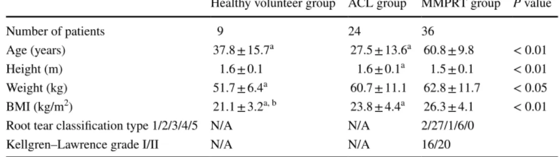
a 0-mm gap. Standard sequences of the Oasis included a sagittal proton density-weighted sequence [repetition time (TR)/echo time (TE), 1718/12] using a driven equilibrium pulse. We used a picture archiving and communication system (FUJIFILM Holdings Corporation, Tokyo, Japan) to measure the MTS and MTPD according to the method described by Hudek et al. [13] and Hashemi et al. [11], as described in detail below. Values were rounded to one decimal place. All measurements were performed by two board-certified orthopaedic surgeons on a best agreement basis. To determine the inter-observer reproducibility, additional measurements were performed independently by the two reviewers on 25 randomly selected MRI scans, and interclass correlation coefficients (ICCs) were calcu- lated. The inter-observer/intra-observer reliabilities for the measurements were satisfactory with mean ICC values of 0.91/0.95 for MTS and 0.89/0.90 for MTPD. MTS was measured on sagittal proton density sequences according to the method described by Hudek et al. [13] (Figs. 1, 2).
This method has been reported to be the most repeatable method to measure sagittal tibial slopes on MRI and is independent of the proximal tibial length [18]. First, the proximal anatomical axis of the tibia was defined on the central sagittal slice in which the attachment of the poste- rior cruciate ligament and the intercondylar eminence were visualized, and both the anterior and posterior tibial cor- tices appeared in a concave shape. Within this slice, two circles were placed into the proximal tibia. The proximal circle was fit within the proximal, anterior, and posterior cortical borders. The distal circle was fit within the ante- rior and posterior cortices with the centre of the circle positioned on the perimeter of the proximal circle. A line drawn through the centres of both circles defined the prox- imal anatomical axis. Then, the sagittal slice showing the mediolateral centres of the medial plateau was identified.
On this image, a tangent to the medial plateau connect- ing the uppermost superior anterior and posterior cortex edges excluding articular cartilage was drawn. MTS was defined as the angle from the orthogonal to the proximal anatomical axis and the tangent to the medial plateau.
Since no clear definition exists for an abnormal LTS [29], only MTS was measured in the present study. MTPD was measured by drawing a line connecting the superior and inferior crests of the tibial plateau in the same plane in which MTS was measured. A line parallel to this line was then drawn tangential to the lowest point of the concavity, representing the lowest boundary of the subchondral bone.
The perpendicular distance between the two lines was then measured and was used to represent MTPD.
The study was approved by the Institutional Review Board at Okayama University (approval no. 1857), and informed consent was provided from all patients and volunteers.
Statistical analysis
Data are reported as mean ± standard deviation. Statistical analysis and sample size/power calculation were performed using EZR software (Saitama Medical Center Jichi Medi- cal University, Tochigi, Japan), which is a graphical user interface for R (The R Foundation for Statistical Comput- ing, Vienna, Austria). Differences in MRI measurements or subjects’ demographics among the three groups were evalu- ated using the Mann–Whitney U test or one-way analysis of variance with the post hoc Tukey HSD test. Statistical significance was set at P < 0.05. Two orthopaedic surgeons independently measured MTS and MTPD. Each observer performed each measurement twice, at 2-week intervals.
Inter-observer and intra-observer reliabilities were assessed using ICC. ICC values > 0.9 were considered excellent, values between 0.8 and 0.9 were considered good, and
Fig. 1 Magnetic resonance images of a normal left knee. a Determi- nation of the tibial axis (long-dashed line) according to the method described by Hudek et al. [13]. The solid line is perpendicular to the tibial axis. b Measurement of the medial tibial slope (1.5°). An angle is formed by the solid line perpendicular to the tibial axis and the dashed line connecting the uppermost superior-anterior and pos- terior cortex edges. c Measurement of the medial tibial plateau depth (3.5 mm). The perpendicular distance was measured from the dashed line to the lowest point of the concavity (dotted line)
values < 0.8 were considered poor. A multivariate logis- tic regression analysis was performed to evaluate the odds ratios of the preoperative prognostic factors using the step- wise method by computation via analysis for MMPRT. To determine the number of test samples, sample size calcula- tion was performed under a significance level of 0.05 and a power of 0.80, and the resulting sample sizes were 6, 8, and 24 in groups V, A, and M, respectively.
Results
The mean age and body mass index (BMI) of the healthy volunteer group (group V) were 37.8 ± 15.7 years and 21.1 ± 3.2 kg/m2. respectively, and those of the ACL recon- struction group (group A) were 27.5 ± 13.6 years and
23.8 ± 4.4 kg/m2, respectively. A significantly higher age and BMI were observed in the MMPRT repair group (group M) than in groups V and A (60.8 ± 9.8 years and 26.3 ± 4.1 kg/
m2, respectively).
The mean MTS in group V was 3.5° ± 1.4° and that in group A was 4.0° ± 1.9°. A significantly higher MTS was observed in group M than in groups V and A (7.2° ± 1.9°).
The mean MTPD in group V was 2.1 ± 0.7 mm and that in group A was 2.2 ± 0.6 mm. A significantly lower MTPD was observed in group M than in groups V and A (1.2 ± 0.5 mm).
In summary, group M showed a significantly steeper MTS and a shallower MTP concavity than did groups V and A.
Group comparisons of MRI measurements are shown in Figs. 3 and 4. In the multivariate logistic regression analysis, BMI, MTS, and MTPD were significant factors associated with MMPRT (Table 2).
Discussion
The most important finding of this study was that a steeper MTS and a shallower MTPD were observed in patients with MMPRT than in healthy volunteers and patients with ACL injuries. To date, many studies about bone morpho- logical factors influencing cartilage, menisci, and ACL have been reported [28]. The geometry of the tibial plateau has a direct influence on the biomechanics of the tibiofem- oral joint in terms of translation, location of instantane- ous centre of rotation, screw-home mechanism, and strain biomechanics of the knee ligament [3, 7]. An important characteristic of the tibial plateau is its posterior slope
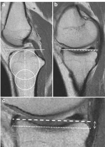
Fig. 2 Magnetic resonance images of a knee with a medial meniscus posterior root tear. a Determination of the tibial axis (long-dashed line) according to the method described by Hudek et al. [13] The solid line is perpendicular to the tibial axis. b Measurement of the medial tibial slope (10°). An angle is formed by the solid line perpen- dicular to the tibial axis and the dashed line connecting the uppermost superior-anterior and posterior cortex edges. c Measurement of the medial tibial plateau depth (0.9 mm). The perpendicular distance was measured from the dashed line to the lowest point of the concavity (dotted line)
0 2 4 6 8 10
Volunteer ACL MMPRT
Medial tibial slope(˚)
* *
Fig. 3 Magnetic resonance image-based medial tibial slope. The medial tibial slope in the medial meniscus posterior root tear (MMPRT) group was significantly steeper than that in the volunteer group and the anterior cruciate ligament (ACL) group. *P < 0.01
(with the anterior elevation being higher than the posterior elevation); when this characteristic is considered in associ- ation with a large compressive joint-reaction force such as that produced during weight-bearing activities, the force may have an anteriorly directed shear force component that acts to produce a corresponding anteriorly directed translation of the tibia [7, 10]. Biomechanical studies have shown that a steep tibial slope leads to increased ante- rior tibial translation under load, which may increase the forces acting on the menisci [1, 7, 10, 20, 21, 25]. Fur- thermore, a biconcave medial tibial plateau was reported to be more frequently associated with a complex medial meniscus tear [2] and shallower MTS and LTS may result in impingement of the posterior horn of the medial menis- cus and anterior horn of the lateral meniscus, respectively, although no significant association was observed between MTPD and meniscal tears [15]. However, only one study has analysed the association between tibial geometry and MMPRT. This study demonstrated that MMPRT and a higher posterior tibial slope were more closely associated
with the development of spontaneous osteonecrosis of the knee than with that of osteoarthritis [31].
In the present study, only women were selected for stand- ardization because women have a higher MTS than men, and MMPRT is more common in women. Subsequently, we evaluated MTS and MTPD using the same method by Hudek et al. and Hashemi et al. [11, 13] because the reproducibility is good [18]. Although our results of MTS and MTPD in groups V and A as the control groups were somewhat differ- ent from those of previous studies that used different meas- urement methods or modalities, such as radiographs or CT [32] [23], they were consistent with the results of previous studies that used the same measurement method [17]. Sig- nificantly steeper MTS and shallower MTP concavity were observed in group M than in group V (P < 0.01) and group A (P < 0.01). Moreover, in the multivariate logistic regression analysis, BMI, MTS, and MTPD were significant factors leading to MMPRT. This was consistent with previous stud- ies showing that MMPRT is more common in middle-aged women with a high BMI [14].
The present findings suggest that biomechanical altera- tion to the posterior rollback of the femur has been likely caused by a steep MTS and a shallow MTPD, resulting in impingement of the posterior horn of the medial meniscus and predisposing it to MMPRT. This association can be well explained by biomechanical and clinical data: from a biomechanical view, the tibial slope produces an anteriorly directed shear force when a compressive load is applied to the knee joint, resulting in an anterior translation of the tibia relative to the femur [1, 7, 10, 20, 21, 25]. However, the biomechanical effect of the tibial slope might be even more complex, as characterization of the tibial plateau surface geometry with a single slope represents an insuf- ficient approximation of its three dimensionality [11]. If knees with similar articular cartilage thickness profiles and meniscal geometries were compared, a deeper MTPD will constrain the femoral condyle to a greater extent and may result in increased resistance to the displacement of the tibia relative to the femur [11]. Conversely, the combina- tion of a steep MTS and a shallow MTPD may be associ- ated with a decreased resistance to the displacement of the tibia relative to the femur, placing the knee at an increased
Medial tibial plateau depth (mm)
* *
0 1 2 3
Volunteer ACL MMPRT
Fig. 4 Magnetic resonance image-based medial tibial plateau depth.
The medial tibial plateau depth in the medial meniscus posterior root tear (MMPRT) group was significantly shallower than that in the volunteer group and the anterior cruciate ligament (ACL) group.
*P < 0.01
Table 2 Multivariate logistic regression analysis
Significant variables (P < 0.05) were calculated using the forward stepwise method to evaluate risk factors leading to MMPRT
CI confidence interval, BMI body mass index, MMPRT medial meniscus posterior root tear, MTS medial tibial slope, MTPD medial tibial plateau depth
Dependent variables Significant variables Odds ratio P value 95% CI Subjects with or without
MMPRT, n = 69 BMI (kg/m2) 1.24 0.03 1.02–1.51
MTS (°) 2.76 < 0.01 1.33–5.73
MTPD (increase of 0.1 mm) 0.63 < 0.01 0.45–0.87
risk for MMPRT. Therefore, we believe that a steep MTS and a shallow MTPD may be anatomic risk factors for MMPRT.
The findings of the present study may have several clinical implications. First, the preoperative diagnosis of MMPRT is difficult, and some of these injuries are missed even on preoperative MRI. The results of the present study suggest that a high index of suspicion is especially neces- sary in patients with a steep MTS and a shallow MTPD detected by plain radiographs, CT, or MRI. Ultimately, better knowledge of the risk factors for MMPRT may increase the diagnostic accuracy because of an increased awareness about this injury.
This study has several limitations, which must be con- sidered when interpreting our results. First, the study design was retrospective, and the number of patients was relatively small; therefore, the validity of our find- ings is limited. However, the sample size had acceptable statistical power for the endpoint. Second, age was not integrated, and only female Asian patients with MMPRT and ACL injuries were evaluated; therefore, the results may not allow generalization to all patients presenting with MMPRT. However, age may not greatly affect the results because each parameter was measured using cortex edges excluding articular cartilage. Third, biomechanical studies were not performed in patients with various MTS or MTPD. Finally, other factors that increase the risk for MMPRT, such as coronal slope or lateral tibial slope, may exist and were not examined in this study.
In daily clinical work, a more conservative rehabilita- tion protocol may be beneficial to avoid excessive load on the repaired structure after MMPRT pullout repair.
Conclusions
A steep MTS and a shallow MTPD are considered ana- tomic risk factors for MMPRT. In MMPRT pullout repair, a more conservative rehabilitation protocol may be ben- eficial to avoid excessive load on the repaired structure.
Acknowledgements We thank Editage (www.edita ge.jp) for English language editing.
Author contributions TF designed the study. YuKo, YuKa, and YuO contributed to the analysis and interpretation of data. All authors have contributed to data collection and interpretation and critically reviewed the manuscript. All authors approved the final version of the manu- script and agreed to be accountable for all aspects of the work in ensur- ing that questions related to the accuracy or integrity of any part of the work are appropriately investigated and resolved.
Funding No funding was received in support of this study.
Compliance with ethical standards
Conflict of interest The authors declare that they have no competing interests.
Ethical approval This study was approved by the International Review Board (IRB) of Okayama University (Ethical approval No. 1857).
References
1. Agneskirchner JD, Hurschler C, Stukenborg-Colsman C, Imhoff AB, Lobenhoffer P (2004) Effect of high tibial flexion osteotomy on cartilage pressure and joint kinematics: a biomechanical study in human cadaveric knees. Winner of the AGA-DonJoy Award 2004. Arch Orthop Trauma Surg 124:575–584
2. Barber FA, Getelman MH, Berry KL (2017) Complex medial meniscus tears are associated with a biconcave medial tibial pla- teau. Arthroscopy 33:783–789
3. Beynnon B, Yu J, Huston D, Fleming B, Johnson R, Haugh L et al (1996) A sagittal plane model of the knee and cruciate liga- ments with application of a sensitivity analysis. J Biomech Eng 118:227–239
4. Chung KS, Ha JK, Ra HJ, Kim JG (2016) A meta-analysis of clini- cal and radiographic outcomes of posterior horn medial meniscus root repairs. Knee Surg Sports Traumatol Arthrosc 24:1455–1468 5. Chung KS, Ha JK, Ra HJ, Kim JG (2016) Prognostic factors in
the midterm results of pullout fixation for posterior root tears of the medial meniscus. Arthroscopy 32:1319–1327
6. Cinotti G, Sessa P, Ragusa G, Ripani FR, Postacchini R, Mas- ciangelo R et al (2013) Influence of cartilage and menisci on the sagittal slope of the tibial plateaus. Clin Anat 26:883–892 7. Dejour H, Bonnin M (1994) Tibial translation after anterior cruci-
ate ligament rupture. Two radiological tests compared. J Bone Jt Surg Br 76:745–749
8. Feucht MJ, Salzmann GM, Bode G, Pestka JM, Kuhle J, Sudkamp NP et al (2015) Posterior root tears of the lateral meniscus. Knee Surg Sports Traumatol Arthrosc 23:119–125
9. Furumatsu T, Okazaki Y, Okazaki Y, Hino T, Kamatsuki Y, Mas- uda S et al (2019) Injury patterns of medial meniscus posterior root tears. Orthop Traumatol Surg Res 105:107–111
10. Giffin JR, Vogrin TM, Zantop T, Woo SL, Harner CD (2004) Effects of increasing tibial slope on the biomechanics of the knee.
Am J Sports Med 32:376–382
11. Hashemi J, Chandrashekar N, Gill B, Beynnon BD, Slauterbeck JR, Schutt RC Jr et al (2008) The geometry of the tibial plateau and its influence on the biomechanics of the tibiofemoral joint. J Bone Jt Surg Am 90:2724–2734
12. Hashemi J, Chandrashekar N, Mansouri H, Gill B, Slauterbeck JR, Schutt RC Jr et al (2010) Shallow medial tibial plateau and steep medial and lateral tibial slopes: new risk factors for anterior cruciate ligament injuries. Am J Sports Med 38:54–62
13. Hudek R, Schmutz S, Regenfelder F, Fuchs B, Koch PP (2009) Novel measurement technique of the tibial slope on conventional MRI. Clin Orthop Relat Res 467:2066–2072
14. Hwang BY, Kim SJ, Lee SW, Lee HE, Lee CK, Hunter DJ et al (2012) Risk factors for medial meniscus posterior root tear. Am J Sports Med 40:1606–1610
15. Khan N, McMahon P, Obaid H (2014) Bony morphology of the knee and non-traumatic meniscal tears: is there a role for meniscal impingement? Skeletal Radiol 43:955–962
16. Kim SB, Ha JK, Lee SW, Kim DW, Shim JC, Kim JG et al (2011) Medial meniscus root tear refixation: comparison of clinical,
radiologic, and arthroscopic findings with medial meniscectomy.
Arthroscopy 27:346–354
17. Kolbe R, Schmidt-Hebbel A, Forkel P, Pogorzelski J, Imhoff AB, Feucht MJ (2018) Steep lateral tibial slope and lateral-to-medial slope asymmetry are risk factors for concomitant posterolateral meniscus root tears in anterior cruciate ligament injuries. Knee Surg Sports Traumatol Arthrosc. https ://doi.org/10.1007/s0016 7-018-5279-6
18. Lipps DB, Wilson AM, Ashton-Miller JA, Wojtys EM (2012) Evaluation of different methods for measuring lateral tibial slope using magnetic resonance imaging. Am J Sports Med 40:2731–2736
19. Luczkiewicz P, Daszkiewicz K, Witkowski W, Chroscielewski J, Zarzycki W (2015) Influence of meniscus shape in the cross sectional plane on the knee contact mechanics. J Biomech 48:1356–1363
20. Marouane H, Shirazi-Adl A, Hashemi J (2015) Quantification of the role of tibial posterior slope in knee joint mechanics and ACL force in simulated gait. J Biomech 48:1899–1905
21. Meyer EG, Haut RC (2005) Excessive compression of the human tibio-femoral joint causes ACL rupture. J Biomech 38:2311–2316 22. Moatshe G, Chahla J, Slette E, Engebretsen L, Laprade RF (2016)
Posterior meniscal root injuries. Acta Orthop 87:452–458 23. Nagai K, Tashiro Y, Herbst E, Gale T, Wang JH, Irrgang JJ et al
(2018) Steeper posterior tibial slope correlates with greater tibial tunnel widening after anterior cruciate ligament reconstruction.
Knee Surg Sports Traumatol Arthrosc 26:3717–3723
24. Okazaki Y, Furumatsu T, Kodama Y, Kamatsuki Y, Masuda S, Ozaki T (2019) Description of a surgical technique of medial meniscus root repair: a fixation technique with two simple stiches under an expected initial tension. Eur J Orthop Surg Traumatol 29:705–709
25. Shelburne KB, Kim HJ, Sterett WI, Pandy MG (2011) Effect of posterior tibial slope on knee biomechanics during functional activity. J Orthop Res 29:223–231
26. Song GY, Liu X, Zhang H, Wang QQ, Zhang J, Li Y et al (2016) Increased medial meniscal slope is associated with greater risk of ramp lesion in noncontact anterior cruciate ligament injury. Am J Sports Med 44:2039–2046
27. Song GY, Zhang H, Zhang J, Liu X, Xue Z, Qian Y et al (2018) Greater static anterior tibial subluxation of the lateral compart- ment after an acute anterior cruciate ligament injury is associ- ated with an increased posterior tibial slope. Am J Sports Med 46:1617–1623
28. Takahashi K, Hashimoto S, Kiuchi S, Watanabe A, Nakamura H, Ikuta F et al (2018) Bone morphological factors influencing cartilage degeneration in the knee. Mod Rheumatol 28:351–357 29. Weinberg DS, Williamson DF, Gebhart JJ, Knapik DM, Voos JE
(2017) Differences in medial and lateral posterior tibial slope: an osteological review of 1090 Tibiae comparing age, sex, and race.
Am J Sports Med 45:106–113
30. Wu J, Huang JM, Zhao B, Cao JG, Chen X (2016) Risk fac- tors comparison for radial and horizontal tears. J Knee Surg 29:679–683
31. Yamagami R, Taketomi S, Inui H, Tahara K, Tanaka S (2017) The role of medial meniscus posterior root tear and proximal tibial morphology in the development of spontaneous osteonecrosis and osteoarthritis of the knee. Knee 24:390–395
32. Zhang Y, Chen Y, Qiang M, Zhang K, Li H, Jiang Y et al (2018) Comparison between three-dimensional CT and conventional radiography in proximal tibia morphology. Medicine (Baltimore) 97:e11632
Publisher’s Note Springer Nature remains neutral with regard to jurisdictional claims in published maps and institutional affiliations.