Endoscopic detection and differentiation of esophageal lesions using a deep neural network
Masayasu Ohmori, MD1,2, Ryu Ishihara, MD, PhD1, Kazuharu Aoyama, PhD3, Kentaro Nakagawa,
MD1, Hiroyoshi Iwagami, MD1, Noriko Matsuura, MD1, Satoki Shichijo, MD, PhD1, Katsumi
Yamamoto, MD, PhD4, Koji Nagaike, MD5, Masanori Nakahara, MD, PhD6, Takuya Inoue, MD,
PhD7, Kenji Aoi, MD8, Hiroyuki Okada, MD, PhD2, Tomohiro Tada, MD, PhD3,9,10,
1 Department of Gastrointestinal Oncology, Osaka International Cancer Institute, Osaka, Japan
2 Department of Gastroenterology and Hepatology, Okayama University Graduate School of
Medicine, Dentistry and Pharmaceutical Sciences, Okayama, Japan
3 AI Medical Service Inc., Tokyo, Japan
4 Department of Gastroenterology, Japan Community Healthcare Organization, Osaka Hospital,
Osaka, Japan
5 Department of Gastroenterology, Suita Municipal Hospital, Osaka, Japan
6 Department of Gastroenterology, Ikeda Municipal Hospital, Osaka, Japan
7 Department of Gastroenterology, Osaka General Medical Center, Osaka, Japan
8 Department of Gastroenterology, Kaiduka City Hospital, Osaka, Japan
9 Tada Tomohiro Institute of Gastroenterology and Proctology, Saitama, Japan
10 Department of Surgical Oncology, Graduate School of Medicine, The University of Tokyo, Tokyo,
Japan
Correspondence to: Ryu Ishihara, M.D., Ph.D.
Department of Gastrointestinal Oncology, Osaka International Cancer Institute, 3-1-69 Otemae,
Chuo-ku, Osaka 541-8567, Japan.
E-mail: [email protected]
ABSTRACT
Background and Aims: Diagnosing esophageal squamous cell carcinoma (SCC) depends on individual
physician expertise and may be subject to interobserver variability. Therefore, we developed a
computerized image-analysis system to detect and differentiate esophageal SCC.
Methods: A total of 9591 non-magnified endoscopic (ME) and 7844 ME images of pathologically-
confirmed superficial esophageal SCCs and 1692 non-ME and 3435 ME images from non-cancerous
lesions or normal esophagus were used as training image data. Validation was performed using 255
non-ME white-light images (WLI), 268 non-ME narrow-band images/blue-laser images (NBI/BLI),
and 204 ME-NBI/BLI images from 135 patients. The same validation test data were diagnosed by 15
board-certified specialists (experienced endoscopists).
Results: Regarding diagnosis by non-ME with NBI/BLI, the sensitivity, specificity, and accuracy were
100%, 63%, and 77%, respectively, for the AI system and 92%, 69%, and 78%, respectively, for the
experienced endoscopists. Regarding diagnosis by non-ME with WLI, the sensitivity, specificity, and
accuracy were 90%, 76%, and 81%, respectively, for the AI system and 87%, 67%, and 75%,
respectively, for the experienced endoscopists. Regarding diagnosis by ME, the sensitivity, specificity,
and accuracy were 98%, 56%, and 77%, respectively, for the AI system and 83%, 70%, and 76%,
respectively for the experienced endoscopists. There was no significant difference in the diagnostic
performance between the AI system and the experienced endoscopists.
Conclusions: Our AI system showed high sensitivity for detecting SCC by non-ME and high accuracy
for differentiating SCC from non-cancerous lesions by ME.
INTRODUCTION
Esophageal cancer is the seventh most common cancer and the sixth most common cause of cancer-
related death, with an estimated 572,000 new cases and 508,000 deaths in 2018 worldwide.1 Although
the incidence of esophageal adenocarcinoma is increasing rapidly in Europe and North America,
squamous cell carcinoma (SCC) remains the most common tumor type accounting for 80% of the all
esophageal cancers.1 The overall survival of patients with advanced esophageal SCC remains poor,
but the prognosis can be favorable if this cancer is detected as a mucosal or submucosal cancer. 2-7
Countermeasures for esophageal SCC have accordingly focused on surveillance endoscopy and early
detection, particularly in high-risk populations.
The current use of white-light imaging (WLI) endoscopy as a screening tool is limited because of
its poor performance in identifying esophageal SCC.8,9 In contrast, equipment-based image-enhanced
endoscopy, represented by narrow-band imaging (NBI) and blue-laser imaging (BLI), is reportedly
useful for the early detection of esophageal SCC.10-16 Although non-magnifying endoscopy (non-ME)
with NBI has demonstrated high sensitivity for the detection of esophageal lesions, its performance in
characterizing these lesions is limited. 13,17 However, recent technological advances have stimulated
the development of numerous optical methods, and image enhancement and ME notably allow the
accurate diagnosis of esophageal SCCs in vivo and are thus referred to as optical biopsy techniques.
Endoscopic optical biopsy has the advantage of providing a noninvasive and real-time diagnosis
without the additional cost of physical biopsy. However magnified observation is complicated,
depends on individual expertise, and may be subject to inter-observer variability.
The application of computer-aided diagnosis represents a potential solution to mitigate both the
variability and complexity of endoscopic diagnosis. Deep learning is a comprehensive term covering
a wide range of machine learning models, typically based on convolutional neural networks (CNNs),
which aim to learn based on multilevel representations of data useful for making classifications. This
technology has demonstrated good performances in visual tasks,18 and has been applied in various
medical fields including skin cancer classification, diagnosis of diabetic retinopathy, and detection of
gastrointestinal cancers.19-22 We therefore aimed to develop a computerized image analysis system
using deep learning for the detection and differentiation of esophageal SCC.
METHODS
Preparation of training data sets
We developed a deep learning-based artificial intelligence (AI) system for the detection and
differentiation of superficial esophageal SCC. The system was trained using endoscopic images
taken during daily endoscopic examinations at Osaka International Cancer Institute. The endoscopic
procedures were carried out using the following equipment: GIF-RQ260Z, GIF-FQ260Z, GIF-
Q240Z, GIF-H290Z, GIF-HQ290, GIF-H260Z, GIF-XP290N, GIF-Q260J, or GIF-H290 (Olympus
Co., Tokyo, Japan) and video processors CV260 (Olympus Co.); EVIS LUCERA CV-260/CLV-260
and EVIS LUCERA ELITE CV-290/CLV-290SL (Olympus Co.); or EG-L590ZW, EG-L600ZW, or
EG-L600ZW7 (Fujifilm Co., Tokyo, Japan) and the video endoscopic system LASEREO (Fujifilm
Co.). A black soft hood was mounted on the tip of the endoscope to maintain an adequate distance
between the tip of the endoscope and the mucosal surface during magnifying observations. The
structure enhancement was set to B-mode level 8 for NBI and level 5-6 for BLI.
Initial routine inspection with non-ME with WLI or NBI/BLI was carried out to detect lesions
suspicious for esophageal SCC. ME with NBI/BLI was subsequently conducted to differentiate the
lesions by evaluating the appearance of the superficial vascular architecture, especially changes in
intrapapillary capillary loops. Iodine staining was then carried out followed by biopsy from
unstained areas. We obtained endoscopic images of superficial esophageal SCCs taken in our facility
between December 2005 and December 2016, and images of non-cancerous lesions or normal
esophagus between January 2009 and January 2019. We excluded the other histological types
because the number of these cancers was too low to educate the AI system. Histologic assessments
of biopsies or resected specimens were conducted according to the Japanese classification of
esophageal cancer.23
The training images included WLI, NBI/BLI, and chromoendoscopic imaging with iodine
solution. Patients with a history of chemotherapy or radiation to the esophagus, lesions adjacent to
the ulcer or ulcer scar, poor-quality images resulting from less insufflation of air, bleeding, halation,
blur, defocus, or mucus were excluded. After selection, 9,591 non-ME and 7,844 ME images from
804 pathologically proven superficial esophageal SCCs, 564 non-ME and 2,744 ME images of non-
cancerous lesions, and 1128 non-ME and 691 ME images of normal esophagus were collected as the
training image data set. These images were saved in JPEG format and marked manually using
rectangular frames by 10 endoscopists. The marking was reconfirmed by a board-certified trainer
(R.I.) at the Japan Gastroenterological Endoscopy Society.
Construction of an AI system
Deep learning is the process of training a neural network (a large mathematical function with
millions of parameters) to perform a given task. The neural network used in this study is a CNN that
uses a function that first combines nearby pixels into local features, then aggregates those features
into global features. The learning process for CNN can be supervised, semi-supervised, or
unsupervised. We used supervised learning, in our study. The mathematical algorithm we used was a
Single Shot MultiBox Detector (https://arxiv.org/abs/1512.02325) that consists of 16 layers.
The CNN was trained by the dataset and was validated using the Caffe deep learning framework,
which is one of the most popular and widely used frameworks, originally developed at the Berkeley
Vision and Learning Center. All the layers of the CNN were fine-tuned from weights from ImageNet
using stochastic gradient descent as a back-propagation method with a global learning rate of 0.0001,
80 epochs, and a batch size of 32. For the dataset, we included endoscopic images from various time
periods, shooting conditions, and resolutions when considering the generalizability of the system.
Evaluation of the AI system
Evaluation of the deep learning-based AI system was conducted using an independent validation
dataset of images of superficial esophageal SCC, non-cancerous lesions, and normal esophageal
mucosa. Images were collected from patients who underwent esophagogastroduodenoscopy at Osaka
International Cancer Institute from January 2017 to December 2018. The inclusion criteria for cancer
images were superficial esophageal SCC treated with endoscopic resection or esophagectomy, with
pathologic proof of SCC. The inclusion criteria for non-cancer images were lesions with pathologic
proof of non-cancer or lesions stained by iodine staining. Non-cancerous lesions included lesions
showing esophagitis, vessel abnormalities, mild pigmentation, glycogenic acanthosis, white spots or
other abnormalities. Barrett’s esophagus lesions were not included in this study. A total of 255 non-
ME WLI images, 268 non-ME NBI/BLI images, and 204 ME NBI/BLI images from 135 patients were
selected for the validation test data. Four to six representative images, one or two non-ME WLI images,
one or two non-ME NBI/BLI images and one or two ME NBI/BLI images per one patient were
selected and subjected to diagnosis by the AI system.
The trained neural network generated a diagnosis of esophageal SCC or non-cancerous lesions, such
as abnormal vessels or esophagitis, with a continuous value between 0 and 1, corresponding to the
probability of that diagnosis. We analyzed the diagnoses using non-ME with WLI, non-ME with
NBI/BLI, and ME with NBI/BLI. If any image out of one or two images of non-ME WLI, non-ME
NBI/BLI, or ME NBI/BLI was judged as cancer by the AI system, it was regarded as cancer. If no
image out of one or two images of non-ME WLI, non-ME NBI/BLI, or ME NBI/BLI was judged as
cancer by the AI system, it was regarded as non-cancer In addition, we also analyzed the diagnosis
together with the diagnostic flow, i.e. initial detection by non-ME followed by differentiation by ME.
Diagnoses by board-certified endoscopic specialists
The performance of the AI system was compared with that of an invited group of 15 board-certified
specialists (experienced endoscopists) at the Japan Gastroenterological Endoscopy Society, with 8-24
years’ expertise as doctors and experience of 2,500-20,000 endoscopic examinations. The experienced
endoscopists had also been conducting preoperative diagnosis and endoscopic resection of
gastrointestinal cancers routinely. They were provided with the same validation test data, including
the NBI, BLI, and WLI images with and without ME as for the AI system, and were asked to mark the
lesions suspicious for SCC based on non-ME images and to diagnose the lesions in the images as SCC
or non-cancer with ME. If any image out of one or two images of non-ME WLI, non-ME NBI/BLI,
or ME NBI/BLI was judged as cancer by the experienced endoscopist, it was regarded as cancer. If no
image out of one or two images of non-ME WLI, non-ME NBI/BLI, or ME NBI/BLI was judged as
cancer by the experienced endoscopist, it was regarded as non-cancer. They were blinded to the results
of the AI diagnoses.
Outcome measures
For the analyses, each image was different and was treated independently. When all images of the
lesion were diagnosed as non-cancer, the lesion was diagnosed as non-cancer, and if some images of
the lesion were diagnosed as cancer, the lesion was diagnosed as cancer. The main outcome measures
were sensitivity, specificity, positive predictive value, negative predictive value, accuracy, and
diagnostic time. These parameters were calculated as follows: sensitivity = correctly diagnosed
cancers/total cancers; specificity = correctly diagnosed non-cancers/total non-cancers; PPV = total
cancers/total lesions diagnosed as cancers; NPV = total non-cancers/total lesions diagnosed as non-
cancers; and accuracy = correctly diagnosed lesions/total lesions. A two-sided McNemar test with
significance level of 0.05 was used to compare differences in accuracy, sensitivity, and accuracy.
Receiver operating characteristic curves were constructed by varying the operating threshold. Inter-
observer variation in the diagnosis of esophageal SCC was assessed using kappa statistics; a kappa-
value > 0.8 denoted almost perfect agreement, 0.8–0.6, substantial agreement; 0.6–0.4, moderate
agreement; 0.4–0.2, fair agreement; and < 0.2, slight agreement. A kappa-value of 0 indicated
agreement equal to chance, and < 0 suggested disagreement.24 We also examined the diagnostic
abilities of three modalities: non-ME with WLI, non-ME with NBI/BLI, and ME with NBI/BLI. All
calculations were performed using EZR version 1.27 (Saitama Medical Center, Jichi Medical
University, Japan).25
Ethics
This study was approved by the Institutional Review Board of Osaka International Cancer Institute
(no.18149).
RESULTS
Performances of the AI system and experienced endoscopists for detection with non-magnified
images
A total 255 non-ME WLI images and 268 non-ME NBI/BLI images from 135 patients were selected
after excluding patients according to the same exclusion criteria as the training data set. The
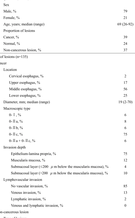
demographics of the selected patients and lesions are summarized in Table 1. The cut-offs for the
diagnosis of cancer by non-ME with WLI and non-ME with NBI/BLI were set at a value of 0.4 for
both. From our previous experience of developing the AI system for endoscopy, the best cut-off value
for cancer ranged from 0.4 to 0.5. In this study, we selected 0.4 because we wanted secure high
sensitivity for diagnosing cancers.
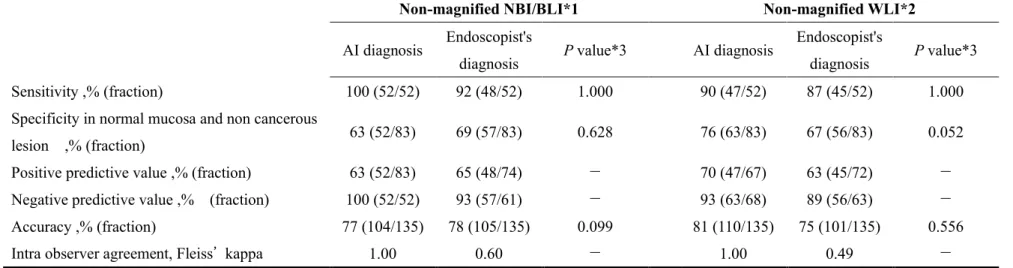
Regarding diagnosis by NBI/BLI, the AI system diagnosed all 52/52 (100%) SCCs as cancers, all
33/33 (100%) normal mucosal areas, and 19/50 (38%) non-cancerous lesions as non-cancers (Figures
1a, 1b, 2a, 2b). The experienced endoscopists diagnosed an average of 48/52 (92%) SCCs as cancers,
31/33 (94%) normal mucosal areas, and 26/50 (52%) non-cancerous lesions as non-cancers (Table 2).
The AI system accurately diagnosed SCC and normal esophagus in all samples, with no false-negative
or -positive results. The sensitivity of the AI system was slightly better than that of the experienced
endoscopists, but its specificity for non-cancerous lesions was lower than that of the experienced
endoscopists, although the difference was not significant by McNemar test (Table 2). The area under
the receiver operating characteristic curve for the validation dataset using non-ME NBI/BLI was 93%
(Figure 3). Interobserver agreement among the experienced endoscopists for detecting SCC was
moderate (Fleiss’ kappa = 0.60, z = 71.2, p < 0.001).
Regarding diagnosis by WLI, the AI system diagnosed 47/52 (90%) SCCs as cancers, 31/33 (94%)
mucosal areas, and 32/50 (64%) non-cancerous lesions as non-cancers (Figures 1c, 1d, 2c, 2d). The
experienced endoscopists diagnosed an average of 45/52 (87%) SCCs as cancers, 27/33 (82%) normal
mucosal areas, and 29/50 (58%) non-cancerous lesions as non-cancers (Table 2). The sensitivity and
specificity of the AI system were both slightly higher than those for the experienced endoscopists,
although the difference was not significant by McNemar test (Table 2). The area under the receiver
operating characteristic curve for the validation dataset using non-ME WLI was 92% (Figure 4).
Interobserver agreement among the experienced endoscopists for detecting SCC was moderate (Fleiss’
kappa = 0.49, z = 58.8, p < 0.001).
Performances of the AI system and experienced endoscopists for differentiation using magnified
images
A total of 204 ME NBI/BLI images from the same patients were selected for the validation test data.
The cut-off for the diagnosis of cancer by ME was set at a value of 0.4. The AI system correctly
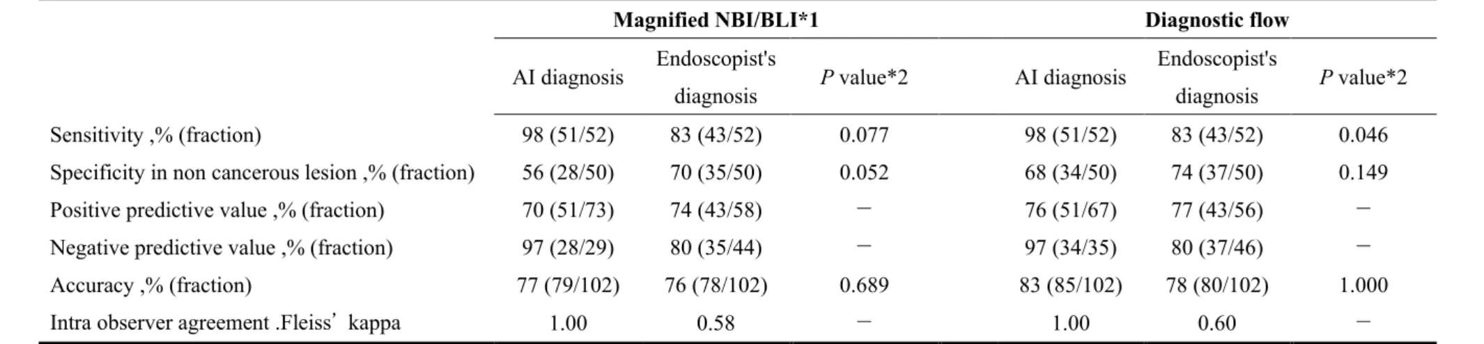
diagnosed 51/52 (98%) SCCs as cancers and 28/50 (56%) non-cancerous lesions as non-cancers
(Figures 1e, 1f, 2e, 2f) (Table 3). The experienced endoscopists diagnosed an average of 43/52 (83%)
SCCs as cancers and 35/50 (70%) non-cancerous lesions as non-cancers (Table 3). The sensitivities,
specificities, and accuracy were 98%, 56%, and 77%, respectively, for the AI system and 83%, 70%,
and 76% respectively, for the experienced endoscopists. There was no significant difference between
the AI system and the experienced endoscopists (Table 3).
Performances of the AI system and experienced endoscopists together with the diagnostic flow
We assume that in clinical practice, the diagnosis of superficial cancer is usually made in a two-step
process consisting of the initial identification of lesions suspicious for cancer (detection) followed by
characterization of the lesions under close examination (differentiation). When diagnosing esophageal
SCC, detection is usually performed using non-ME and differentiation by ME. Using this diagnostic
flow approach (detection followed by differentiation), we evaluated the performance of the AI system
and the experienced endoscopists (Table 3). The AI system diagnosed all the images in 22 seconds,
while experienced endoscopists required an average of 210 minutes (120–600 minutes). All SCCs
were detected by non-ME and 51/52 detected SCCs were correctly diagnosed as SCC by ME using
the AI system, while 50/52 SCCs were detected by non-ME and 43/50 detected SCCs were correctly
diagnosed as SCC by ME by the experienced endoscopists, on average. The sensitivity, specificity,
and accuracy of the diagnosis together with the diagnostic flow, were 98%, 68%, and 83% for the AI
system and 82%, 74%, and 78% for the experienced endoscopists, respectively. There was no
significant difference except for sensitivity between the AI system and the experienced endoscopists
(Table 3).
DISCUSSION
Our AI system showed a good performance for the detection of SCC and its differentiation from
normal mucosa and non-cancerous lesions. The values were comparable to those for experienced
endoscopists. To the best of our knowledge, this AI system is the first to integrate WLI, NBI/BLI,
non-ME, and ME for the detection and differentiation of esophageal SCC.
AI systems have previously been used for the detection of gastrointestinal cancers.21,22 These
reports mainly used cancer and normal images for the validation test, and evaluated the diagnostic
parameters of the AI systems. Using the current validation test with SCC and normal mucosa, our
system showed perfect performance with NBI/BLI, with sensitivity and specificity of 100%. This
performance was better than the sensitivities of 89%–92% and specificity of 44% reported in
previous studies. 21,22 A low specificity for normal esophagus or stomach may increase the need for
further evaluations and biopsies of detected non-cancerous lesions. In the present study, we used
numerous normal esophageal images to educate the AI system, which may have helped to increase
the specificity of the system for normal esophageal mucosa. Considering that most patient
populations who receive endoscopy do not have cancer, the high specificity of the current AI system
may represent a major advantage.
The sensitivities and accuracies of SCC diagnoses by endoscopists and AI systems have been
reported previously.11-16,22,26,27 However, the results of studies regarding the detection and
differentiation of SCC may have a high risk of bias, especially regarding patient selection and it may
therefore not be appropriate to compare the diagnostic accuracy of the current study with these
previous reports. We therefore compared the diagnostic accuracy of our AI system with that of
experienced endoscopists using the same validation test dataset, and found that the AI system had
comparable accuracy to that of the experienced endoscopists. We also assessed the diagnostic
parameters along with the diagnostic flow, including initial detection by non-ME followed by
differentiation by ME, which showed the sensitivity, specificity, and accuracy of the AI system to be
98%, 68%, and 83%, respectively. The utility of the AI system was thus confirmed because its values
were better than those of the experienced endoscopists. In clinical practice, many endoscopic
procedures are performed by endoscopists without sufficient experience to diagnose superficial
esophageal SCC. Considering that the performance for diagnosing superficial esophageal SCC is
influenced by an endoscopist's experience28, this AI system may be a good supporting tool. Given
the speed of AI the confirmatory assessment would add very little time to the evaluation.
Our system comprised non-ME and ME, and was trained on 9,591 non-ME and 7,844 ME images
of SCCs, and 564 non-ME and 2744 ME images of non-cancers. Non-ME systems are designed to
have high sensitivity to detect all lesions suspicious for SCC by training the system mainly using
SCC images, and the high sensitivity of non-ME can minimize undetected errors. ME systems are
designed to have high accuracy for differentiating between SCC and non-cancerous lesions by
training the system using both SCC and non-cancer images. The high accuracy of ME may thus
realize a real-time diagnosis and thus reduce the number and cost of biopsies. Although ME can
realize the real-time diagnosis of SCC, its availability worldwide is limited; however, most images
used in this study were mildly magnified, and such images can be obtained by dual-focus endoscopy
(GIF-HQ190, GIF-HQ290) or endoscopy with close focus (GIF-H170, GIF-H180, GIF-H180J, GIF-
H185, GIF-H190, GIF-2TH180, GIF-1TH190). In addition, our AI system also showed high
performance for detecting SCC by non-ME with NBI/BLI and WLI, which can be applied for
differentiating between SCC and non-cancerous lesions by changing the threshold value of the AI
system. Non-ME with WLI is a conventional and common endoscopic imaging modality available
worldwide, suggesting that the current AI system could be widely applicable.
This study has some limitations, including its retrospective nature using still images and
validation using high-quality images. Our AI system analyzed at a speed of 36 images/second, which
is adequate for analyzing video images, suggesting that real-time detection and differentiation of
SCC will be possible in the near future. Another concern is the applicability of our AI system to
general practice. To improve the general applicability of the AI system, we included images of
various quality and resolution taken from 2005 to 2019 using different endoscopy systems. On the
other hand, we excluded poor-quality images and images of rare cancers such as cancer after
chemoradiation. These rare cancers constitute a very small proportion of all esophageal lesions in
our daily practice and thus, excluding such cancers likely had little effect on the general applicability
of the AI system. A broader ability of the AI system could be achieved using poor-quality images in
the training process, but this could impair the accuracy of the system. Therefore, we believe that the
usefulness of the system to evaluate poor-quality images should be considered after confirming its
performance with large numbers of high-quality images. The use of selected images for the
validation test represents another limitation of this study. However, extracting adequate images and
controlling the number of images were essential to ensure that they could also be feasibility
evaluated by the experienced endoscopists, who were required to evaluate and mark up to 600
images, even after image extraction. As a future study, the performance of the AI system should be
evaluated using real-time videos.
In conclusion, our novel AI system showed high sensitivity for the detection of SCC by non-ME
and high accuracy for differentiating SCC from non-cancerous lesions by ME. This system may thus
provide valuable support for many endoscopists.
Acknowledgements
We thank S. Hiyama (Japan Community Healthcare Organization, Osaka Hospital), T. Kannno (Japan
Community Healthcare Organization, Osaka Hospital), Y. Onishi (Japan Community Healthcare
Organization, Osaka Hospital) M. Sawamura (Japan Community Healthcare Organization, Osaka
Hospital), K. Nagai (Suita Municipal Hospital), N. Dan (Suita Municipal Hospital), Y. Matsumoto
(Ikeda Municipal Hospital), Y. Yamaguchi (Ikeda Municipal Hospital), Y. Masuda (Ikeda Municipal
Hospital), and S. Kawai (Osaka General Medical Center), and Y. Kakita (Kaiduka City Hospital) for
acting as experienced endoscopists. We also thank to M. Kono, H. Fukuda, Y. Shimamoto, T. Iwatsubo,
K. Matsuno, S. Inoue, H. Nakahira, A. Maekawa, T. Kanesaka, from Osaka International Cancer
Institute for data collection, and Susan Furness, PhD, from Edanz Group (www.edanzediting.com/ac)
for editing a draft of this manuscript.
Specific author contributions
Study concept and design (M.O. and R.I.); endoscopic specialists (KY, KN, MN, TI, and KA);
acquisition of data (MO, RI, KN, HI, NM and SS); analysis and interpretation of data (MO, RI, KA,
and TT); drafting of the manuscript (MO, RI, and KA) ; critical review of the article (HO). All authors
have approved the final draft submitted.
Potential competing interests: None.
Financial support: None.
REFERENCES
1. Ferlay J, Colombet M, Soerjomataram I, et al. Estimating the global cancer incidence and
mortality in 2018: GLOBOCAN sources and methods. Int J Cancer 2019;144:1941-53. DOI:
10.1002/ijc.31937
2. Kodama M, Kakegawa T. Treatment of superficial cancer of the esophagus: a summary of
responses to a questionnaire on superficial cancer of the esophagus in Japan. Surgery 1998;123:432-
9.
3. Yamashina T, Ishihara R, Nagai K, et al. Long-term outcome and metastatic risk after endoscopic
resection of superficial esophageal squamous cell carcinoma. Am J Gastroenterol 2013;108:544-51.
DOI: 10.1038/ajg.2013.8
4. Katada C, Muto M, Momma K, et al. Clinical outcome after endoscopic mucosal resection for
esophageal squamous cell carcinoma invading the muscularis mucosae-a multicenter retrospective
cohort study. Endoscopy 2007;39:779-83. DOI: 10.1055/s-2007-966761
5. Shimizu Y, Tsukagoshi H, Fujita M, et al. Long-term outcome after endoscopic mucosal resection
in patients with esophageal squamous cell carcinoma invading the muscularis mucosae or deeper.
Gastrointest Endosc 2002;56:387-90.
6. Igaki H, Kato H, Tachimori Y, et al. Clinicopathologic characteristics and survival of patients with
clinical Stage I squamous cell carcinomas of the thoracic esophagus treated with three-field lymph
node dissection. Eur J Cardiothorac Surg 2001;20:1089-94.
7. Yamamoto S, Ishihara R, Motoori M, et al. Comparison between definitive chemoradiotherapy
and esophagectomy in patients with clinical stage I esophageal squamous cell carcinoma. Am J
Gastroenterol 2011;106:1048-54. DOI: 10.1038/ajg.2011.42
8. Hashimoto CL, Iriya K, Baba ER, et al. Lugol's dye spray chromoendoscopy establishes early
diagnosis of esophageal cancer in patients with primary head and neck cancer. Am J Gastroenterol
2005;100:275-82. DOI: 10.1111/j.1572-0241.2005.30189.x
9. Dawsey SM, Fleischer DE, Wang GQ, et al. Mucosal iodine staining improves endoscopic
visualization of squamous dysplasia and squamous cell carcinoma of the esophagus in Linxian,
China. Cancer 1998;83:220-31.
10. Gono K, Obi T, Yamaguchi M, et al. Appearance of enhanced tissue features in narrow-band
endoscopic imaging. J Biomed Opt 2004;9:568-77. DOI: 10.1117/1.1695563
11. Muto M, Minashi K, Yano T, et al. Early detection of superficial squamous cell carcinoma in
the head and neck region and esophagus by narrow band imaging: a multicenter randomized
controlled trial. J Clin Oncol 2010;28:1566-72. DOI: 10.1200/JCO.2009.25.4680
12. Takenaka R, Kawahara Y, Okada H, et al. Narrow-band imaging provides reliable screening for
esophageal malignancy in patients with head and neck cancers. Am J Gastroenterol 2009;104:2942-
8. DOI: 10.1038/ajg.2009.426
13. Ishihara R, Takeuchi Y, Chatani R, et al. Prospective evaluation of narrow-band imaging
endoscopy for screening of esophageal squamous mucosal high-grade neoplasia in experienced and
less experienced endoscopists. Dis Esophagus 2010;23:480-6. DOI: 10.1111/j.1442-
2050.2009.01039.xka
14. Kaneko K, Oono Y, Yano T, et al. Effect of novel bright image enhanced endoscopy using blue
laser imaging (BLI). Endosc Int Open 2014;2:E212-9. DOI: 10.1055/s-0034-1390707
15. Tomie A, Dohi O, Yagi N, et al. Blue Laser Imaging-Bright Improves Endoscopic Recognition
of Superficial Esophageal Squamous Cell Carcinoma. Gastroenterol Res Pract 2016;2016:6140854.
DOI: 10.1155/2016/6140854
16. Morita FH, Bernardo WM, Ide E, et al. Narrow band imaging versus lugol chromoendoscopy to
diagnose squamous cell carcinoma of the esophagus: a systematic review and meta-analysis. BMC
Cancer 2017;17:54. DOI: 10.1186/s12885-016-3011-9
17. Nagami Y, Tominaga K, Machida H, et al. Usefulness of non-magnifying narrow-band imaging
in screening of early esophageal squamous cell carcinoma: a prospective comparative study using
propensity score matching. Am J Gastroenterol 2014;109:845-54. DOI: 10.1038/ajg.2014.94
18. Russakovsky O, Deng J, Su H, et al. ImageNet Large Scale Visual Recognition Challenge.
International Journal of Computer Vision 2015;115:211-52. DOI: 10.1007/s11263-015-0816-y
19. Esteva A, Kuprel B, Novoa RA, et al. Dermatologist-level classification of skin cancer with
deep neural networks. Nature 2017;542:115-118. DOI: 10.1038/nature21056
20. Gulshan V, Peng L, Coram M, et al. Development and Validation of a Deep Learning Algorithm
for Detection of Diabetic Retinopathy in Retinal Fundus Photographs. JAMA 2016;316:2402-10.
DOI: 10.1001/jama.2016.17216
21. Hirasawa T, Aoyama K, Tanimoto T, et al. Application of artificial intelligence using a
convolutional neural network for detecting gastric cancer in endoscopic images. Gastric Cancer
2018;21:653-660. DOI: 10.1007/s10120-018-0793-2
22. Horie Y, Yoshio T, Aoyama K, et al. Diagnostic outcomes of esophageal cancer by artificial
intelligence using convolutional neural networks. Gastrointest Endosc 2019;89:25-32. DOI:
10.1016/j.gie.2018.07.037
23. Society JE. Japanese Classification of Esophageal Cancer, tenth edition: part I. Esophagus
2009;6:1-25. DOI: 10.1007/s10388-009-0169-0
24. Landis JR, Koch GG. The measurement of observer agreement for categorical data. Biometrics
1977;33:159-74.
25. Kanda Y. Investigation of the freely available easy-to-use software 'EZR' for medical statistics.
Bone Marrow Transplant 2013;48:452-8. DOI: 10.1038/bmt.2012.244
26. Zhao YY, Xue DX, Wang YL, et al. Computer-assisted diagnosis of early esophageal squamous
cell carcinoma using narrow-band imaging magnifying endoscopy. Endoscopy 2019;51:333-41.
DOI: 10.1055/a-0756-8754
27. Kumagai Y, Takubo K, Kawada K, et al. Diagnosis using deep-learning artificial intelligence
based on the endocytoscopic observation of the esophagus. Esophagus 2019;16:180-7. DOI:
10.1007/s10388-018-0651-7
28. Ishihara R, Takeuchi Y, Chatani R, et al. Prospective evaluation of narrow-band imaging
endoscopy for screening of esophageal squamous mucosal high-grade neoplasia in experienced and
less experienced endoscopists. Dis Esophagus. 2010;23:480-6. DOI: 10.1111/j.1442-
2050.2009.01039.x.
Table 1 Demographics of selected patients and lesions Data of patients (n=135)
Sex
Male, % 79
Female, % 21
Age, years; median (range) 69 (26-92)
Proportion of lesions
Cancer, % 39
Normal, % 24
Non-cancerous lesion, % 37
Data of lesions (n=135) Cancer
Location
Cervical esophagus, % 2
Upper esophagus, % 17
Middle esophagus, % 56
Lower esophagus, % 25
Diameter, mm; median (range) 19 (2-70)
Macroscopic type
0-Ⅰ, % 6
0-Ⅱa, % 8
0-Ⅱb, % 6
0-Ⅱc, % 75
0-Ⅱa + 0-Ⅱc, % 6
Invasion depth
Epithelium-lamina propria, % 75
Muscularis mucosa, % 12
Submucosal layer (≤200 μm below the muscularis mucosa), % 4 Submucosal layer (>200 μm below the muscularis mucosa), % 10 Lymphovascular invasion
No vascular invasion, % 85
Venous invasion, % 13
Lymphatic invasion, % 2
Venous and lymphatic invasion, % 0
Non-cancerous lesion Type of lesions
Gastro esophageal reflux disease, % 6
Vessel abnormality, % 78
White spot, % 4
Other, % 12
Table 2 Diagnostic performance for detection with non-magnified images
Non-magnified NBI/BLI*1 Non-magnified WLI*2
AI diagnosis Endoscopist's
diagnosis P value*3 AI diagnosis Endoscopist's
diagnosis P value*3
Sensitivity ,% (fraction) 100 (52/52) 92 (48/52) 1.000 90 (47/52) 87 (45/52) 1.000
Specificity in normal mucosa and non cancerous
lesion ,% (fraction) 63 (52/83) 69 (57/83) 0.628 76 (63/83) 67 (56/83) 0.052
Positive predictive value ,% (fraction) 63 (52/83) 65 (48/74) − 70 (47/67) 63 (45/72) −
Negative predictive value ,% (fraction) 100 (52/52) 93 (57/61) − 93 (63/68) 89 (56/63) −
Accuracy ,% (fraction) 77 (104/135) 78 (105/135) 0.099 81 (110/135) 75 (101/135) 0.556
Intra observer agreement, Fleiss’ kappa 1.00 0.60 − 1.00 0.49 −
*1 NBI: Narrow-band imaging, BLI: blue-laser imaging, *2 WLI: White-light imaging.
*3 Comparison between AI diagnosis and exerienced endoscopists' diagnosis by a majority (McNemar test).
Table 3 Diagnostic performance for differentiation with magnified images
Magnified NBI/BLI*1 Diagnostic flow
AI diagnosis Endoscopist's
diagnosis P value*2 AI diagnosis Endoscopist's
diagnosis P value*2
Sensitivity ,% (fraction) 98 (51/52) 83 (43/52) 0.077 98 (51/52) 83 (43/52) 0.046
Specificity in non cancerous lesion ,% (fraction) 56 (28/50) 70 (35/50) 0.052 68 (34/50) 74 (37/50) 0.149
Positive predictive value ,% (fraction) 70 (51/73) 74 (43/58) − 76 (51/67) 77 (43/56) −
Negative predictive value ,% (fraction) 97 (28/29) 80 (35/44) − 97 (34/35) 80 (37/46) −
Accuracy ,% (fraction) 77 (79/102) 76 (78/102) 0.689 83 (85/102) 78 (80/102) 1.000
Intra observer agreement .Fleiss’ kappa 1.00 0.58 − 1.00 0.60 −
*1 NBI: Narrow-band imaging, BLI: blue-laser imaging imaging.
*2 Comparison between AI diagnosis and exerienced endoscopists' diagnosis by a majority (McNemar test).
Figure legends
Figure 1. a. A case of cancer in the mid-esophagus with non-magnified narrow-band imaging. b. The
AI system correctly detected the lesion by indicating it with a square frame. c. Same case of cancer in
the mid-esophagus with white-light imaging. d. The AI system correctly detected the lesion by
indicating it with a square frame. e. Same case of cancer in the mid-esophagus with magnified narrow-
band imaging. f. The AI system correctly differentiated the lesion as a cancer.
Figure 2. a. A case of lesion in the mid-esophagus with non-magnified narrow-band imaging. b. The
AI system detected the lesion by indicating it with a square frame. c. Same case of non-cancerous
lesion in the mid-esophagus with white-light imaging. d. The AI system detected the lesion by
indicating it with a square frame. e. Same case of non-cancer in the mid-esophagus with magnified
narrow-band imaging. f. The AI system correctly differentiated the lesion as a non-cancer.
Figure 3. Receiver operating characteristic curve for the validation dataset using non-magnified
narrow-band images/blue laser images. The area under the curve was 93%.
Figure 4. Receiver operating characteristic curve for the validation dataset using non-magnified white
light images. The area under the curve was 92%.
Fig.1
Fig.2
Fig.3
Fig.4