Structural Basis of the Sec Translocon and YidC Revealed Through X-ray Crystallography
26
0
0
全文
(2) Crystal structures of Sec translocon and YidC 11. Tsukazaki. Abstract. 12. Protein translocation and membrane integration are fundamental, conserved processes. After or during. 13. ribosomal protein synthesis, precursor proteins containing an N-terminal signal sequence are directed to a. 14. conserved membrane protein complex called the Sec translocon (also known as the Sec translocase) in the. 15. endoplasmic reticulum membrane in eukaryotic cells, or the cytoplasmic membrane in bacteria. The Sec. 16. translocon comprises the Sec61 complex in eukaryotic cells, or the SecY complex in bacteria, and mediates. 17. translocation of substrate proteins across/into the membrane. Several membrane proteins are associated with. 18. the Sec translocon. In Escherichia coli, the membrane protein YidC functions not only as a chaperone for. 19. membrane protein biogenesis along with the Sec translocon, but also as an independent membrane protein. 20. insertase. To understand the molecular mechanism underlying these dynamic processes at the membrane,. 21. high-resolution structural models of these proteins are needed. This review focuses on X-ray crystallographic. 22. analyses of the Sec translocon and YidC and discusses the structural basis for protein translocation and. 23. integration.. 24 25. Key words. 26. protein translocation. 27. protein insertion. 28. X-ray crystallography. 29. membrane protein. 30. Sec translocon. 31. 2.
(3) Crystal structures of Sec translocon and YidC 32. Tsukazaki. Introduction. 33. Membrane or secretory proteins are synthesized by cytoplasmic ribosomes, and their nascent. 34. polypeptides possess a specific membrane-targeted signal sequence for transport across or integration into the. 35. membrane. The transported polypeptides then fold into mature proteins and function at the appropriate. 36. locations. The membrane restricts the passive diffusion of small molecules and ions across the membrane. To. 37. overcome the membrane permeability barrier and environmental factors such as the pH or salt concentration,. 38. sophisticated machineries present in the membrane enable protein translocation across and integration into. 39. the membrane. The Sec translocon is an evolutionarily conserved protein-conducting channel at the. 40. endoplasmic reticulum (ER) membrane of eukaryotic cells, or the cytoplasmic membrane of bacteria and. 41. archaea (Fig. 1A)1,2. The Sec translocon is an essential hetero-ternary protein complex, comprising membrane. 42. proteins Sec61α/γ/β in eukaryotic cells or SecY/E/G in bacteria (Fig. 1B), and is involved in translocation and. 43. integration of nascent ribosomally synthesized, unfolded proteins, in a signal sequence-dependent manner. In. 44. the case of soluble proteins, the N-terminal membrane-targeting signal sequences of precursor proteins are. 45. cleaved during translocation, thus decreasing the size of the mature proteins3. In many membrane proteins,. 46. the first transmembrane region contains the targeting signal and is not cleaved. As shown in Figure 1, protein. 47. translocation via the Sec translocon is classified into co-translational and post-translational translocations.. 48. The basic mechanism of co-translational translocation in bacteria and eukaryotic cells is the same. During. 49. co-translational translocation, the highly hydrophobic signal sequence emerging from the ribosomal exit. 50. tunnel is recognized by the signal-recognition particle (SRP) and is targeted to the membrane. 51. co-translationally, along with the ribosome, owing to interactions between the SRP and the SRP receptor. 52. present in the membrane4. Thereafter, the ribosome directly interacts with the protein-conducting channel (i.e.,. 53. the Sec translocon), which is present in the membrane. Subsequently, protein translocation occurs. 54. simultaneously with protein translation. During post-translational translocation, unfolded proteins are targeted. 55. to the membranes. The Sec62/63 complex and BiP protein are involved in this translocation in eukaryotic. 56. cells. BiP proteins drive protein translocation via a ratchet mechanism, which can move in only one direction. 57. owing to substrate interactions and conformational transitions of BiP by ATP hydrolysis5. In Escherichia coli,. 3.
(4) Crystal structures of Sec translocon and YidC. Tsukazaki. 58. precursor proteins, maintained in an unfolded state by chaperones such as SecB, are directed to the membrane. 59. where the SecA ATPase drives protein translocation6,7, although many chaperones including SecB are not. 60. essential for the viability of E. coli. SecA repeatedly undergoes conformational changes to move the precursor. 61. protein into the Sec translocon, using energy from ATP hydrolysis8. Because the Sec translocon itself is a. 62. passive protein channel, other factors, including Sec62/63, SecA, and BiP, play indispensable roles in protein. 63. translocation, as described above. Data from an electron microscopy study demonstrated that the eukaryotic. 64. Sec translocon is associated with translocon-associated proteins (TRAPs) and the oligosaccharyl-transferase. 65. (OST)9,10. The cryo-electron microscopic density map of a TRAP showed it protruding into the ER space,. 66. probably interacting with the substrate protein during protein translocation. In bacteria and archaea, the. 67. SecD–SecF complex (SecDF), which interacts with Sec YEG, promotes protein translocation11,12. SecDF. 68. repeatedly undergoes drastic conformational changes with the substrate at the trans side of the plasma. 69. membrane using proton motive force, which promotes substrate release into the trans-side space13-15. SecDF. 70. can drive protein translocation at the trans side of the plasma membrane independently of SecA13. Hence,. 71. SecDF is considered a second protein-translocation motor. The bacterial Sec translocon is involved in. 72. membrane protein sorting during co-translational translocation in collaboration with YidC, a membrane. 73. protein16,17. YidC functions as a chaperone, facilitating co-translational membrane protein folding. Moreover,. 74. YidC directly binds to the ribosome and is responsible for membrane insertion of certain single- and. 75. double-spanning membrane proteins. Additionally, MPIase (membrane protein integrase), a glycolipozyme,. 76. also plays an essential role in membrane protein insertion, before YidC inserts proteins into the membrane18,19.. 77. YidC corresponds to plant Alb3 in the thylakoid membrane and eukaryote Oxa1 in the inner mitochondrial. 78. membrane (Fig. 1C). The YidC/Oxa1/Alb3 family proteins, containing five conserved transmembrane. 79. α-helices, are involved in membrane protein insertion and assembly of the respiratory chain-related complex20.. 80. YidC of gram-negative bacteria possesses additional transmembrane and periplasmic regions (P1) at its. 81. N-terminus. This review is focused on studies of the crystal structures of the Sec translocon and YidC, aimed. 82. at elucidating the mechanisms underlying protein transport across/into the membrane at the atomic level, and. 83. provides a detailed and comprehensive description of these membrane proteins.. 4.
(5) Crystal structures of Sec translocon and YidC. Tsukazaki. 84 85. Architecture of Sec translocon. 86. Molecular modeling based on the first reported crystal structure of the Sec translocon in 2004 (Fig.. 87. 2A left, B)21 provided many insights into the mechanisms of protein translocation across and integration into. 88. the membrane, via the Sec translocon. Subsequently, different types of functional analyses were performed to. 89. elucidate the molecular mechanism underlying the action of Sec translocon1,2,22. The available crystal. 90. structure models of Sec translocons in the Protein Data Bank (PDB)21,23-28 are summarized in Table 1. The. 91. first reported structure of the Sec translocon is from an archaeon, Methanocaldococcus jannaschii21, and is. 92. designated as SecYEβ in this review. As of March 20, 2019, the highest-resolution structure of the Sec. 93. translocon was reported in 2014 for Thermus thermophilus SecYEG (2.7-Å resolution; Fig. 2A, right)27.. 94. Some reports do not include SecG/β, a non-essential subunit29-32, because it easily dissociates from the. 95. essential SecYE complex and does not influence its stability. Purified T. thermophilus SecYEG, generated by. 96. overexpressing SecG with an additional plasmid in SecYEG-overexpressing cells, was crystallized in the. 97. lipidic cubic phase, facilitating determination of a high-resolution structure of the Sec translocon27. Both the. 98. M. jannaschii SecYEβ and T. thermophilus SecYEG crystal structures represent the resting states of the Sec. 99. translocon (Fig. 2A, B). Ten transmembrane α-helices of SecY compose the core of the Sec translocon, which. 100. is stabilized by the cytoplasmic α-helix parallel to the membrane and the tilted transmembrane α-helix of. 101. single-membrane-spanning SecE (triple membrane-spanning, in the case of E. coli). SecG, containing two. 102. transmembrane α-helices, is peripherally located adjacent to transmembrane region 1 (TM1) and TM4 of. 103. SecY. The single-membrane-spanning protein, Secβ, an alternative component of SecG, is located in a. 104. position similar to that of SecG. The N-terminal and C-terminal halves, TM1–5 and TM6–10, respectively,. 105. are arranged in a pseudosymmetrical manner and linked by a cytoplasmic loop, called a hinge. The protruding. 106. cytoplasmic region 4 (C4) between the TM6–7 regions and C5 between the TM8–9 regions provide major. 107. interaction sites for cytosolic factors, including SecA and ribosomes. The interior channel of SecY is. 108. hourglass-shaped, its center containing a constricted region called the pore ring (Fig. 2C). The narrow point is. 109. formed by six hydrophobic amino acid residues, primarily including Ile, at the middle regions of TM2, 5, 7,. 5.
(6) Crystal structures of Sec translocon and YidC. Tsukazaki. 110. and 10, and does not permit secretion of substrate proteins via the Sec channel, based on the crystal structures.. 111. Furthermore, the trans-side funnel of the hourglass-shaped space is occupied at the exterior side by a short. 112. α-helix, called a plug, between TM1 and TM2, resulting in a completely sealed SecY channel. Although. 113. previous structural studies on the Sec translocon revealed that the cytoplasmic funnel of SecY is not occupied,. 114. Tanaka et al. reported a high-resolution structure of SecYEG wherein the cytoplasmic loop of SecG covers. 115. the cytoplasmic side of the channel27, thus restricting membrane permeability in a manner similar to that of. 116. the plug domain (Fig. 2D). Therefore, the SecG loop can function as a cytoplasmic cap for the SecY channel.. 117. The cytoplasmic N-terminal region of Secβ is disordered in structural models; however, it could be located. 118. near the cytoplasmic funnel in the resting state, similar to the SecG loop. The mechanism underlying this. 119. covering process from each side of the pore ring may be universally conserved. The boundary area between. 120. TM1–5 and TM6–10 of SecY on the opposite side of the tilted SecE transmembrane α-helix is called a lateral. 121. gate, comprising TM2, 3, 7, and 8, which are binding sites for the signal sequences33. The Sec translocon in. 122. the resting states is not wide enough for protein transport. Therefore, the pore ring, plug, cap, and lateral gate. 123. regions have been predicted to undergo conformational changes and/or are dislocated, thereby enabling. 124. protein translocation across and integration into the membrane via the Sec translocon. The variable models of. 125. the Sec translocon have been experimentally verified27,34-37. Recent structural X-ray crystallographic21,23-28,. 126. electron microscopic38-44, and functional45,46 analyses strongly suggested that the oligomeric state of the Sec. 127. translocon is one heterotrimeric unit, although an efficient functional state comprising two or more units. 128. cannot be excluded47,48. Several crystal structures of the SecY complex imply that interactions between SecA,. 129. Fab, or a peptide mimicking a part of the signal peptide and the protruded cytoplasmic regions of SecY. 130. (which are intrinsically flexible) induce conformational changes in the lateral gate (Fig. 2E). Similar to these. 131. crystal structures, binding of cytosolic factors to the Sec translocon would trigger structural changes to easily. 132. interact with precursor proteins initially during protein translocation. In the SecA-bound conformation of. 133. SecY, the plug domain is dislocated outwards, thereby expanding the inner space of SecY. This structural. 134. change may lower the energy barrier to protein translocation via SecY.. 135. 6.
(7) Crystal structures of Sec translocon and YidC 136. Tsukazaki. Sec translocon in the protein translocation state. 137. An outstanding report shows the crystal structure of SecYE and precursor segment-inserted SecA at. 138. 3.7-Å resolution (Fig. 3A, B)26. For the structural analysis, the polypeptide was artificially introduced into a. 139. loop of SecA as a fusion protein, accompanied by the generation of an intermolecular disulfide bond between. 140. the peptide and SecY at the trans side of the plasma membrane to stabilize the protein-translocation. 141. intermediate. The lateral gate is the most widely open in the available crystal structures. The signal peptide of. 142. the substrate is located at the expanded lateral gate, surrounded by TM2, 3, and 7, presumably oriented. 143. toward the hydrophobic regions of the lipid bilayer. During insertion, the signal peptide can be laterally. 144. released from the expanded gate to the membrane via hydrophobic interactions. The part of the substrate. 145. being transported is located along the center of the Sec translocon, and the pore ring is larger than that in. 146. other crystal structures. Four of six residues of the pore were found to tightly interact with the transported. 147. peptide, simultaneously blocking membrane permeability like a gasket. Hence, even during protein. 148. translocation, SecY can maintain the membrane barrier simultaneously.. 149 150. Recent structural studies of Sec translocon. 151. Recent structural studies on the ribosome–Sec translocon complex by electron microscopy at medium. 152. resolution revealed densities corresponding to the α-helices and conformational changes in transmembrane. 153. regions and the localization of precursor proteins, providing insights into protein translocation across and. 154. integration into the membrane38-44. Furthermore, samples can be directly observed by electron microscopy. 155. without crystallization steps, which are needed for X-ray crystallography. Electron microscopic images of the. 156. ribosome–nascent chain complex (RNC) probably include various intermediate states of SecY complexes,. 157. thus providing several snapshots of co-translational translocation. The cryo-electron microscopic structure of. 158. Sec61 and RNC, including a signal peptide, elucidated that the signal peptide is observed in a manner similar. 159. to that of the SecYE–SecA–signal peptide complex (Fig. 3C, left)42. A different cryo-electron microscopic. 160. imaging analysis of SecY and RNC, including two newly synthesized transmembrane α-helices, revealed that. 161. two transmembrane regions are peripherally located near the lateral gate of SecY (Fig. 3C, right)38. This. 7.
(8) Crystal structures of Sec translocon and YidC. Tsukazaki. 162. structure is considered to represent the intermediate state after the substrate is sorted into the membrane via. 163. the Sec translocon. Although the electron density of the translocating peptide was unclear upon electron. 164. microscopic analyses, probably owing to its unfolded conformation, polypeptides are thought to traverse the. 165. central pore of the Sec translocon. Cryo-electron microscopic analysis has revealed several snapshots of the. 166. active Sec translocon during protein translocation. Because the Sec translocon contains highly motile regions,. 167. including the plug, cap, and cytoplasmic regions, we cannot accurately refine the structural models of the Sec. 168. translocon at atomic resolution using the current electron density data at limited resolution, thus preventing. 169. an accurate understanding of the transition states of the Sec translocon. Future structural analyses are required. 170. at a higher resolution. Highly flexible regions may not be visible even in high-resolution structures. 171. determined by X-ray crystallography, but cryo-electron microscopic analysis may elucidate several forms of. 172. such flexible regions because recently developed programs can analyze several states separately. Structural. 173. studies of the Sec translocon have indicated that the passive Sec translocon has a flexible structure, which. 174. appropriately changes to direct the transportation of proteins to the trans side of the plasma membrane or into. 175. the membrane, in response to interactions with cytosolic factors and precursor proteins. The fundamental. 176. concepts underlying transitions of the Sec translocon (including changes in pore size, opening and closing the. 177. lateral gate, and plug dislocation) were reported with the first crystal structure of the Sec translocon21 and. 178. have been supported by structure-based functional studies for more than a decade1.. 179 180. Overall structures of YidC. 181. The YidC core comprises five conserved transmembrane α-helices (cTM1–5) (Fig. 1C). The. 182. arrangements of the transmembrane α-helices of YidC were predicted based on an evolutionary co-variation. 183. analysis49; however, the detailed interactions, arrangements, and tilting angles of the transmembrane regions. 184. remain unknown. Crystal structures of YidC derived from three species were published (Table 1) in the past. 185. five years50-53. All reported crystal structures for YidC displayed monomeric states (Fig. 4A–C), concurrent. 186. with recent functional and structural reports that YidC functions as a monomer54-56, although functional. 187. dimeric states of YidC have been previously proposed57,58. The first transmembrane α-helix (1st TM) of E.. 8.
(9) Crystal structures of Sec translocon and YidC. Tsukazaki. 188. coli YidC, which functions as a signal sequence, was disordered even in the recent higher-resolution crystal. 189. structure (Fig. 4A)50. Furthermore, the 1st TM was reported to interact with SecY and SecG59 and to be. 190. involved in substrate binding60,61; however, the significance of this interaction is not yet clear. The first. 191. periplasmic regions (P1) of E. coli and T. maritima (Fig. 1C) do not share the same architecture, suggesting. 192. that the P1 region is not essential in E. coli62. However, a part of the P1 region of E. coli interacts with Sec. 193. components and YidC63,64, potentially contributing to the formation of the Sec holo-translocon complex65. The. 194. N-terminal extension of cTM1, called EH1, is a conserved amphiphilic helix parallel to the membrane surface.. 195. The hydrophobic half of EH1 is embedded in the membrane. EH1 may function as a float to stabilize YidC. 196. localization in the membrane. The five conserved transmembrane α-helices create a hydrophilic cavity (Fig.. 197. 4B). A comparison of the transmembrane regions of the reported crystal structures of YidC revealed that. 198. hydrophilic cavities of the same size are evolutionarily conserved among YidC family proteins (Fig. 4D).. 199 200. Detailed structures and functions of YidC. 201. The cavity is positively charged and exposed to the cytoplasm and the membrane, whereas the. 202. trans side of the plasma membrane is entirely closed by tightly packed hydrophobic residues (Fig. 4B). At the. 203. center of the cavity, a conserved positively charged residue, Arg, in B. halodurans, T. maritima, and E. coli,. 204. primarily contributes to the characterized positive charge of the cavity (Fig. 4C). The positive charge in B.. 205. halodurans was shown to be essential for cell growth and insertion of MifM, a substrate of YidC51. In. 206. contrast, the positive charge in E. coli is important, but replaceable52,66. The difference in positive charge. 207. requirements may be related to the importance of the functions of substrate proteins of YidC in each species.. 208. Systematic mutational analysis revealed that the hydrophilicity of this region is also an important factor. 209. influencing YidC activity67. Short, rigid loops of the trans side, showing lower B-factors upon. 210. crystallographic analysis, structurally support the closed extracellular side of the transmembrane region. On. 211. the opposite side, the C1 loop forms a hairpin loop comprising two hydrophilic α-helices, which protrude. 212. from the transmembrane region to the cytoplasm. It has been suggested that the C1 region contains sites for. 213. interaction with substrates59. The arrangements of the C1 regions in the crystal structures are not the same, i.e.,. 9.
(10) Crystal structures of Sec translocon and YidC. Tsukazaki. 214. they are flexible, concurrent with higher B-factors in the C1 region compared to other core regions (Fig. 4E).. 215. In the case of T. maritima YidC, the cytoplasmic loops, including the C1 loop, were disordered. Although the. 216. C2 loop was disordered in reported crystal structure models (except for the structure reported most recently),. 217. the 2.8-Å resolution data from E. coli YidC helped characterize the C2 loop (Fig. 4C, E). The C2 loop is. 218. located near the entrance of the hydrophilic cavity. The B-factors of the C2 loop are even higher than those of. 219. the C1 loop, implying that the C2 region is most flexible in the core of YidC. The C2 loop at the cavity. 220. entrance may function as a cover to prevent excessive exposure of hydrophilic regions in the membrane. The. 221. fundamental role of C2 may be similar to that of the SecG loop27. The crystal structure of B. halodurans YidC. 222. (Form II) (Fig. 4C, right) only shows the C-terminal region, which interacts with the C1 region. Because the. 223. C-terminal, C1, and C2 regions were reported to interact with the ribosome54,68,69, the cytoplasmic regions. 224. may bind the ribosome cooperatively. Functional analysis using deletion mutations supported the importance. 225. of the loops of the core region51. Because YidC contains a hydrophilic cavity facing the membrane interior,. 226. YidC may preferably not exist stably in the hydrophobic membrane. Three molecular dynamics simulations. 227. of YidC revealed that YidC can stably exist in the membrane with some cytoplasmic fluctuations50,51,70.. 228. During the simulations, the cavity of YidC was filled with approximately 20 water molecules. One of the. 229. important functions of the transmembrane region of YidC is to generate a pool of water molecules at the. 230. membrane. Together, the structure derived from all conserved regions from EH1 to cTM5 seem crucial for. 231. YidC activity. The conserved, positively charged cavity of YidC could reflect the importance of electrostatic. 232. interactions. A certain type of YidC substrate is negatively charged. In the case of the. 233. single-membrane-spanning substrate MifM, three positively charged residues are positioned at the N-terminal. 234. region. When the negatively charged residues were mutated, the MifM-insertion activity of YidC decreased,. 235. thus increasing the possibility that the interaction between the positive charge in the hydrophilic cavity and. 236. the negative charges of substrates is important for YidC-dependent membrane protein insertion51.. 237. Subsequently, a site-specific photo-crosslinking analysis elucidated direct interactions between the cavity and. 238. MifM51. Hence, a membrane-insertion model of a simple membrane protein such as a once-spanning or. 239. twice-spanning transmembrane protein resulting from YidC activity has been proposed (Fig. 5A). Initially. 10.
(11) Crystal structures of Sec translocon and YidC. Tsukazaki. 240. during protein insertion by YidC, the flexible C1 and C2 regions may recognize and interact with the. 241. substrate, and then the substrate is temporally captured by the YidC cavity, which is mediated by electrostatic. 242. and hydrophilic interactions between the substrate and YidC. Thereafter, the captured substrate protein is. 243. sorted from the cavity into the membrane via hydrophobic interactions with membrane lipids. The. 244. non-uniform distribution of electrostatic charges resulting from the membrane potential derived from the. 245. proton motive force further influences protein sorting.. 246 247. Conserved mechanism of membrane protein insertion. 248. Cryo-electron microscopic analysis of the RNC and YidC complex showed the transmembrane. 249. segments of a substrate in the front of the entrance of the cavity exposed to the membrane49. This state is. 250. assumed to be adopted immediately after the substrate is released from the YidC cavity. A. 251. molecular-dynamics simulation suggested that the thickness of the membrane surrounding YidC was reduced. 252. by the existence of YidC, thus decreasing the local energy barrier of protein translation across the. 253. membrane70. A similar reduction in thickness was previously reported based on a molecular-dynamics. 254. simulation of the outer-membrane protein BamA71, which functions as a membrane protein insertase for the. 255. outer membrane. Owing to the lack of energy sources such as ATP at the outer membrane, protein insertion is. 256. achieved via a delicate balance involving molecular interactions, collision frequency, and concentration.. 257. YidC-like proteins identified in Archaea and in the ER membrane of eukaryotes contain three transmembrane. 258. α-helices, corresponding to cTM1, 2, and 5 in YidC, which are proposed to form a hydrophilic surface similar. 259. to that of YidC72,73. The functional roles of YidC family proteins and the YidC-like proteins may be conserved. 260. in each membrane as primitive machinery.. 261 262. Collaborative functional model of SecYEG and YidC for membrane protein insertion. 263. YidC functions not only as an insertase, but also as a membrane chaperone for integrating certain. 264. types of multi-membrane-spanning proteins into the membrane in collaboration with SecYEG74,75.. 265. Cross-linking experiments revealed that cTM3 and cTM5 mainly interact with substrates60,76, and the lateral. 11.
(12) Crystal structures of Sec translocon and YidC. Tsukazaki. 266. gate of SecY interacts with YidC64. Therefore, the entrance for the lipid bilayer of YidC should face the lateral. 267. gate of SecYEG. To elucidate the molecular mechanism underlying collaborative protein integration, the. 268. detailed three-dimensional structure of the complex of YidC and SecYEG warrants elucidation. Based on the. 269. cryo-electron microscopic structure of the Sec holo-translocon complex at ~10-Å resolution77, it is difficult to. 270. discuss the detailed interactions and conformational transitions of the components. Further high-resolution. 271. structural analysis studies are required. The cryo-electron microscopic structures of RNC in complex with. 272. SecYEG or YidC have been reported previously, as described above. Therefore, future studies may. 273. potentially reveal the structure of the RNC–YidC–SecYEG complex at improved resolution. As the. 274. hydrophilic cavity of YidC faces the membrane, YidC can shelter the hydrophilic region of the newly. 275. synthesized membrane protein being sorted from the lateral gate of the Sec translocon. The number of YidC. 276. molecules in the cell would be greater than that of SecYEG78, presumably enabling several YidCs to function. 277. simultaneously as chaperones for Sec-dependent membrane integration/maturation, which is important for. 278. membrane protein biogenesis. The mechanism underlying substrate recognition by YidC as a chaperone. 279. during membrane protein folding is in complete contrast with that of soluble chaperones, which typically. 280. provide hydrophobic surfaces to prevent misfolding of soluble proteins (Fig. 4B). The positively charged. 281. YidC cavity preferentially interacts with and transports negatively charged regions of substrates to the. 282. opposite side, such that YidC is more likely to be involved in the positive inside rule of membrane proteins79.. 283. Several YidC substrates have been identified17; however, identification of other YidC substrates is necessary. 284. to further clarify the details regarding YidC-mediated capture and release of substrates into the membrane.. 285. Unidentified substrates with high stability even in an aqueous buffer are preferable for functional analysis,. 286. especially for in vitro experiments with purified proteins.. 287 288. Concluding remarks. 289. Considering recent developments in electron microscopy, crystallization procedures, and data-collection. 290. systems, determination of high-resolution structures as snapshots during protein translocation is promising. In. 291. high-resolution cryo-electron microscopic structural analyses of the Sec translocon and YidC in. 12.
(13) Crystal structures of Sec translocon and YidC. Tsukazaki. 292. co-translational translocation studies, the samples contained ribosomes, the size of which allowed the. 293. construction of molecular models. In contrast, X-ray crystallography is considered desirable for structural. 294. analysis in post-translational translocation studies, because the Sec translocon complex lacks a ribosome.. 295. However, the latest structural studies of the post-translational Saccharomyces cerevisiae Sec61 complex. 296. (consisting of Sec61α/γ/β, Sec62/63, and Sec71/72) by cryo-electron microscopy and single-particle analysis. 297. was reported at a maximum resolution of 3.4 Å80 and 4.1 Å81. It is noteworthy that the authors built molecular. 298. models of Sec translocon without a ribosome structure. Similar to that of SecY in Fig. 2E, the lateral gate of. 299. Sec61 is opened by cytosolic interactions. In addition, a Sec61β–Sec63 fusion protein was used for the. 300. former80 study to stabilize the post-translational Sec translocon complex. A bacterial SecY–SecA fusion. 301. protein, possessing protein-translocation activity, can be embedded into nanodisc particles82. Therefore,. 302. further structural analysis of the Sec translocon in the post-translational pathway, without a ribosome, can be. 303. performed by electron microscopy in combination with some fusion proteins to uncover several detailed. 304. intermediate architectures. In the near future, electron microscopy will be considered one of the more. 305. powerful tools for structural analysis at atomic resolution, even for post-translational protein translocation. 306. studies. Moreover, time-dependent structural analyses are also required to further the current understanding of. 307. protein transport. Single-molecule analysis helps resolve the underlying mechanism83,84, and high-speed. 308. atomic force microscopic observations of one unit may provide an overall view of structural changes. 309. occurring during protein translocation in real time85. Numerous interesting questions regarding a. 310. comprehensive understanding of protein transport remain to be answered.. 311. 13.
(14) Crystal structures of Sec translocon and YidC. Tsukazaki. 312. ACKNOWLEDGMENTS. 313. I thank K. Abe for providing secretarial assistance. This review was supported by the JSPS/MEXT. 314. KAKENHI (grant numbers JP26119007, JP18H02405, and JP18KK0197).. 315. 14.
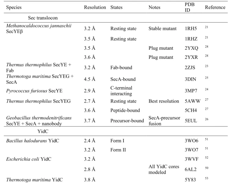
(15) Crystal structures of Sec translocon and YidC 316. Tsukazaki. Table 1 | Crystal structures of the Sec translocon and YidC Species. Resolution States. Notes. PDB ID. Reference. 3.2 Å. Resting state. Stable mutant. 1RH5. 21. 3.5 Å. Resting state. 1RHZ. 21. Sec translocon Methanocaldococcus jannaschii SecYEβ. Thermus thermophilus SecYE + Fab Thermotoga maritima SecYEG + SecA. 3.5 Å. Plug mutant. 2YXQ. 28. 3.6 Å. Plug mutant. 2YXR. 28. 3.2 Å. Fab-bound. 2ZJS. 23. 4.5 Å. SecA-bound. 3DIN. 25. 3MP7. 24. 5AWW. 27. 5CH4. 27. SecA-precursor 5EUL fusion. 26. Pyrococcus furiosus SecYE. 2.9 Å. Thermus thermophilus SecYEG. 2.7 Å. C-terminal interacting Resting state. 3.6 Å. Peptide-bound. 3.7 Å. Precursor-bound. 2.4 Å. Form I. 3WO6. 51. 3.2 Å. Form II. 3WO7. 51. 3WVF. 52. 6AL2. 50. 5Y83. 53. Geobacillus thermodenitrificans SecYE + SecA + nanobody. Best resolution. YidC Bacillus halodurans YidC. Escherichia coli YidC. 3.2 Å All YidC cores modeled. 2.8 Å Thermotoga maritima YidC. 3.8 Å. 317 318. 15.
(16) Crystal structures of Sec translocon and YidC. Tsukazaki. 319. Figure legends. 320. Figure 1 | Bacterial protein translocation and integration via the Sec translocon and YidC.. 321. A, Protein translocation across and integration into the membrane. During post-translational translocation,. 322. precursor proteins with an N-terminal signal sequence are targeted to and translocated across the membrane,. 323. which is driven by Sec62/63 complex and BiP in eukaryotes and SecA ATPase and SecDF in bacteria. During. 324. co-translational translocation, the ribosome–nascent chain complex (RNC) is directed to the membrane by the. 325. interaction between signal recognition particle (SRP) and its receptor, and interacts with SecYEG or YidC.. 326. Subsequent membrane protein integration via the interior of the Sec translocon and/or YidC occurs. 327. co-translationally. B, Schematic representation of Sec translocon components. C, Schematic representation of. 328. the YidC/Oxa1/Alb3 protein family.. 329 330. Figure 2 | Crystal structures of the Sec translocon.. 331. A, The Sec translocon in the resting state. Crystal structures of SecYEβ from Methanocaldococcus jannaschii. 332. (PDB ID 1RH5) (left) and SecYEG from Thermus thermophilus (PDB ID 5AWW) (right). B, Schematic. 333. representation of the Sec translocon. C, Magnified views of the pore ring of the structures in A from the. 334. cytoplasm. D, Cut-away models of the surface representation of T. thermophilus SecYEG without the plug. 335. and cap regions. The plug and cap regions are represented by the ribbon model with a stick model for the side. 336. chains. E, Crystal structures of the Sec translocons in which the cytoplasmic region interacts with other. 337. molecules in the crystals (PDB ID 3MP7, 5CH4, 2ZJS, and 3DIN).. 338 339. Figure 3 | Structures of the Sec translocon in the intermediate stages of protein transport.. 340. A, Crystal structures of the SecYEG–SecA complex with part of the precursor protein expressed as a fusion. 341. protein (PDB ID 5EUL). B, Magnified views of the pore ring of the structure in A from the cytoplasmic side .. 342. C, Electron microscopic structures of the Sec translocon of the RNC complex during protein transport (PDB. 343. ID 3JC2 and 5ABB).. 344. 16.
(17) Crystal structures of Sec translocon and YidC. Tsukazaki. 345. Figure 4 | Crystal structures of YidC.. 346. A, Crystal structure of E. coli YidC at 2.8-Å resolution, elucidating all YidC core regions (PDB ID 6AL2).. 347. The cTM numbers are shown. B, Cut-away model of the E. coli YidC structure. C, Gallery of crystal. 348. structures of YidC (PDB ID 6AL2, 5Y83, 3WO6, and 3WO7). D, Superimposition of the core region of B.. 349. halodurans, T. maritima, and E. coli YidCs. E, Magnified view of the C1 loop region.. 350 351. Figure 5 | Functional model of YidC.. 352. A, Membrane-insertion model of a single-membrane-spanning protein via YidC, independently of SecYEG.. 353. YidC temporally captures the precursor protein at the positively charged cavity. Thereafter, the substrate. 354. protein is sorted primarily via hydrophobic interactions. B, Chaperone activity model of YidC. YidC protects. 355. a hydrophilic region sorted from the lateral gate of the Sec translocon until its interacting region emerges. 356. from the gate, promoting correct folding of substrate proteins.. 357. 17.
(18) Crystal structures of Sec translocon and YidC 358 359 360 361 362 363 364 365 366 367 368 369 370 371 372 373 374 375 376 377 378 379 380 381 382 383 384 385 386 387 388 389 390 391 392 393 394 395 396 397 398 399 400 401 402 403 404 405 406 407 408 409 410 411 412 413 414. Tsukazaki. References 1 2 3 4 5 6 7 8 9 10 11 12 13 14 15 16 17 18 19 20 21 22 23 24 25 26 27. Rapoport, T. A., Li, L. & Park, E. Structural and Mechanistic Insights into Protein Translocation. Annu Rev Cell Dev Biol, doi:10.1146/annurev-cellbio-100616-060439 (2017). Tsirigotaki, A., De Geyter, J., Sostaric, N., Economou, A. & Karamanou, S. Protein export through the bacterial Sec pathway. Nat Rev Microbiol 15, 21-36, doi:10.1038/nrmicro.2016.161 (2017). Blobel, G. & Dobberstein, B. Transfer of proteins across membranes. J Cell Biol 67, 835-862 (1975). Akopian, D., Shen, K., Zhang, X. & Shan, S. O. Signal recognition particle: an essential protein-targeting machine. Annu Rev Biochem 82, 693-721, doi:10.1146/annurev-biochem-072711-164732 (2013). Matlack, K. E., Misselwitz, B., Plath, K. & Rapoport, T. A. BiP acts as a molecular ratchet during posttranslational transport of prepro-alpha factor across the ER membrane. Cell 97, 553-564 (1999). Chatzi, K. E., Sardis, M. F., Economou, A. & Karamanou, S. SecA-mediated targeting and translocation of secretory proteins. Biochim Biophys Acta 1843, 1466-1474, doi:10.1016/j.bbamcr.2014.02.014 (2014). Hartl, F. U., Lecker, S., Schiebel, E., Hendrick, J. P. & Wickner, W. The binding cascade of SecB to SecA to SecY/E mediates preprotein targeting to the E. coli plasma membrane. Cell 63, 269-279 (1990). Economou, A. & Wickner, W. SecA promotes preprotein translocation by undergoing ATP-driven cycles of membrane insertion and deinsertion. Cell 78, 835-843 (1994). Braunger, K. et al. Structural basis for coupling protein transport and N-glycosylation at the mammalian endoplasmic reticulum. Science, doi:10.1126/science.aar7899 (2018). Pfeffer, S. et al. Dissecting the molecular organization of the translocon-associated protein complex. Nat Commun 8, 14516, doi:10.1038/ncomms14516 (2017). Tsukazaki, T. Structure-based working model of SecDF, a proton-driven bacterial protein translocation factor. FEMS Microbiol Lett 365, doi:10.1093/femsle/fny112 (2018). Duong, F. & Wickner, W. Distinct catalytic roles of the SecYE, SecG and SecDFyajC subunits of preprotein translocase holoenzyme. Embo J 16, 2756-2768 (1997). Tsukazaki, T. et al. Structure and function of a membrane component SecDF that enhances protein export. Nature 474, 235-238, doi:10.1038/nature09980 (2011). Furukawa, A., Nakayama, S., Yoshikaie, K., Tanaka, Y. & Tsukazaki, T. Remote Coupled Drastic beta-Barrel to beta-Sheet Transition of the Protein Translocation Motor. Structure 26, 485-489 e482, doi:10.1016/j.str.2018.01.002 (2018). Furukawa, A. et al. Tunnel Formation Inferred from the I-Form Structures of the Proton-Driven Protein Secretion Motor SecDF. Cell Rep 19, 895-901, doi:10.1016/j.celrep.2017.04.030 (2017). Kiefer, D. & Kuhn, A. YidC-mediated membrane insertion. FEMS Microbiol Lett 365, doi:10.1093/femsle/fny106 (2018). Hennon, S. W., Soman, R., Zhu, L. & Dalbey, R. E. YidC/Alb3/Oxa1 Family of Insertases. J Biol Chem 290, 14866-14874, doi:10.1074/jbc.R115.638171 (2015). Nishikawa, H., Sasaki, M. & Nishiyama, K. I. Membrane insertion of F0 c subunit of F0F1 ATPase depends on glycolipozyme MPIase and is stimulated by YidC. Biochem Biophys Res Commun 487, 477-482, doi:10.1016/j.bbrc.2017.04.095 (2017). Nishiyama, K. et al. MPIase is a glycolipozyme essential for membrane protein integration. Nat Commun 3, 1260, doi:10.1038/ncomms2267 (2012). Wang, P. & Dalbey, R. E. Inserting membrane proteins: the YidC/Oxa1/Alb3 machinery in bacteria, mitochondria, and chloroplasts. Biochim Biophys Acta 1808, 866-875, doi:10.1016/j.bbamem.2010.08.014 (2011). van den Berg, B. et al. X-ray structure of a protein-conducting channel. Nature 427, 36-44, doi:10.1038/nature02218 (2004). du Plessis, D. J., Nouwen, N. & Driessen, A. J. The Sec translocase. Biochim. Biophys. Acta 1808, 851-865, doi:10.1016/j.bbamem.2010.08.016 (2011). Tsukazaki, T. et al. Conformational transition of Sec machinery inferred from bacterial SecYE structures. Nature 455, 988-991 (2008). Egea, P. F. & Stroud, R. M. Lateral opening of a translocon upon entry of protein suggests the mechanism of insertion into membranes. Proc Natl Acad Sci U S A 107, 17182-17187, doi:10.1073/pnas.1012556107 (2010). Zimmer, J., Nam, Y. & Rapoport, T. A. Structure of a complex of the ATPase SecA and the protein-translocation channel. Nature 455, 936-943, doi: 10.1038/nature07335 (2008). Li, L. et al. Crystal structure of a substrate-engaged SecY protein-translocation channel. Nature 531, 395-399, doi:10.1038/nature17163 (2016). Tanaka, Y. et al. Crystal Structures of SecYEG in Lipidic Cubic Phase Elucidate a Precise Resting and a. 18.
(19) Crystal structures of Sec translocon and YidC 415 416 417 418 419 420 421 422 423 424 425 426 427 428 429 430 431 432 433 434 435 436 437 438 439 440 441 442 443 444 445 446 447 448 449 450 451 452 453 454 455 456 457 458 459 460 461 462 463 464 465 466 467 468 469 470 471. 28 29 30 31 32 33 34 35 36 37 38 39 40 41 42 43 44 45 46 47 48 49 50 51 52. Tsukazaki. Peptide-Bound State. Cell Rep 13, 1561-1568, doi:10.1016/j.celrep.2015.10.025 (2015). Li, W. et al. The plug domain of the SecY protein stabilizes the closed state of the translocation channel and maintains a membrane seal. Mol Cell 26, 511-521 (2007). Flower, A. M., Hines, L. L. & Pfennig, P. L. SecG is an auxiliary component of the protein export apparatus of Escherichia coli. Mol Gen Genet 263, 131-136 (2000). Nishiyama, K., Hanada, M. & Tokuda, H. Disruption of the gene encoding p12 (SecG) reveals the direct involvement and important function of SecG in the protein translocation of Escherichia coli at low temperature. EMBO J 13, 3272-3277 (1994). Bost, S. & Belin, D. A new genetic selection identifies essential residues in SecG, a component of the Escherichia coli protein export machinery. Embo J 14, 4412-4421 (1995). Brundage, L., Hendrick, J. P., Schiebel, E., Driessen, A. J. & Wickner, W. The purified E. coli integral membrane protein SecY/E is sufficient for reconstitution of SecA-dependent precursor protein translocation. Cell 62, 649-657 (1990). Plath, K., Mothes, W., Wilkinson, B. M., Stirling, C. J. & Rapoport, T. A. Signal sequence recognition in posttranslational protein transport across the yeast ER membrane. Cell 94, 795-807 (1998). Tam, P. C., Maillard, A. P., Chan, K. K. & Duong, F. Investigating the SecY plug movement at the SecYEG translocation channel. EMBO J 24, 3380-3388 (2005). Allen, W. J. et al. Two-way communication between SecY and SecA suggests a Brownian ratchet mechanism for protein translocation. elife 5, doi:10.7554/eLife.15598 (2016). Cannon, K. S., Or, E., Clemons, W. M., Jr., Shibata, Y. & Rapoport, T. A. Disulfide bridge formation between SecY and a translocating polypeptide localizes the translocation pore to the center of SecY. J Cell Biol 169, 219-225 (2005). Ge, Y., Draycheva, A., Bornemann, T., Rodnina, M. V. & Wintermeyer, W. Lateral opening of the bacterial translocon on ribosome binding and signal peptide insertion. Nat Commun 5, 5263, doi:10.1038/ncomms6263 (2014). Bischoff, L., Wickles, S., Berninghausen, O., van der Sluis, E. O. & Beckmann, R. Visualization of a polytopic membrane protein during SecY-mediated membrane insertion. Nat Commun 5, 4103, doi:10.1038/ncomms5103 (2014). Gogala, M. et al. Structures of the Sec61 complex engaged in nascent peptide translocation or membrane insertion. Nature 506, 107-110, doi:10.1038/nature12950 (2014). Park, E. et al. Structure of the SecY channel during initiation of protein translocation. Nature 506, 102-106, doi:10.1038/nature12720 (2014). Voorhees, R. M., Fernandez, I. S., Scheres, S. H. & Hegde, R. S. Structure of the mammalian ribosome-Sec61 complex to 3.4 A resolution. Cell 157, 1632-1643, doi:10.1016/j.cell.2014.05.024 (2014). Voorhees, R. M. & Hegde, R. S. Structure of the Sec61 channel opened by a signal sequence. Science 351, 88-91, doi:10.1126/science.aad4992 (2016). Pfeffer, S. et al. Structure of the native Sec61 protein-conducting channel. Nat Commun 6, 8403, doi:10.1038/ncomms9403 (2015). Jomaa, A., Boehringer, D., Leibundgut, M. & Ban, N. Structures of the E. coli translating ribosome with SRP and its receptor and with the translocon. Nat Commun 7, 10471, doi:10.1038/ncomms10471 (2016). Kedrov, A., Kusters, I., Krasnikov, V. V. & Driessen, A. J. A single copy of SecYEG is sufficient for preprotein translocation. The EMBO journal 30, 4387-4397, doi:10.1038/emboj.2011.314 (2011). Park, E. & Rapoport, T. A. Bacterial protein translocation requires only one copy of the SecY complex in vivo. J Cell Biol 198, 881-893, doi:10.1083/jcb.201205140 (2012). Dalal, K., Chan, C. S., Sligar, S. G. & Duong, F. Two copies of the SecY channel and acidic lipids are necessary to activate the SecA translocation ATPase. Proceedings of the National Academy of Sciences of the United States of America 109, 4104-4109, doi:10.1073/pnas.1117783109 (2012). Osborne, A. R. & Rapoport, T. A. Protein translocation is mediated by oligomers of the SecY complex with one SecY copy forming the channel. Cell 129, 97-110 (2007). Kedrov, A. et al. Structural Dynamics of the YidC:Ribosome Complex during Membrane Protein Biogenesis. Cell Rep 17, 2943-2954, doi:10.1016/j.celrep.2016.11.059 (2016). Tanaka, Y. et al. 2.8-A crystal structure of Escherichia coli YidC revealing all core regions, including flexible C2 loop. Biochem Biophys Res Commun 505, 141-145, doi:10.1016/j.bbrc.2018.09.043 (2018). Kumazaki, K. et al. Structural basis of Sec-independent membrane protein insertion by YidC. Nature 509, 516-520, doi:10.1038/nature13167 (2014). Kumazaki, K. et al. Crystal structure of Escherichia coli YidC, a membrane protein chaperone and insertase. Sci Rep 4, 7299, doi:10.1038/srep07299 (2014).. 19.
(20) Crystal structures of Sec translocon and YidC 472 473 474 475 476 477 478 479 480 481 482 483 484 485 486 487 488 489 490 491 492 493 494 495 496 497 498 499 500 501 502 503 504 505 506 507 508 509 510 511 512 513 514 515 516 517 518 519 520 521 522 523 524 525 526 527 528. 53 54 55 56 57 58 59 60 61 62 63 64 65 66 67 68 69 70 71 72 73 74 75 76 77 78. Tsukazaki. Xin, Y. et al. Structure of YidC from Thermotoga maritima and its implications for YidC-mediated membrane protein insertion. FASEB J 32, 2411-2421, doi:10.1096/fj.201700893RR (2018). Kedrov, A. et al. Elucidating the native architecture of the YidC: ribosome complex. J Mol Biol 425, 4112-4124, doi:10.1016/j.jmb.2013.07.042 (2013). Wickles, S. et al. A structural model of the active ribosome-bound membrane protein insertase YidC. elife 3, e03035, doi:10.7554/eLife.03035 (2014). Spann, D., Pross, E., Chen, Y., Dalbey, R. E. & Kuhn, A. Each protomer of a dimeric YidC functions as a single membrane insertase. Sci Rep 8, 589, doi:10.1038/s41598-017-18830-9 (2018). Kohler, R. et al. YidC and Oxa1 form dimeric insertion pores on the translating ribosome. Mol Cell 34, 344-353, doi:10.1016/j.molcel.2009.04.019 (2009). Lotz, M., Haase, W., Kuhlbrandt, W. & Collinson, I. Projection structure of yidC: a conserved mediator of membrane protein assembly. J Mol Biol 375, 901-907, doi:10.1016/j.jmb.2007.10.089 (2008). Petriman, N. A. et al. The interaction network of the YidC insertase with the SecYEG translocon, SRP and the SRP receptor FtsY. Sci Rep 8, 578, doi:10.1038/s41598-017-19019-w (2018). Klenner, C. & Kuhn, A. Dynamic disulfide scanning of the membrane-inserting Pf3 coat protein reveals multiple YidC substrate contacts. J Biol Chem 287, 3769-3776, doi:10.1074/jbc.M111.307223 (2012). Klenner, C., Yuan, J., Dalbey, R. E. & Kuhn, A. The Pf3 coat protein contacts TM1 and TM3 of YidC during membrane biogenesis. FEBS Lett 582, 3967-3972, doi:10.1016/j.febslet.2008.10.044 (2008). Jiang, F. et al. Defining the regions of Escherichia coli YidC that contribute to activity. J Biol Chem 278, 48965-48972 (2003). Xie, K., Kiefer, D., Nagler, G., Dalbey, R. E. & Kuhn, A. Different regions of the nonconserved large periplasmic domain of Escherichia coli YidC are involved in the SecF interaction and membrane insertase activity. Biochemistry 45, 13401-13408 (2006). Sachelaru, I. et al. YidC occupies the lateral gate of the SecYEG translocon and is sequentially displaced by a nascent membrane protein. J Biol Chem 288, 16295-16307, doi:10.1074/jbc.M112.446583 (2013). Schulze, R. J. et al. Membrane protein insertion and proton-motive-force-dependent secretion through the bacterial holo-translocon SecYEG-SecDF-YajC-YidC. Proc Natl Acad Sci U S A 111, 4844-4849, doi:10.1073/pnas.1315901111 (2014). Chen, Y., Soman, R., Shanmugam, S. K., Kuhn, A. & Dalbey, R. E. The role of the strictly conserved positively charged residue differs among the Gram-positive, Gram-negative, and chloroplast YidC homologs. J Biol Chem 289, 35656-35667, doi:10.1074/jbc.M114.595082 (2014). Shimokawa-Chiba, N. et al. Hydrophilic microenvironment required for the channel-independent insertase function of YidC protein. Proc Natl Acad Sci U S A 112, 5063-5068, doi:10.1073/pnas.1423817112 (2015). Geng, Y. et al. Role of the Cytosolic Loop C2 and the C Terminus of YidC in Ribosome Binding and Insertion Activity. J Biol Chem 290, 17250-17261, doi:10.1074/jbc.M115.650309 (2015). Seitl, I., Wickles, S., Beckmann, R., Kuhn, A. & Kiefer, D. The C-terminal regions of YidC from Rhodopirellula baltica and Oceanicaulis alexandrii bind to ribosomes and partially substitute for SRP receptor function in Escherichia coli. Mol Microbiol 91, 408-421, doi:10.1111/mmi.12465 (2014). Chen, Y. et al. YidC Insertase of Escherichia coli: Water Accessibility and Membrane Shaping. Structure 25, 1403-1414 e1403, doi:10.1016/j.str.2017.07.008 (2017). Noinaj, N. et al. Structural insight into the biogenesis of beta-barrel membrane proteins. Nature 501, 385-390, doi:10.1038/nature12521 (2013). Anghel, S. A., McGilvray, P. T., Hegde, R. S. & Keenan, R. J. Identification of Oxa1 Homologs Operating in the Eukaryotic Endoplasmic Reticulum. Cell Rep 21, 3708-3716, doi:10.1016/j.celrep.2017.12.006 (2017). Borowska, M. T., Dominik, P. K., Anghel, S. A., Kossiakoff, A. A. & Keenan, R. J. A YidC-like Protein in the Archaeal Plasma Membrane. Structure 23, 1715-1724, doi:10.1016/j.str.2015.06.025 (2015). Nagamori, S., Smirnova, I. N. & Kaback, H. R. Role of YidC in folding of polytopic membrane proteins. J Cell Biol 165, 53-62, doi:10.1083/jcb.200402067 (2004). Serdiuk, T. et al. YidC assists the stepwise and stochastic folding of membrane proteins. Nat Chem Biol 12, 911-917, doi:10.1038/nchembio.2169 (2016). Yu, Z., Koningstein, G., Pop, A. & Luirink, J. The conserved third transmembrane segment of YidC contacts nascent Escherichia coli inner membrane proteins. J Biol Chem 283, 34635-34642, doi:10.1074/jbc.M804344200 (2008). Botte, M. et al. A central cavity within the holo-translocon suggests a mechanism for membrane protein insertion. Sci Rep 6, 38399, doi:10.1038/srep38399 (2016). Urbanus, M. L. et al. Targeting, insertion, and localization of Escherichia coli YidC. J Biol Chem 277, 12718-12723 (2002).. 20.
(21) Crystal structures of Sec translocon and YidC 529 530 531 532 533 534 535 536 537 538 539 540 541 542 543. 79 80 81 82 83 84 85. Tsukazaki. von Heijne, G. The distribution of positively charged residues in bacterial inner membrane proteins correlates with the trans-membrane topology. EMBO J 5, 3021-3027 (1986). Itskanov, S. & Park, E. Structure of the posttranslational Sec protein-translocation channel complex from yeast. Science, doi:10.1126/science.aav6740 (2018). Wu, X., Cabanos, C. & Rapoport, T. A. Structure of the post-translational protein translocation machinery of the ER membrane. Nature 566, 136-139, doi:10.1038/s41586-018-0856-x (2019). Sugano, Y., Furukawa, A., Nureki, O., Tanaka, Y. & Tsukazaki, T. SecY-SecA fusion protein retains the ability to mediate protein transport. PLoS One 12, e0183434, doi:10.1371/journal.pone.0183434 (2017). Kedrov, A., Kusters, I. & Driessen, A. J. Single-molecule studies of bacterial protein translocation. Biochemistry 52, 6740-6754, doi:10.1021/bi400913x (2013). Taufik, I., Kedrov, A., Exterkate, M. & Driessen, A. J. Monitoring the activity of single translocons. J Mol Biol 425, 4145-4153, doi:10.1016/j.jmb.2013.08.012 (2013). Haruyama, T. et al. Single-Unit Imaging of Membrane Protein-Embedded Nanodiscs from Two Oriented Sides by High-Speed Atomic Force Microscopy. Structure, doi:10.1016/j.str.2018.09.005 (2018).. 21.
(22) Figure. A. Click here to access/download;Figure;190320_Figs_3.pdf. Bacterial protein translocation Post-translational translocation. Co-translational translocation Mature protein. Mature protein. SecDF. YidC. YidC SecYEG. SecYEG. SecA ATPase. Mature membrane protein. Ribosome Nascent chain. Ribosome Signal peptide. Cytoplasm. Precursor protein. B. Sec translocon components SecY/Sec61α. SecE/Sec61γ. SecG. Plu. N. C. N. C. Sec61β. g 1. 2 3 4 5 6 7 8. 9. N C4. 1. 2. 1. N. C. Cytoplasm. C. 1. 10. C. Additional 2 TMs in E. coli. C5. YidC/Oxa1/Alb3 family Gram-negative bacteria. Gram-positive bacteria. Mitochondria. Chloroplasts. P1. 1. N. C1. 1. 2 3 4 5. C2. C. C1. 2 3 4 5. C2. 1. 2 3 4 5. C C1. Cytoplasm. C2. 1. C. Matrix. Tsukazaki Fig. 1. C1. 2 3 4 5. C2. C Stroma.
(23) Resting state (SecYEβ at 3.2 Å). B. Resting state (SecYEG at 2.7 Å) Lateral gate. Lateral gate Plug SecY TM6-10. 7. Secβ. SecG. 8. 2. SecY TM6-10. 8. SecE. SecY TM1-5. Pore ring. SecE SecY TM1-5. C5 C4. Lateral gate Cap. Cap. Cytoplasm. 1RH5. C. SecY TM1-5. 7. Pore ring. 2. SecG. Plug. SecE. A. 5AWW Plug. TM1-5. TM6-10. TM1-5. Plug. TM6-10. D. Plug. Pore ring. SecG V79. I81 I260. Secβ. SecY TM6-10. L406. I275. Cap. I170 I174. I75. SecY TM6-10. I77. T184. I188. I403. SecY TM1-5. SecE. SecG Cap 1RH5. E. C-terminal interacting SecYE at 2.9 Å. 5AWW. Peptide bound SecYEG at 3.6 Å. Fab bound SecYE at 3.2 Å. SecA bound SecYEG at 4.5 Å Dislocated plug Extended pore. TM8 TM2. helix. Peptide. Fab 3MP7. 5CH4. 2ZJS. Tsukazaki Fig. 2. SecA ATPase 3DIN.
(24) A. B. Protein translocation intermediate stats SecYE-SecA at 3.7 Å Dislocated Plug. Signal peptide at Lateral gate Preprotein. Signal peptide at Lateral gate. SecY TM6-10. 7. Extended Pore ring. SecY TM1-5. 8. 2 I275. SecE. I78 I404. V74. I183 I179. SecE. SecA ATPase. C. 5EUL. EM structures of co-translational translocation. SecY TM6-10. Sec61α TM6-10. Membrane protein substrate Sec61α TM1-5. Membrane protein substrate Sec61β. SecY TM1-5. SecE. Ribosome. Ribosome 3JC2. Tsukazaki Fig. 3. 5ABB.
(25) A. B. P1. 2. cTM1-5 (E. coli TM2-6). 5. Hydrophobic core. Hydrophobic core. EH1 1. 4 3 Core region C2. Hydrophilic cavity. Hydrophilic cavity. 180˚. C1 Cytoplasm. C. Arg. C2. C1. C1. EcYidC 2.8 Å 6AL2. TmYidC 3.8 Å 5Y83. D. C1. BhYidC (Form I) 2.4 Å 3WO6. BhYidC (Form II) 3.2 Å 3WO7. E C2. C2. C1. C1. EcYidC BhYidC (Form I) BhYidC (Form II) TmYidC. EcYidC BhYidC (Form I) BhYidC (Form II). Tsukazaki Fig. 4. C-terminal region.
(26) Hydrophilic cavity. PMF dependent. +. Hydrophobic interaction. +. –+. le xib Fle /C2 1 C YidC. – Substrate. Δψ. –. Membrane. A. +. – Cytoplasm. Lateral gate. Substrate. Sec translocon. YidC. –. +. Mature. +. – +. +–. Membrane. B. Cytoplasm. Ribosome. Tsukazaki Fig. 5.
(27)
図
関連したドキュメント
As an application, we give semantics of modal proofs (a.k.a., programs) in categories of augmented simplicial sets and of topological spaces, and prove a completeness result in
of the conference on ergodic theory and related topics, II (Georgenthal, 1986), Teubner-Texte Math. Misiurewicz , Dimension of invariant measures for maps with ex- ponent zero,
The edges terminating in a correspond to the generators, i.e., the south-west cor- ners of the respective Ferrers diagram, whereas the edges originating in a correspond to the
H ernández , Positive and free boundary solutions to singular nonlinear elliptic problems with absorption; An overview and open problems, in: Proceedings of the Variational
Keywords: Convex order ; Fréchet distribution ; Median ; Mittag-Leffler distribution ; Mittag- Leffler function ; Stable distribution ; Stochastic order.. AMS MSC 2010: Primary 60E05
In [9], it was shown that under diffusive scaling, the random set of coalescing random walk paths with one walker starting from every point on the space-time lattice Z × Z converges
The main novelty of this paper is to provide proofs of natural prop- erties of the branches that build the solution diagram for both smooth and non- smooth double-well potentials,
Inside this class, we identify a new subclass of Liouvillian integrable systems, under suitable conditions such Liouvillian integrable systems can have at most one limit cycle, and
