oxide-enhanced dynamic magnetic resonance imaging and comparison with contrast-enhanced computed tomography in endoleak detection after endovascular aneurysm repair
Shigeo Ichihashi, MD,
aNagaaki Marugami, MD,
aToshihiro Tanaka, MD,
aShinichi Iwakoshi, MD,
aNorio Kurumatani, MD,
bSatoru Kitano, MD,
aAkihiro Nogi, RT,
aand Kimihiko Kichikawa, MD,
aNara, Japan
Objective: Contrast-enhanced computed tomography (CE-CT) has been commonly used for follow-up imaging after endovascular aneurysm repair (EVAR), but it is dif fi cult to use on patients with renal insuf fi ciency. Superparamagnetic iron oxide (SPIO) particles, contrast medium for magnetic resonance imaging (MRI) that has been widely used for detection of the liver tumor, rarely affects renal function. The present study examined SPIO-enhanced dynamic MRI as a potential alternative to CE-CT for detection of endoleaks after EVAR.
Methods: Institutional review board approval was obtained for this prospective study. Twenty-three consecutive patients with normal renal function were evaluated using both CE-CT and SPIO-enhanced MRI within 2 weeks after EVAR. The median interval between the two modalities was 2 days. SPIO-enhanced MRI was performed at 1.5 T with T1-weighted, SPIO-enhanced dynamic, and postcontrast T1-weighted gradient echo sequences. The CE-CT protocol consisted of triple scans. Two experienced, blinded observers evaluated all images. Consensus reading of CE-CT and SPIO-MRI was defined as the reference standard. Interobserver, intraobserver, and intermodality agreement for endoleak detection was assessed by k statistics.
Results: A total of 11 type II endoleaks originating from either the lumbar or inferior mesenteric artery were detected.
Eight were able to be detected by CE-CT (8/11:73%) and 10 (10/11:91%) by SPIO-enhanced MRI. Interobserver ( k [ 0.91; 95% CI, 0.74-1.00) and intraobserver agreement for MRI ( k [ 1.00) were excellent. Intermodality agreement for endoleak detection was moderate ( k [ 0.63; 95% CI, 0.32-0.94; and k [ 0.62; 95% CI, 0.29-0.95 for observers A and B, respectively).
Conclusions: SPIO-enhanced MRI could represent a useful alternative to CE-CT, as it offers excellent interobserver, intraobserver agreement, and could detect more endoleaks than CE-CT. (J Vasc Surg 2013;58:66-72.)
During the last decade, endovascular aneurysm repair (EVAR) has become the important modality of treatment for abdominal aortic aneurysms (AAA). Although previous multiple randomized controlled trials showed lower 30-day mortality with EVAR, compared with open AAA repair, the procedure has also been associated with postoperative complications such as endoleaks, and a significantly higher rate of secondary interventions.
1,2Because endoleaks may cause enlargement of aneurysm sac and, thus, increase the risk of rupture after EVAR, long-term surveillance of
endoleaks is necessary to allow for secondary interventions to maintain optimal outcomes.
3Contrast-enhanced computed tomography (CE-CT) has been commonly used for postoperative surveillance of EVAR because, in part, of its rapid acquisition time and high diagnostic value. However, it is also associated with the cumulative risks of radiation exposure and, most of all, contrast nephrotoxicity. With increasing reliance on diag- nostic CT using iodinated intravenous contrast, the inci- dence of contrast-induced nephropathy is likely to increase.
In a previous article evaluating contrast-induced nephro- pathy after percutaneous coronary intervention, more severe renal impairment was found to be correlated with a greater risk of developing contrast-induced nephropathy,
4so that the risk is highest in patients with chronic kidney disease (CKD).
Dynamic contrast-enhanced magnetic resonance im- aging (MRI) techniques using gadolinium diethylenetri- amine penta-acetic acid (Gd-DTPA) as a contrast agent have been shown to be very sensitive for depicting small endoleaks
5and can have additional value for their classifica- tion.
6However, the association between nephrogenic
From the Department of Radiology
aand Department of Community Health and Epidemiology,
bNara Medical University.
Author con
flict of interest: none.
Reprint requests: Shigeo Ichihashi, MD, Department of Radiology, Nara Medical University, 840 Shijo-cho, Kashihara, Nara 634-8521, Japan (e-mail:
[email protected]).The editors and reviewers of this article have no relevant
financial relationshipsto disclose per the JVS policy that requires reviewers to decline review of any manuscript for which they may have a conflict of interest.
0741-5214/$36.00
Copyright
Ó2013 by the Society for Vascular Surgery.
http://dx.doi.org/10.1016/j.jvs.2012.12.061
66
systemic fibrosis and gadolinium-based contrast agents has been reported in patients with CKD.
7,8Contrast-enhanced ultrasound (CE-US) has also been shown to be sensitive for detecting endoleaks and to allow for better classification of endoleaks compared with CE-CT.
9However, diagnostic accuracy of CE-US depends largely on operator skill, and scan and study qualities are generally poor in obese patients.
Superparamagnetic iron oxide (SPIO) particles were originally developed as contrast medium for MRI of the liver, where they are administered to improve tumor detec- tion with T2-weighted imaging (T2WI). The SPIO parti- cles improve contrast by being taken up by the Kupffer cells of normal liver tissues but not by metastases or primary liver tumors. Intravenously injected SPIO particles also shorten the T1 relaxation time, and thus, vascularized structures show signal enhancement on T1-weighted imaging (T1WI),
10,11so SPIO-enhanced MRI may have the potential to detect endoleaks. SPIO is different from Gd-DTPA. SPIO is metabolized as iron, the rate of urinary excretion is less than 0.5%, and it is extremely rare that SPIO complicates renal function.
12,13If SPIO-enhanced MRI is proven to be effective for endoleak detection, it should be useful for monitoring patients with CKD after EVAR. The purpose of this study was to examine SPIO- enhanced MRI as a potential alternative to CE-CT for detection of endoleaks after EVAR.
METHODS
Patients. Twenty-three consecutive patients who underwent implantation of nitinol aortic stent grafts for AAA were recruited to this prospective study. No patients had contraindications of use of SPIO (ie, history of iron allergy or hemochromatosis), and administration of SPIO could be performed in every patient. The median patient age was 79 (range, 71-83) years (male, n ¼ 20; female, n ¼ 3). The median body weight was 61 kg (range, 45- 71 kg). The median sac diameter was 48.5 mm (range, 44.5-53.7 mm). Excluder stent grafts (Gore Medical, Newark, Del) were implanted in 10 patients and Endurant stent grafts (Medtronic, Fridley, Minn) were implanted in 13. CE-CT and SPIO-enhanced MRI were performed for endoleak detection within 2 weeks after EVAR. The time period between CE-CT and SPIO-enhanced MRA was not more than 1 week. Written informed consent was obtained from all patients. The institutional review board approved this prospective study.
CE-CT. All examinations were performed with a dual- source 64 slice CT scanner (Somatom Definition; Siemens Medical Solutions, Erlangen, Germany). A triple-phase CT protocol consisting of a nonenhanced, arterial and a delayed venous phase encompassing the abdomen and pelvis from the level of the cardiac apex to the femoral head was performed. After standard nonenhanced data acquisi- tion, arterial phase scanning was performed using a bolus- tracking technique. A total of 100-150 mL of nonionic iodinated contrast medium was administered at a flow rate of 4 mL/s. All injections were followed by a chaser bolus of
30 mL of saline solution. The arterial phase data acquisition was initiated 6 seconds after the attenuation reached a predefined threshold of 120 HU. This delay allowed the patient to receive breathing instructions and facilitated maximal aortic enhancement. Delayed phase dual-energy CT scanning was performed with a standard delay of 70 seconds after the beginning of the contrast material injection.
SPIO-enhanced MRI. All scans were performed at 1.5 T (Siemens Medical Solutions). Before injection of SPIO, native turbo fast low angle shot magnetic resonance imaging (FLASH) and volumetric interpolated breath- hold examination (VIBE) sequences were performed under breath-hold conditions (Table I). Resovist (Fujifilm RI Pharma Co, Tokyo, Japan), a bolus injectable SPIO, was used as the contrast medium. Depending on patient weight, a dose of 0.6-1.4 mL (0.016 mL/kg body weight) of Resovist was injected at 1.0 mL/s followed by a 20 mL saline flush administered at the same rate using an elec- tronic power injector (Spectris Solaris; Medrad, Warren- dale, Pa). After injection, the fluoroscopic triggering technique using two-dimensional sagittal gradient refo- cused images (CareBolus technique) was obtained rapidly through the thoracic aorta. When the contrast bolus arrived at the descending aorta, the turbo FLASH sequence was started for the vascular phase. The VIBE sequence was started 2 minutes after injection. These series were also evaluated after subtraction processing. Subtrac- tion imaging is a technique whereby an unenhanced T1- weighted sequence is digitally subtracted from the identical sequence performed after SPIO administration.
By performing this operation, the native T1 signal is removed and any signal remaining on the subtracted images is due solely to enhancement (ie, endoleaks). MR angiography (MRA) images were also obtained by maximal intensity projection processing on subtraction images.
Image analysis. Analysis of CE-CT and SPIO- enhanced MRI was performed by two independent radi- ologists (S.I. and N.M., with 9 and 16 years of experience in vascular imaging, respectively) who were blinded to previous angiographic results. Both readers independently reviewed all images and evaluated the presence and types of endoleaks. The order in which the different reading sessions were performed was selected randomly by another radiologist (T.T.) who was not involved in the reading sessions. The classification of endoleaks was performed according to the criteria proposed by White et al
14: type I, II, III, IV, and unknown (meaning the exact site of inflow could not be identified). One observer (S.I.) evaluated CE- CT and SPIO-enhanced MRI twice for intraobserver comparison with an interval between the first and second evaluations of more than 1 month. During the second evaluation, the observer was blinded to the results of the first evaluation. In addition to the endoleak evaluation, patencies of the endografts and renal arteries were also evaluated by a single observer (S.I.) referring to the MRA.
Dynamic scans were considered technically unsuccessful if
diagnostic evaluation was seriously compromised by patient motion, fold-over artifacts, or misregistration artifacts.
Statistical analysis. Categorical variables are expressed as frequencies or percentages. Interobserver, intraobserver, and intermodality agreements for endoleak detection were assessed by k statistics as follows: k values of 0.00-0.40 were considered to indicate poor agreement; 0.41-0.75, fair to good agreement; and 0.76-1.00, excellent agree- ment.
15When disagreements were encountered between the two readers about the existence of endoleaks, a discus- sion was held until consensus could be reached. Consensus reading of MRI and multidetector computed tomography studies was defined as reference standard, similar to pre- vious studies.
5,16All statistical tests were performed using SPSS v. 20 software (IBM, New York, NY).
RESULTS
The median interval between CE-CT and SPIO- enhanced MRI was 2 days (0-7 days). All patients completed the protocol, and no adverse events were recorded. All MRI images could be evaluated without significant motion or misregistration artifacts, and there were no unsuccessful scans.
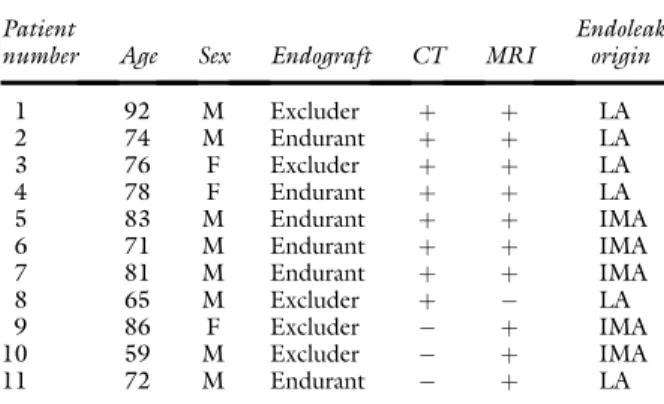
At consensus reading, endoleaks were detected in 11 patients in total by CT and/or MRI (11/23:47%). All endoleaks were type II and originated from either the lumbar (n ¼ 6) or inferior mesenteric artery (n ¼ 5).
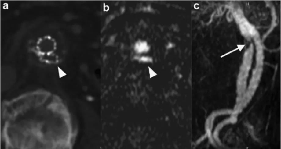
CE-CT was able to detect eight of the endoleaks (8/11:73%) and SPIO-enhanced MRI was able to detect 10 (10/11:91%) (Table II; Fig 1). All 10 endoleaks detected by SPIO-enhanced MRI were detected by subtraction dynamic images, whereas three endoleaks were not detected by nonsubtraction conventional dynamic images. One type II endoleak from the lumbar artery and two type II endoleaks from the inferior mesen- teric artery visualized by SPIO-enhanced MRI could not be detected by CE-CT (Fig 2). These endoleaks broadened with time on dynamic scan and connected with the lumbar artery or the inferior mesenteric artery, which were thought to be origin of the endoleaks. On the other hand, one type II endoleak from the lumbar artery visualized by CE-CT could not be detected by SPIO-enhanced MRI. A consensus reading was required for one patient in both
CE-CT and SPIO-enhanced MRI. Misregistration artifacts were seen in three cases, although these artifacts did not affect the image interpretations. Interobserver ( k ¼ 0.91;
95% CI, 0.74-1.00) and intraobserver agreement for MRI ( k ¼ 1.00) were excellent. Intermodality agreement for endoleak detection was moderate ( k ¼ 0.63; 95% CI, 0.32-0.94; and k ¼ 0.62; 95% CI, 0.29-0.95 for observers A and B, respectively).
Graft and renal artery patency could be evaluated in all patients (Fig 3). All renal arteries were patent; however, in one patient, after implantation of the Excluder endograft, stenosis of the ipsilateral limb because of compression by the contralateral limb was clearly detected by MRA (Fig 4). In this patient, additional kissing balloon dilation was performed as a secondary intervention.
DISCUSSION
Endoleaks are frequent problems after EVAR. Previous CE-CT-based studies have shown that 20%-40% of patients had an initial endoleak, many of which were type II.
17In the present study, type II endoleaks were able to be detected in eight patients by CE-CT and in 10 patients by SPIO-enhanced MRI. Sensitivity of CE-CT for endo- leak detection has been reported to be 82%-83%.
9,18On the other hand, contrast-enhanced MRI using Gd-DTPA has been shown to be significantly more sensitive for endo- leak detection than CE-CT because of the high sensitivity, intrinsic three dimensionality, and excellent soft tissue contrast.
5Resovist, the bolus injectable SPIO, shortens both T1 and T2 relaxation times, but the shortening of T2 is much greater than that of T1.
19The positive T1 contrast effect is less pronounced compared with Gd-DTPA.
20-22Unlike Gd-DTPA, however, Resovist is characterized by a biexponential blood half-life of 3.9-5.8 minutes and a blood pool effect.
22Therefore, it was hypothesized that Resovist may have the potential to allow evaluation of endoleak detection after EVAR. The present study showed Table II. Summary of the type II endoleaks detected by CT and/or MRI
Patient
number Age Sex Endograft CT MRI
Endoleak origin
1 92 M Excluder þ þ LA
2 74 M Endurant þ þ LA
3 76 F Excluder þ þ LA
4 78 F Endurant þ þ LA
5 83 M Endurant þ þ IMA
6 71 M Endurant þ þ IMA
7 81 M Endurant þ þ IMA
8 65 M Excluder þ LA
9 86 F Excluder þ IMA
10 59 M Excluder þ IMA
11 72 M Endurant þ LA
CT,
Computed tomography;
F,female;
IMA,inferior mesenteric artery;
LA,lumbar artery,
M,male;
MRI,magnetic resonance imaging;
þ,detected;
,undetected.
Table I. MRI scan parameter
Parameter Turbo FLASH VIBE
TR, milliseconds 2.51 2.95
TE, milliseconds 0.88 1.1
FA,
25 10
Slice thickness, mm 1.5 3
FOV, mm 350 350
Matrix 147 256 176 256
Slice per slab 128 60
Scan time, seconds 16 25
FA,
Flip angle;
FLASH,fast low angle shot magnetic resonance imaging;
FOV,field of view;MRI,