Fukushima Medical University
福島県立医科大学 学術機関リポジトリ
This document is downloaded at: 2021-11-07T23:24:29Z
Title Clinical evaluation of dendritic cells vaccination for advanced cancer patients at fukushima medical university
Author(s)
Nakamura, Izumi; Kanazawa, Masashi; Sato, Yu; Irisawa, Atsushi; Takagi, Tadayuki; Ogata, Takashi; Kashimura, Shogo;
Kenjo, Akira; Suzuki, Hiroyuki; Shibata, Masahiko; Shimura, Tatsuo; Ohira, Hiromasa; Goto, Mitsukazu; Takenoshita, Seiichi; Ohto, Hitoshi
Citation Fukushima Journal of Medical Science. 58(1): 40-48
Issue Date 2012
URL http://ir.fmu.ac.jp/dspace/handle/123456789/322
Rights © 2012 The Fukushima Society of Medical Science
DOI 10.5387/fms.58.40
Text Version publisher
40
中村 泉,金澤匡司,佐藤 雄,入澤篤志,高木忠之,尾形 隆,樫村省吾,見城 明,鈴木弘行,柴田昌彦,
志村龍男,大平弘正,後藤満一,竹之下誠一,大戸 斉
Corresponding author : Izumi Nakamura E-mail address : [email protected] https://www.jstage.jst.go.jp/browse/fms http://www.fmu.ac.jp/home/lib/F-igaku/
[Original Article]
CLINICAL EVALUATION OF DENDRITIC CELLS VACCINATION FOR ADVANCED CANCER PATIENTS AT FUKUSHIMA
MEDICAL UNIVERSITY
IZUMI NAKAMURA
1), MASASHI KANAZAWA
1), YU SATO
1), ATSUSHI IRISAWA
2), TADAYUKI TAKAGI
2), TAKASHI OGATA
2), SHOGO KASHIMURA
3),
AKIRA KENJO
3), HIROYUKI SUZUKI
3), MASAHIKO SHIBATA
4), TATSUO SHIMURA
4), HIROMASA OHIRA
2), MITSUKAZU GOTO
3),
SEIICHI TAKENOSHITA
1)and HITOSHI OHTO
5)1)Department of Organ Regulatory Surgery, 2)Department of Gastroenterology and Rheumatology,
3)Department of Organ Regererative Surgery, 4)Department of Tumor Host Bioscience, 5)Department of Blood Transfusion and Transplantation Immunology
(Received October 5, 2011, accepted February 2, 2012)
Abstract: Dendritic cells (DCs) are powerful antigen-presenting cells (APCs) that have attracted attention in recent years from the viewpoint of DC vaccine therapy against cancer. However, the existence of a strongly immunosuppressed state in cancer-bearing individuals inhibits DC matura- tion, which is one of the problems facing anti-cancer DC vaccine therapy. Isolated DCs loaded with tumor antigen ex vivo and administered as a cellular vaccine have been found to induce protective and therapeutic anti-tumor immunity in experimental animals. In clinical trials of DC vaccination for cancer patients, induction of anti-tumor immune responses and tumor regression has been observed. In this study, eighty-one advanced cancer patients unsuccessfully treated by established treatment in individual cases were selected between January 2002 and May 2007 at Fukushima Medical University. The usefulness of DC therapy was investigated by intradermal injection of peptide pulsed DCs for an overall objective response rate of 28.0%. Furthermore, direct injection of immature DCs into tumor extracted an overall objective response rate of 35.7%, and especially 40.0% for advanced pancreatic cancer by using endoscopic ultrasound-guided fine-needle injection technique as a novel approach. These results indicate that DC-based vaccination could be a prom- ising treatment modality for various cancers, however multiple hurdles must be cleared before the development of an affordable DC-based vaccination can be used worldwide.
Key words: dendritic cell, vaccination, advance cancer
INTRODUCTION
Human tumors express a number of protein antigens that can be recognized by T cells, thus pro- viding potential targets for cancer immunother- apy. Dendritic cells (DCs) are rare leukocytes that are uniquely potent in their ability to present anti- gens to T cells, and this property has prompted their
recent application to therapeutic cancer vac- cines. Isolated DCs loaded with tumor antigen ex vivo and administered as a cellular vaccine have been found to induce protective and therapeutic anti-tumor immunity in experimental animals. In pilot clinical trials of DC vaccination for patients with non-Hodgkin’s lymphoma and melanoma, induction of anti-tumor immune responses and
CLINICAL EVALUATION OF DENDRITIC CELLS VACCINATION FOR ADVANCED CANCER PATIENTS AT FUKUSHIMA 41
tumor regression has been observed1). Additional trials of DC vaccination for a variety of human can- cers are under way, and methods for targeting tumor antigens to DCs in vivo are also being explo- red. Exploitation of the antigen-presenting proper- ties of DCs thus offers promise for the development of effective cancer immunotherapies. In this study, the usefulness of DC therapy was investigated by intradermal injection of peptide pulsed DCs with/
without chemotherapy, which are direct injection of immature DCs into tumor as well as endoscopic ultrasound-guided fine-needle injection (EUS-FNI) of immature DCs into advanced cancers.
MATERIAL AND METHODS
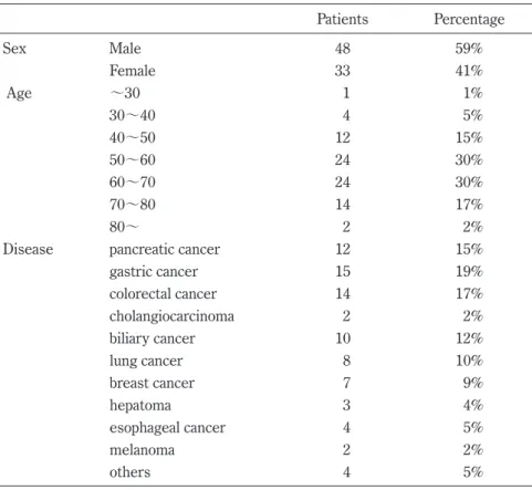
In this study, eighty-one advanced cancer pati- ents unsuccessfully treated by established treatment in individual cases were selected between January 2002 and May 2007 at Fukushima Medical University. The study protocol conformed to the ethical guidelines of the 1975 declaration of Helsinki and was approved by the ethical committee of Fukushima Medical University. All patients pro- vided informed consent before treatment. Demo- graphic characteristics of the individuals treated with DCs vaccines are shown in Table 1. Of the
eighty-one patients treated, 12 had pancreatic can- cer, 15 had gastric cancer, 14 had colorectal cancer, 10 had biliary cancer, 8 had lung cancer, 7 had breast cancer, 3 had hepatoma, 2 had cholangiocarcinoma, 4 had esophageal cancer, 2 had melanoma and 4 had other types of cancer (Table 1).
Isolation of peripheral blood monocytes and DCs induction
Forty ml of heparinized peripheral blood was collected from the patients’ vein and separated using the Ficoll density gradient centrifugation method.
1×10⁸ peripheral blood mononuclear cells (PBMCs) were obtained from the buffy coat. 1×10⁹ PBMCs were obtained from the plasmapheresis using the same method. Of the eighty-one patients, 11 cases with blood collected from the patients’ vein and 70 cases with PBMCs from plasmapheresis were selected corresponding to the individual conditions (Table 2). The PBMCs were cultured in RPMI medium, the monocyte fraction from the adhesion cells were co-cultured with 100 ng/ml GM-CSF and 50 ng/ml IL-4 for 7 days to induce DCs. The sus- pension of DCs was confirmed with FITC-or PE-conjugated monoclonal antibodies against CD40, CD80, CD86, HLA-DR cell surface antigen, and then analyzed using FACS Calibur flow cytometer
Table 1. Patient Characteristics
Patients Percentage
Sex Male 48 59%
Female 33 41%
Age 〜30 1 1%
30〜40 4 5%
40〜50 12 15%
50〜60 24 30%
60〜70 24 30%
70〜80 14 17%
80〜 2 2%
Disease pancreatic cancer 12 15%
gastric cancer 15 19%
colorectal cancer 14 17%
cholangiocarcinoma 2 2%
biliary cancer 10 12%
lung cancer 8 10%
breast cancer 7 9%
hepatoma 3 4%
esophageal cancer 4 5%
melanoma 2 2%
others 4 5%
(FACScan). 1×10⁷, 1×10⁸ DCs were obtained from the 40 ml peripheral blood and plasmapheresis, respectively. Culture was manufactured under GMP grade quality except TNF-α, and before the administration to the patients, DCs were finally checked under endotoxin and β-D glucan.
Maturation of DCs
TNF-α was added to the final concentration of 10 ng/ml on day 7 after initiating culture, followed by conformation with monoclonal antibody against CD83 using FACScan.
Peptide pulse of maturation DCs
Pulsed peptides were selected by high serum CEA or CA19-9 levels, pathological findings of anti- gen-specific examination such as HER2, MUC-1, AFP with individual HLA typing. The mature DCs were stimulated for 4 hr twice with 10 ug/ml sele- cted peptide for each individual patient.
Method of administration a) Peptide pulsed DC therapy
Synthetic peptide approaches require the identification of tumor-specific antigens for indi- vidual tumors. Intra dermal injection near the regional lymph node of peptide pulsed DCs with/without chemotherapy was performed.
b) Injection of immature DCs into tumor
Injected intra-tumor DC is expected to work phagocytosis of the tumor as a localized effect, the consequent cytotoxic T lymphocytes (CTLs) induction in the tumor and the regional lymph nodes, resulting in a systemic effect. Injection of immature DCs into tumor was performed by means of simple syringe or endoscope approach as well as endoscopic ultrasound-guided fine- needle injection (EUS-FNI) as a novel appro- ach. 10 advanced patients (7 pancreatic, 3 oth- ers) received intratumoral injection of a hundred million immature DCs at 2 to 3 sites using
EUS-FNI. DC was administered on days 1 and 15. The cycles were repeated every 28 days as much as possible. Physical and laboratory examinations assessed the potential vaccine toxicity.
Evaluation of DC therapy
Clinical end points were tumor response, and survival as well as individual tumor marker level such as CEA or CA19-9. Standard clinical res- ponse criteria were applied. Complete response (CR) was complete regression of all lesions lasting at least 1 month. Partial response (PR) was a greater than 50% decrease of lesions lasting more than 1 month. Mixed response (MR) was a regres- sion of main pancreatic tumors, whereas other lesions remained stable or progressed. Stable dis- ease (SD) was defined as a less than 25% size reduc- tion with no newly developed lesions for more than 1 month. Progressive disease (PD) was defined as an increase of more than 25% in the sites of primary or metastatic lesions or the occurrence of new lesions.
RESULTS
1) Intradermal injection of peptide pulsed DCs The vaccine treatments received by these patients are shown in Table 2. Peptide vaccines alone were administered to 53 (20 with/ 33 without chemotherapy) individuals using peptides derived from one of the following : cancer-testes antigens such as CEA, MUC-1, HER2 or AFP (Table 3). Of the thirty-three recipients of a peptide vaccine with- out chemotherapy, only 1 case showed a MR with a regression of main tumor and 3 cases showed a SD, for an overall objective response rate of 12.0%. Of the twenty recipients of a peptide vaccine with che- motherapy, 2 cases showed a PR and 9 cases showed a SD, for an overall objective response rate of 55.0%
(Table 4). Tumor markers have decreased to 2 stage IV cases of PR respectively. These two cases got survival duration for one year or more (Table 5). No remarkable complication associated with intradermal injection of the DCs, except 2 cases of mild fever, was noted.
2) Injection of immature DCs into tumor
Immature DCs were administrated into tumor in 28 individuals by using 18 direct injections and 10 EUS guided FNI (Table 2). Of the twenty-eight participants who were injected with immature DCs Table 2. Method of DC therapy
Extraction Apheresis 70
Peripheral blood 11
Method Peptide pulsed DCs 53
with chemotherapy 20 without chemotherapy 33
DC injection 28
by direct injection 18 by EUS guided FNI 10
CLINICAL EVALUATION OF DENDRITIC CELLS VACCINATION FOR ADVANCED CANCER PATIENTS AT FUKUSHIMA 43
into tumor, six cases obtained a MR and four obtained a SD. Thus, the overall objective res- ponse rate for the treatment was 35.7% (Table 6).
Fig. 1 shows a case of bile duct cancer intratumoral DCs injection response. CA19-9 and total bilirubin has been reduced by 5 times of DC injection without any other therapy.
Of the ten participants with advanced pancreatic cancer, 7 were administered into primary tumor using by EUS-FNI. Seven patients had been unsuccessfully treated (gemcitabine) for unresect- able pancreatic cancer : stage IV patients with mul- tiple liver metastasis and/or artery involvement and/
or with malignant ascites. All patients had histo- Table 3. Peptide vaccine immunization and immature DCs
Disease Number Peptide pulsed DCs Injection of
immature DCs
CEA MUC-1 HER-2 AFP
pancreatic cancer 2 1 2 10
gastric cancer 12 7 6 2 3
colorectal cancer 12 10 4 6 2
cholangiocarcinoma 0 2
billiary duct cancer 8 1 8 3 2
lung cancer 8 8 0
breast cancer 5 2 3 1 2
hepatoma 0 3
esophageal cancer 3 2 1 1
melanoma 0 2
others 3 2 1 1 1
Table 4. Peptide pulsed DC Therapy (53 cases)
Total (53) with chemotherapy (20) without chemoterapy (33) Number Percentage Number Percentage Number Percentage
PR 2 3.70% 2 10% 0 0%
MR 1 1.90% 0 0% 1 3%
SD 12 23% 9 45% 3 9%
PD 38 71.40% 9 45% 29 88%
Table 5. Objective responses to DC vaccine treatment Case Age Sex Disease Stage Metastasis Treatment
Clinical efficacy Survival Duration (Month) Tumor
Marker Image 1 45 Female breast cancer IV contralatetal breast,
lymph node Peptide NST-439↓ PR 13
2 62 Male lung cancer IV sacrum Peptide CEA↓ PR 12
3 54 Male biliary cancer IVb peritoneum, ureter Peptide − MR 3 4 54 Male pancreatic cancer IVb liver, peritoneum Injection CA19-9 ↓ MR 6 5 54 Male pancreatic cancer IVb liver Injection CA19-9 ↓ MR 14 6 61 Male pancreatic cancer IVb peritoneum Injection CA19-9 ↓ MR 16
7 61 Male malignant melanoma IV lymph node Injection − MR 8
8 63 Female gastric cancer IV lymph node Injection CA19-9 ·
CEA ↓ MR 6
9 68 Male esophageal cancer IVa lymph node Injection − MR 20
logically proven intraductal adenocarcinoma by EUS-FNI. Five of seven patients received irradia- tion therapy before initial EUS-FNI of DCs to induce apoptosis and necrosis and produce tumor- associated antigens for DC cross-presentation. No complication associated with the EUS-FNI proce- dure was noted. The CA-19-9 levels decreased in 4 patients ; 2 showed a MR with regression of pri- mary tumor. All DCs injections were tolerated with one case of Grade 1 fever and one case of red- ness of injection site.
DISCUSSION
New therapies for advanced cancer are needed to improve its prognosis. Immunotherapy, a novel approach, has been investigated for advanced can- cer2). DCs, used in DC vaccine therapy against cancer, are potent antigen-presenting cells for induction of primary T-cell dependent immune responses3). Many strategies for delivering anti- gens into DCs have been established in murine models and clinically tried. Synthetic peptide approaches require identification of tumor-specific antigens for individual tumors and demonstration of their recognition by CTLs.
Many clinical trials of DC-based vaccinations
using autologous DCs and tumor-associated anti- gens have been conducted to assess the ability of these vaccines to induce clinical responses in cancer patients. In a review by Engell-Noerregaard et al.4), 57 of 626 malignant melanoma patients (9%) showed objective response (20 CR and 37 PR) when treated with DC-based vaccinations, but no significant cor- relations were noted between those objective response and the tested parameters. Many initial vaccine studies used DCs charged with one or few tumor antigens, whereas the potential of DCs rests with their still untapped capacity to elicit a strong and broad immune attack to lessen the chance of tumor escape. In fact, the antitumor effect of active immunotherapy is a relatively simple imple- mentation but has so far been weak5). Until now, the results obtained from reported clinical trials have not been sufficient enough for immunotherapy to become one of the standard cancer therapies.
In this study, peptide-pulsed DCs into intrader- mal tissue were administered to 53 individuals using peptides including CEA, HER2, MUC-1 and AFP. Of the recipients of the peptide vaccine, there was an overall objective response rate of 28.0% (15/
53 cases), whereas, participants who were injected with immature DCs into tumor, had an overall objec- tive response rate of 35.8%. The first problem of DC therapy is that most injected DCs remain at the injection site ; only a few migrate to the draining lymphoid tissue, just 1% in mice and human6). Secondly, cancer has many unknown antigens, anti- genic peptides and CTL epitopes, presented by human advanced cancer, remain largely unidenti- fied. We have intra-tumorally injected unloaded DCs into advanced cancer cells ; they acquire and process tumor antigens in situ, migrate to regional lymphoid organs, and initiate a strong tumor-specific immune response. Therefore intra-tumoral injec- tion appears preferable to intravenous or intrader- mal injections. Furthermore, the third problem is the existence of an immunosuppressive state in can- cer individuals which leads to anergy and failure in CTLs induction and DC migration to the target Table 6. Intratumoral DC injection (28 cases)
Total (28) EUS-FNI (10) Others (18)
Number Percentage Number Percentage Number Percentage
PR 0 0% 0 0% 0 0%
MR 6 21.40% 2 20% 4 18%
SD 4 14.30% 3 30% 1 12%
PD 18 64.30% 5 50% 13 70%
Fig. 1. Intratumoral DC injection for Biliary Cancer After DC injection, CA19-9 and total bilirubin were improved without other therapy.
Fig. 1 Intratumoral DC injection for Biliary Cancer
CLINICAL EVALUATION OF DENDRITIC CELLS VACCINATION FOR ADVANCED CANCER PATIENTS AT FUKUSHIMA 45
organ. It has been reported that injected intra- tumor DCs are expected to work phagocytosis of the tumor as a localized effect. Consequently CTLs induction in the tumor and regional lymph nodes results in a systemic effect. Actually, we have undergone intra-tumor DCs injection therapy per- formed by means of EUS in 2 advanced gastric can- cer cases. As a result, tumor makers and ascites of one case were on the decrease. The results indi- cate this method can be useful in advanced cancer patients7). Interventional EUS can approach pan- creatic cancer cells using EUS-FNI technique2). We performed EUS-FNI of DCs to investigate the feasibility, safety, and clinical response of EUS-FNI of unpulsed immature DCs into the pancreatic can- cer refractory to systemic administration of gem- citabine. This is a pilot clinical study to inject unpulsed immature DCs into advanced pancreatic cancer cells. Median survival was 9.9 months, although previous studies reported 8 to 9 months as the mean for chemotherapy patients ; patients with peritonitis carcinomatosa face shorter survival.
The poor prognosis associated with pancreatic can- cer suggests that the response and survival data reported here for intratumoral injection of DCs are encouraging. These data indicate that intratumoral injection of unpulsed DCs into the pancreatic cancer using EUS-FNI is safe and can induce some clinical responses in patients with advanced diseases.
It is shown that the therapeutic effect improves immunotherapy using DCs with chemotherapy, leads to tumor regression and enhanced sur- vival8). Chemotherapy present with endogenous antigen by the cytotoxic effect and causes the immu- nity induction. We confirmed that peptide pulsed DC therapy with chemotherapy has a clinical signifi- cance without chemotherapy. Of the thirty-three recipients of peptide vaccine without chemotherapy, only one case showed a MR and three showed a SD, for an overall objective response rate of 12.0%. Of the twenty recipients of peptide vaccine with che- motherapy, two showed a PR and nine showed a SD, for an overall objective response rate of 55.0%.
The selection of appropriate medicine is also important. In basic examinations, we demonstra- ted that about 60% of gastric cancer cells led to apoptosis with both paclitaxel (TXL) and docetaxel (TXT). Expression of Toll-like receptor (TLR)-4 mRNA in DCs was up-regulated by TXT, not by TXL. CTLs were induced by DCs pulsed with tumor cell lysate and apoptotic cells showed a simi- lar killing activity to target cells. These results suggest that TXT appears to be an optimal antican-
cer agent for combination therapy with chemother- apy and tumor specific immunotherapy using den- dritic cells in gastric cancer9).
Vaccine studies need to be accompanied by in- depth immune monitoring to define assays for pro- tective lymphocytes, the presence of which corre- lates with tumor regression and/or improved survival. CTL assay and delayed-type hypersensi- tivity (DTH) were used in monitoring cases of pep- tide pulsed DC subcutaneous vaccination ther- apy. CTL assay and DTH using CEA652 (9) that carried out at our facilities were verified10). One out of ten cases (10%) was SD, and 3 out of 10 cases were the induction of the CTL precursor observed for CTL assay in the peripheral blood of patients after the completion of 1 cycle (administered three times). In two cases, positive conversion was observed for DTH reaction. For the SD case, both CTL induction and positive conversion for DTH was observed. A reduction of the CEA value was obse- rved in 3 patients among the 9 cases of PD. While the usefulness of CTL assay and DTH in monitoring cannot be determined solely by verification at this time, the possibility of observing many clinical reac- tions could be shown for CEA652 (9) in cases where positive conversion was observed. de Vries et al.
reported a correlation between the presence of pep- tide-related T cells in DTH-infiltrating lymphocytes (DIL) of Stage IV melanoma patients and a pro- longed PFS. The median PFS of 8 patients with tumor-reactive T cells in the DIL was 17 months, whereas that of 18 patients without them was only 2 months (P=0.0012)11). In addition, a correlation was found between DTH-positive responses against tumor lysate and post-vaccination survival in the other trial12). The post-vaccination survival of 11 patients with DTH responses was 17.2 months, whereas that of 9 patients without them was 8.6 months (P=0.026). These results suggest that T cell responses at skin sites play an important role for OS, but not for tumor reduction. However, only a portion of patients showed reactions and thus overall benefits might not be provided by the pres- ent protocols of the DC-based vaccine.
DC vaccines are being tried only in late-stage cancer patients who are immunosuppresed as a result of extensive radiation, chemotherapy and/or large tumor burdens. Patient immune response is important in immunotherapy13). We administered DCs to one postoperative advanced pancreatic can- cer patient as adjuvant chemotherapy. This patient survived for over six years. In addition, we con- ducted a phase I study of preoperative intratumoral
DCs based immunotherapy for advanced resectable pancreatic cancer patients. Based on our previous findings, DCs and OK-432 were directly injected into the tumor in 5 patients by EUS-FNI. In this series, there were no major complications expect in one patient undergoing distal pancreatectomy and reconstruction of common hepatic artery developed ruptured pseudoaneurysm due to pancreatic fis- tula. Histological lytic necrosis of tumor was found in association with marked inflammatory response surrounding/within the tumor. Giant cells indicat- ing generation of DTH response were also found.
These histological features were never found in pan- creatic cancer patients without DC immunotherapy.
Lymphocyte phenotype in regional lymph nodes was CD4 dominant (CD4>CD8) and lymphocytic cyto- toxity was enhanced in a dose-dependent manner.
One patient died of a hepatic metastasis 16 months post operation, but the remaining 4 patients are dis- ease-free from 2 months to 2 years.
The identification of human tumor antigens using various molecular biological and immunologi- cal techniques enabled us to develop immunother- apy in a more scientific way. More emphasis is needed on DC quality, by testing DC for their capac- ity to induce helper and killer T cells with a high avidity for tumor antigens but few Treg cells.
Treatment of head and neck squamous cell carci- noma patients with 1,25(OH)2D3 reduced levels of immune inhibitory CD34+ cells while increasing maturation of dendritic cells14). Since oral adminis- tration of PSK during DC therapy possibly induces DC1 and Th1 dominance resulting in enhancement of CTLs induction, the vaccination effect is expected to persist even in the immunosuppressive state15,16). Peptides derived from over-expressed p53 protein are presented by class I MHC molecules may act as tumor associated epitopes and thereby act as target for vaccination. Due to the diversity of p53 mutations, immunogenic peptides representing wild-type sequences are preferable as basis for a broad spectrum p53-targeting cancer vaccine.
Furthermore, single amino acid modified p53 pep- tides can increase HLA-A2 binding capacity and induction of p53-specific cytotoxic T cells. A sig- nificant fraction of breast cancer patients with pro- gressive disease obtained disease stabilization dur- ing treatment with p53-peptide loaded DCs17). Targeting the mechanisms of tumoral immune toler- ance with small-molecule inhibitors and siRNA will be expected for regulation of DCs in vivo in future18). MAGRIT provides help in investigating the efficacy of MAGE-A3 antigen-specific cancer immunothera-
peutic agents in preventing cancer relapse, when administered after tumor resection, in patients with MAGE-A3-positive stages IB, II, and IIIA non- small-cell lung cancer19).
Recently, it has been shown that targeting acti- vated tyrosine kinases can mediate striking clinical benefits in several cancers. In 2002, a mutation at the V600E amino acid of the BRAF serine/threonine kinase was described as present in over 50% of mel- anomas. The mutation appeared to confer a depen- dency by the melanoma cancer cell on its activation of the MAP kinase pathway. The frequency and specificity of this mutation suggests that it may be a potential target for immunotherapy20).
One reason for the poor immunogenicity of many tumors may be that they cannot provide sig- nals for CD28-mediated costimulation necessary to fully activate T cells. It has recently become appar- ent that cytotoxic T-lym phocyte antigen-4 (CTLA- 4) is a negative regulator of T cell activa- tion. Blockade of the inhibitory effe cts of CTLA-4 can allow for, and potentiate, effective immune responses against tumor cells21,22).
The combination of programmed death-1 (PD-1) blockade with granulocyte macrophage colony- stimulating factor (GM-CSF)-secreting tumor cell immunotherapy leads to significantly improved anti- tumor responses by augmenting the tumor-reactive T-cell responses induced by cellular immunother- apy23).
One of the main problems in treating cancer by vaccine or immunotherapy is that tumors often evade the body’s immune response. Herber DL et al. reported that a substantial proportion of DCs in tumor-bearing mice and people with cancer have high amounts of triglycerides as compared with DCs from tumor-free mice and healthy individuals. In this studies, DCs with high lipid content were not able to effectively stimulate allogeneic T cells or present tumor-associated antigens. Lipid-laden DCs had a reduced capacity to process antigens.
Pharmacological normalization of lipid abundance in DCs with an inhibitor of acetyl-CoA carboxylase restored the functional activity of DCs and substan- tially enhanced the effects of cancer vaccines24). A new study of prostate tumors has shown that a gene, FOXO3, suppresses activation of cells related to immunity and thus leads to a reduced immune res- ponse against a growing cancer25).
Basic research is making progress, and for clini- cal use, some individuals respond dramatically.
The appearance of the cancer antigens and the indi- vidualized treatment that considers patient immune
CLINICAL EVALUATION OF DENDRITIC CELLS VACCINATION FOR ADVANCED CANCER PATIENTS AT FUKUSHIMA 47
response are important in the immunotherapy4). The identification of human tumor antigens using various molecular biological and immunological techniques enabled us to develop immunotherapy in a more scientific way, along with up-to-date immune intervention techniques based on the understanding of the mechanism at each step of immunological tumor rejection. Collectively, these results indicate that DC-based vaccination could be a promising treatment modality for various cancers, but multiple hurdles must be cleared before the development of an affordable DC-based vaccination can be used worldwide.
Innovatively, the field of cancer immunotherapy received an important boost last year with the FDA’s approval of the first therapeutic cancer vaccine, sip- uleucel-T (Provenge). The vaccine was approved for use in some men with metastatic prostate cancer based on the results of a phase III randomized trial called IMPACT that demonstrated a more than 4-month median improvement in overall survival compared with a placebo vaccine. Sipuleucel-T is being tested in several other clinical trials, including a phase III study called PROTECT, that involves men with early-stage, nonmetastatic prostate can- cer. We would like to expect future development.
REFERENCES
1. Timmerman JM, Levy R. Dendritic cell vaccines for cancer immunotherapy. Annu Rev Med, 50: 507-529, 1999.
2. Chang KJ, Nguyen PT, Thompson JA, Thompson JA, Kurosaki TT, Casey LR, Leung EC, Granger GA. Phase I clinical traial of allogenic mixed lym- phocyte culture (cytoimplant) delivered by endo- scopic ultrasound-guided fine-needle injection in patients with advanced pancreatic carcinoma.
Cancer, 88: 1325-1335, 2000.
3. Schnurr M, Chen Q, Shin A, Chen W, Toy T, Jenderek C, Green S, Miloradovic L, Drane D, Davis ID, Villadangos J, Shortman K, Maraskovsky E, Cebon J. Tumor antigen processing and pre- sentation depend critically on dendrite cell type and the mode of antigen delivery. Blood, 105: 2465-2472, 2005.
4. Engell-Noerregaard L, Hansen TH, Andersen MH, Thor Straten P, Svane IM. Review of clinical studies on dendritic cell-based vaccination of pati- ents with malignant melanoma : assessment of correlation between clinical response and vaccine parameters. Cancer Immunol Immunother, 58: 1-14, 2009.
5. Steven AR, James CY, Nicholas PR. Cancer immunotherapy : moving beyond current vac-
cines. Nature Medicine, 10: 909-915, 2004.
6. De Vries IJ, Krooshoop DJ, Scharenborg NM, Lesterhuis WJ, Diepstra JH, Van Muijen GN, Strijk SP, Ruers TJ, Boerman OC, Oyen WJ, Adema GJ, Punt CJ, Figdor CG. Effective migration of anti- gen-pulsed dendritic cells to lymph nodes in mela- noma patients is determined by their maturation state. Cancer Res, 63: 12-17, 2003.
7. Kanazawa M, Yoshihara K, Abe H, Iwadate M, Watanabe K, Suzuki S, Endoh Y, Ohki S, Takita K, Sekikawa K, Takenoshita S, Takagi T, Irisawa A, Sato Y, Ogata T, Ohto H. Case report on intra- tumor injection therapy of dendritic cell in advan- ced gastric cancer. (in Japanese) Jpn J Cancer Chemotherapy 31: 1773-1776, 2004.
8. Koike N, Pilon-Thomas S, Mulé JJ. Nonmye- loablative chemotherapy followed by T-cell adop- tive transfer and dendritic cell-based vaccination results in rejection of established melanoma. J Immunother, 31: 402-412, 2009.
9. Kashimura S, Terashima M, Soeta N, Otani S, Hoshino M, Kanzaki N, Matsuyama S, Hoshino Y, Kogure M, Gotoh M, Ogata T, Ohto H. Experime- ntal study for a combination chemo immunotherapy using dendritic cells. (in Japanese) Jpn J Cancer Chemotherapy 31: 1631-1633, 2004.
10. Iwadate M, Kanazawa M, Satou Y, Monma T, Sassa M, Suzuki S, Endoh Y, Nakamura I, Ohki S, Sekikawa K, Takenoshita S, Takagi T, Irisawa A, Oohira H, Ogata T, Ohto H. Verification of CTL assay and DTH used in monitoring of peptide- pulsed dendritic cell therapy. Jpn J Cancer Chemother, 34: 1920-1922, 2007.
11. De Vries IJ, Bernsen MR, Lesterhuis WJ, Schare- nborg NM, Strijk SP, Gerritsen MJ, Ruiter DJ, Figdor CG, Punt CJ, Adema GJ. Immunomo- nitoring tumor-specific T cells in delayed-type hypersensitivity skin biopsies after dendritic cell vaccination correlates with clinical outcome. J Clin Oncol, 23: 5779-5787, 2005.
12. Escobar A, López M, Serrano A, Ramirez M, Pérez C, Aguirre A, González R, Alfaro J, Larrondo M, Fodor M, Ferrada C, Salazar-Onfray F. Dendritic cell immunizations alone or combined with low doses of interleukin-2 induce specific immune responses in melanoma patients. Clin Exp Immunol, 142: 555-568, 2005.
13. Kawakami Y, Fujita T, Matsuzaki Y, Sakurai T, Tsukamoto M, Toda M, Sumimoto H. Identifi- cation of human tumor antigens and its implica- tions for diagnosis and treatment of cancer.
Cancer Sci, 95: 784-791, 2004.
14. Kulbersh JS, Day TA, Gillespie MB, Young MR.
1alpha,25-Dihydroxyvitamin D(3) to skew intratu- moral levels of immune inhibitory CD34(+) pro- genitor cells into dendritic cells. Otolaryngology-
Head and Neck Surgery, 140: 235-240, 2009.
15. Kanazawa M, Yoshihara K, Abe H, Iwadate M, Watanabe K, Suzuki S, Endoh Y, Takita K, Sekikawa K, Takenoshita S, Ogata T, Ohto H.
Effects of PSK on T and Dendritic cells differentia- tion in gastric or colorectal cancer patients. Anti- cancer Research, 25: 443-450, 2005.
16. Shibata M, Nezu T, Kanou H, Nagata Y, Kimura T, Takekawa M, Ando K, Fukuzawa M. Immunomo- dulatory effects of low dose cis-diaminedichloro- platinum (cisplatin) combinated with UFT and PSK in patients with advanced colorectal can- cer. Cancer Invest, 20: 166-173, 2002.
17. Svane IM, et al. Journal of Clinical Oncology, 2006 ASCO Annual Meeting Proceedings Part I, 24: 2577, 2006.
18. Muller AJ, Scherle PA. Targeting the mecha- nisms of tumoral immune tolerance with small- molecule inhibitors. Nat Rev Cancer, 6: 613- 625, 2006.
19. Tyagi P, Mirakhur B. MAGRIT : the largest-ever phase III lung cancer trial aims to establish a novel tumor-specific approach to therapy. Clin Lung Cancer, 10: 371-374, 2009.
20. Sumimoto H, Imabayashi F, Iwata T, Kawakami Y. The BRAF-MAPK signaling pathway is essen- tial for cancer immune evasion in human mela-
noma cells. J Exp Med, 203: 1651-1656, 2006.
21. Leach DR, Krummel MF, Allison JP. Enhan- cement of antitumor immunity by CTLA-4 block- ade. Science, 22: 1734-1736, 1996.
22. O’Day SJ, Hamid O, Urba WJ. Targeting cytotoxic T-lymphocyte antigen-4 (CTLA-4) : a novel strat- egy for the treatment of melanoma and other malignancies. Cancer, 15: 2614-2627, 2007.
23. Li B, VanRoey M, Wang C, Chen TH, Korman A, Jooss K. Anti-programmed death-1 synergizes with granulocyte macrophage colony-stimulating factor—secreting tumor cell immunotherapy pro- viding therapeutic benefit to mice with established tumors. Clin Cancer Res, 15: 1623-1634, 2009.
24. Herber DL, Cao W, Nefedova Y, Novitskiy SV, Nagaraj S, Tyurin VA, Corzo A, Cho HI, Celis E, Lennox B, Knight SC, Padhya T, McCaffrey TV, McCaffrey JC, Antonia S, Fishman M, Ferris RL, Kagan VE, Gabrilovich DI. Lipid accumulation and dendritic cell dysfunction in cancer. Nat Med, 16: 880-886, 2010.
25. Watkins SK, Zhu Z, Ribo ldi E, Shafer-Weaver KA, Stagliano KER, Sklavos MM, Ambs S, Yagita H, Hurwitz AA. FOXO3 programs tumor-associated DCs to become tolerogenic in human and murine prostate cancer. J Clin Invest, 121: 1361-1372, 2011.