Fukushima Medical University
福島県立医科大学 学術機関リポジトリ
This document is downloaded at: 2021-11-07T23:11:54Z
Title An electronic radial scanning echoendoscope is superior to a mechanical radial scanning echoendoscope in ultrasound image quality for gastrointestinal tract and pancreaticobiliary lesions
Author(s)
Hikichi, Takuto; Irisawa, Atsushi; Takagi, Tadayuki;
Shibukawa, Goro; Wakatsuki, Takeru; Imamura, Hidemichi;
Takahashi, Yuta; Sato, Ai; Sato, Masaki; Ikeda, Tsunehiko;
Suzuki, Rei; Watanabe, Ko; Nakamura, Jun; Ohira, Hiromasa;
Obara, Katsutoshi
Citation Fukushima Journal of Medical Science. 56(2): 99-106
Issue Date 2010-12
URL http://ir.fmu.ac.jp/dspace/handle/123456789/267
Rights © 2010 The Fukushima Society of Medical Science
DOI 10.5387/fms.56.99
Text Version publisher
99
引地拓人,入澤篤志,高木忠之,渋川悟朗,若槻 尊,今村秀道,高橋裕太,佐藤 愛,佐藤匡記,池田恒 彦,鈴木 玲,渡辺 晃,中村 純,大平弘正,小原勝敏
Corresponding author : Takuto Hikichi, MD, PhD. E-mail address : [email protected] http://www.jstage.jst.go.jp/browse/fms http://fmu.ac.jp/home/lib/F-igaku/
[Original Article]
AN ELECTRONIC RADIAL SCANNING ECHOENDOSCOPE IS SUPERIOR TO A MECHANICAL RADIAL SCANNING ECHOENDOSCOPE IN ULTRASOUND IMAGE QUALITY
FOR GASTROINTESTINAL TRACT AND PANCREATICOBILIARY LESIONS
TAKUTO HIKICHI
1), ATSUSHI IRISAWA
2), TADAYUKI TAKAGI
2), GORO SHIBUKAWA
2), TAKERU WAKATSUKI
2), HIDEMICHI IMAMURA
2), YUTA TAKAHASHI
2), AI SATO
2),
MASAKI SATO
2), TSUNEHIKO IKEDA
2), REI SUZUKI
2), KO WATANABE
2), JUN NAKAMURA
2), HIROMASA OHIRA
2)and KATSUTOSHI OBARA
1)1)Department of Endoscopy, Fukushima Medical University Hospital, Fukushima, Japan, 2)Department of Gastroenterology and Rheumatology, Division of Medicine, School of Medicine, Fukushima Medical University, Fukushima, Japan
(Received June 1, 2010, accepted July 30, 2010)
Abstract: [Purpose] To objectively compare the electronic radial scanning echoendoscope (ER- ES) with the mechanical radial scanning echoendoscope (MR-ES) in the quality of endoscopic ultra- sonography (EUS) images of gastrointestinal tract and pancreaticobiliary lesions. [Methods]
Studied were 56 patients : 20 with gastric cancer, 20 with gallbladder lesions, and 16 with intra- ductal papillary-mucinous neoplasms of the pancreas (IPMN). They were subjected to EUS, half of them using the ER-ES, and the other half using the MR-ES. EUS images thus obtained were eval- uated concerning four items in patients with gastric cancer and those with gallbladder lesions, and three items in patients with IPMN. Six endosonographers blindly assessed two EUS images per patient on a visual analogue scale. In each of the three groups of patients, the evaluators median scores for the ER-ES and MR-ES for each item were compared. [Results] The median scores for the ER-ES were significantly higher than those for the MR-ES in all items in patients with gastric cancer and those with IPMN, and in one item in patients with gallbladder lesions. [Conclusion]
The ER-ES is certainly superior to the MR-ES in the quality of EUS images of gastrointestinal tract and pancreaticobiliary lesions.
Key words: EUS, electronic radial scanning echoendoscope, electronic EUS, mechanical radial scanning echoendoscope, EUS-FNA
INTRODUCTION
Endoscopic ultrasonography (EUS) that uses a scanning echoendoscope (ES) allows high
-resolution imaging of gastrointestinal (GI) tract and pancreati- cobiliary lesions from a short distance
1−9). Al th- ough EUS
-guided fine needle aspiration (EUS
-FNA)
is usually performed with the linear scanning echo-
endoscope (L
-ES) to obtain histopathological evi-
dence of the abnormalities in various digestive
organs
10−19), the radial scanning echoendoscope
(R
-ES) has an advantage over the L
-ES in allowing
visualization of the entire GI tract
20). However, the
conventional mechanical radial scanning echoendo-
100 T. HIKICHI et al.
scope (MR
-ES) has some problems : images have artifacts caused by multiple ring echoes due to the tip cap covering the transducer, and because of its low frame rate, images are deteriorated by echoic reduction and affected by heartbeat and breathing.
Recently, the electronic radial scanning echoen- doscope (ER
-ES) has been developed to make up for those faults of the MR
-ES. Papanikolaou
et al.reported the excellent image quality of the ER
-ES with a scanning range of 360 degrees ; however, they used normal structures, not lesions
21). Therefore, the aim of this study was to compare the ER
-ES and MR
-ES, each with a scanning range of 360 degrees, in the quality of EUS images of gastro- intestinal tract and pancreaticobiliary lesions.
METHODS
Patients
A total of 1,117 patients underwent EUS at Fukushima Medical University Hospital during the period from April 2005 to October 2006. The total included 66 patients with gastric cancer (invasion depth of the submucosal [SM] or deeper layers), 102 with gallbladder lesions (small lesions, such as stones, polyps, and debris), and 40 with intraductal papillary
-mucinous neoplasms of the pancreas (IPMN) of branch duct type (invasive carcinoma excluded). From those patients, we randomly picked 20, 20, and 16 patients, respectively, and sub- jected them to EUS, half of them using the ER
-ES, and the other half using the MR
-ES.
Informed consent was obtained from each patient for the clinically indicated EUS.
Instruments
The ER
-ES used was the GF
-UE260
-AL5 (Olympus Inc., Tokyo, Japan), and the MR
-ES, the GF
-UM2000 (Olympus Inc., Tokyo, Japan). In the GF
-UE260
-AL5, the maximum diameter of the tip is 13.8 mm, the diameter of the shaft, 11.8 mm, the field of view, 100 degrees, and the direction of view, 55 degrees forward oblique. The scanning range is 360 degrees. The scope is compatible with the US processor SSD
-5500 (Aloka Co. Ltd., Tokyo, Japan).
EUS images are visualized by standard B
-mode imaging and tissue harmonic imaging (THI), and color and power Doppler functions are provided.
The variable US transducer frequencies are 5.0, 6.0, 7.5, and 10 MHz in standard B
-mode imaging, and 3.75, and 5.0 MHz in THI.
In the GF
-UM2000, the maximum diameter of
the tip is 12.7 mm, the diameter of the shaft, 10.5 mm, the field of view, 100 degrees, and the direction of view, 50 degrees forward oblique. The scanning range is 360 degrees. The scope is compatible with the EU
-M2000 (Olympus Inc., Tokyo, Japan) on an EUS trolley. EUS images are visualized only by standard B
-mode imaging, and the frequencies are 5.0, 7.5, 12, and 20 MHz.
The basic scanning method was a balloon method, and the GI lumen was appropriately filled with water. The frequency was appropriately changed in both the ER
-ES and MR
-ES, and obser- vation with the ER
-ES was appropriately made using either THI or Doppler function.
Evaluation items
In patients with gastric cancer, subjected to EUS using either the ER
-ES or MR
-ES, the items evaluated were 1) absence of artifacts caused by multiple ring echoes, 2) visibility of a tumor echo, 3) visibility of the deepest part of a tumor, and 4) visi- bility of the whole echo of a tumor and its surround- ing organs (Table 1).
In patients with gallbladder lesions, subjected to EUS using either the ER
-ES or MR
-ES, the items evaluated were 1) absence of artifacts caused by multiple ring echoes, 2) visibility of small lesions in the gallbladder, 3) absence of artifacts in the gall- bladder lumen, and 4) visibility of a high echo of the outer layer (liver bed side) of the gallbladder wall (Table 2).
In patients with IPMN, subjected to EUS using either the ER
-ES or MR
-ES, the items evaluated were 1) absence of artifacts caused by multiple ring echoes, 2) visibility of all lesions, and 3) absence of artifacts in the dilated branched pancreatic duct (Table 3).
Evaluators
Six endosonographers having at least eight years’ experience with EUS served as evaluators.
They each received digitized files (JPEG format) of two EUS images per patient that one of the authors had selected randomly, and assessed them on a visual analogue scale (VAS) from 1 (presence of arti- facts or unclearness of visibility) to 5 (absence of artifacts or clearness of visibility) without knowl- edge of patient identity or the type of echoendo- scope used (Table 1
-3).
Statistics
In each of the three groups of patients, the eval-
uators’ median scores for the ER
-ES and MR
-ES for
each item were compared. Data were analyzed using Statcel 2 (OMS Publication, Tokorozawa, Japan). Differences were evaluated using the Wilcoxon rank sum test. P value of less than 0.05 was considered significant.
RESULTS
For patients with gastric cancer, the median scores for the ER
-ES were significantly higher than those for the MR
-ES in all items evalua- ted : absence of artifacts caused by multiple ring echoes (P=0.028), visibility of a tumor echo (P=0.043), visibility of the deepest part of a tumor (P=0.043), and visibility of the whole echo of a tumor and its surrounding organs (P=0.043) (Table 4, Fig. 1, 2).
For patients with gallbladder lesions, the median scores for the ER
-ES were significantly higher than those for the MR
-ES in absence of arti- facts caused by multiple ring echoes (P=0.043), but
not in visibility of small lesions in the gallbladder (P=0.068), absence of artifacts in the gallbladder lumen (P=0.109), or visibility of a high echo of the outer layer (liver bed side) of the gallbladder wall (P=0.180), although no evaluator gave the MR
-ES higher scores (Table 4, Fig. 3, 4).
For patients with IPMN, the median scores for the ER
-ES were significantly higher than those for the MR
-ES in all items evaluated : absence of arti- facts caused by multiple ring echoes (P=0.028), vis- ibility of all lesions (P=0.043), and absence of arti- facts in the dilated branched pancreatic duct (P=0.043) (Table 4, Fig. 5, 6).
DISCUSSION
The recently developed ER
-ES offers several advantages over the MR
-ES, which has been the only R
-ES in the past. Unlike the MR
-ES, which has only a single transducer, the ER
-ES has small transducers all around the tip of the scope. These
Table 1. Score of echo image in gastric carcinoma1) Artifacts caused by multiple ring echoes 5 points : It is none.
4 points : It is not anxious though it is seen a little.
3 points : It is cannot be said that it is not anxious or anxious.
2 points : It is a little anxious.
1 point : It is very anxious.
2) Tumor echo 5 points : It is clear.
4 points : There is not a part of influence in the interpretation of judgment though it is not distinct.
3 points : It cannot be said either. It is cannot be said that it is distinct or not.
2 points : It is not distinct and interpretation of judgment.
1 point : The interpretation of judgment cannot be done.
3) Deepest part of a tumor echo 5 points : It is clear.
4 points : There is not a part of influence in the interpretation of judgment though it is not distinct.
3 points : It is cannot be said that it is distinct or not.
2 points : It is not distinct and interpretation of judgment.
1 point : The interpretation of judgment cannot be done.
4) Whole echo of a tumor and its surrounding organs 5 points : The deep attenuation is few and its image is clear.
4 points : It is comparatively clear though part is not distinct.
3 points : It is cannot be said that it is distinct or not.
2 points : Attenuation is a little strong.
1 point : Surroundings cannot be evaluated by attenuation.
102 T. HIKICHI et al.
transducers rotate with time lags at a rate of 23 frames per second, producing more real
-time 360
-degree images. Image quality is excellent, because there is hardly any gap between the beginning and end of an image when the transducers rotate.
Furthermore, with the ER
-ES the ring echoes are fewer because it has no tip cap, and the short dis- tance images are clearer. In resolution and pene- tration, ER
-ES images are better at equal frequency.
Still another advantage of the ER
-ES is that it can be used in combination with THI and color and power Doppler functions by connecting it to a US processor. When it is combined with THI, the clar- ity of EUS images improves further, and when it is combined with Doppler functions, blood vessels can be not only identified but also distinguished from other structures, such as lymph nodes and bile ducts, and the blood flow in a tumor can be visual- ized.
Anderson
et al. reported a comparison of theER
-ES and MR
-ES for the first time
22). In 2002, they performed EUS using the ER
-ES and MR
-ES in 14 patients, including 6 with chronic pancreatitis, and found the superiority of ER
-ES images in 12 of them. In 2003, Niwa et al. compared excision spec- imens of the laminar structures of the gastric wall and gallbladder wall in pigs and humans using the ER
-ES and MR
-ES, and reported that the two structures were similar, and that the standard B mode images from the two instruments were equal in quality
23). In 2004, Niwa et al. used the ER
-ES and MR
-ES to obtain standard B mode images of 52 patients with pancreatic lesions
24), and three endo- sonographers evaluated both artifacts caused by multiple ring echoes in 30 patients with cystic dis- ease, and penetration in 22 patients with solid tumors. They concluded that the ER
-ES was superior in both groups of patients. However, the ER
-ES used by all these authors had a scanning range of 270 degrees. In April 2005, the ER
-ES
Table 2. Score of echo image in gallbladder lesion1) Artifacts caused by multiple ring echoes 5 points : It is none.
4 points : It is not anxious though it is seen a little.
3 points : It is cannot be said that it is not anxious or anxious.
2 points : It is a little anxious.
1 point : It is very anxious.
2) Lesion’s echo 5 points : It is clear.
4 points : There is not a part of influence in the interpretation of judgment though it is not distinct.
3 points : It is cannot be said that it is distinct or not.
2 points : It is not distinct and interpretation of judgment.
1 point : The interpretation of judgment cannot be done.
3) Artifacts in the gallbladder lumen
5 points : There is no artifact and the omission is good.
4 points : It is not anxious though there is a part of artifact.
3 points : It is not anxious though artifact is strong or not.
2 points : The artifact is strong, and it influences the interpretation of judgment.
1 point : Artifact is too strong, and on the inside is not understood.
4) High echo of the outer layer (liver bed side) of the gallbladder wall 5 points : It is clear.
4 points : There is not a part of influence in the interpretation of judgment though it is not distinct.
3 points : It is cannot be said that it is distinct or not.
2 points : The part that cannot be chased a little indistinctly is abundant. The part that cannot be chased a little indistinctly is abundant.
1 point : It is not possible to chase it indistinctly at all.
with a scanning range of 360 degrees was intro- duced, and Papanikolaou et al. reported its excellent image quality in normal structures in the pancreas,
common bile duct, gastric wall, and esophageal wall
21). We wished to objectively evaluate the ER
-ES not with normal structures but with lesions,
Table 3. Score of echo image in a IPMN1) Artifacts caused by multiple ring echoes 5 points : It is none.
4 points : It is not anxious though it is seen a little.
3 points : It is cannot be said that it is not anxious or anxious.
2 points : It is a little anxious.
1 point : It is very anxious.
2) Lesion’s echo 5 points : It is clear.
4 points : There is not a part of influence in the interpretation of judgment though it is not distinct.
3 points : It cannot be said either. It is cannot be said that it is distinct or not.
2 points : It is not distinct and interpretation of judgment.
1 point : The interpretation of judgment cannot be done.
3) Artifact in the dilated branched pancreatic duct 5 points : There is no artifact and the omission is good.
4 points : It is not anxious though there is a part of artifact.
3 points : It is not anxious though artifact is strong or not.
2 points : The artifact is strong, and it influences the interpretation of judgment.
1 point : Artifact is too strong, and on the inside is not understood.
Table 4. Evaluation of EUS images obtained by ER-ES and MR-ES
ER-ES MR-ES P-value
Gastric carcinoma
Artifacts caused by multiple ring echoes 5, 5, 5, 5, 4.5, 5 4, 2.5, 4, 3, 3, 3 0.028 Tumor echo 5, 5, 5, 4.5, 4.5, 5 4, 4, 5, 2, 4, 3 0.043 Deepest part of a tumor echo 4, 5, 5, 4.5, 5, 5 4, 3, 4, 4, 4, 3.5 0.043 Whole echo of a tumor and 4, 5, 5, 4, 4, 4 3, 2.5, 4, 3.5, 4, 2 0.043 its surrounding organs
Gallbladder lesion
Artifacts caused by multiple ring echoes 4, 4, 5, 5, 5, 4.5 4, 2, 4, 2, 3, 4 0.043 Lesion’s echo 4, 5, 5, 4.5, 5, 5 4, 3.5, 4.5, 3.5, 5, 4 0.068 Artifacts in the gallbladder lumen 5, 5, 4.5, 4, 3, 5 4, 4, 4.5, 4, 3, 4 0.109 High echo of the outer layer 4, 5, 4.5, 4, 4, 5 4, 4, 4.5, 4, 4, 4 0.180 (liver bed side) of the gallbladder wall
IPMN
Artifacts caused by multiple ring echoes 5, 5, 5, 5, 5, 5 4, 2, 4, 2, 3, 4 0.028 Lesion’s echo 4, 4, 5, 4, 5, 5 3.5, 3, 5, 3.5, 3, 4 0.043 Artifacts in the dilated branched pancreatic duct 4, 4, 5, 4, 3.5, 5 3.5, 3, 4, 4, 3, 3.5 0.043 Subjective assessment on a visual analogue scale (VAS) from 1 (presence of artifacts or unclearness of visibility) to 5 (absence of artifacts or clearness of visibility).
Each score is median of each evaluator.
104 T. HIKICHI et al.
which prompted us to undertake this study.
We chose gastric cancer, gallbladder lesions, and branch
-duct type IPMN for this study for the follow- ing reasons. EUS
-FNA, for which the L
-ES is required, is now widely used to diagnose digestive diseases and others ; however, diagnosis based on EUS morphology is more important than diagnosis
by EUS
-FNA in some diseases. The three types of diseases we chose are among them. Gastric cancer is one of the most common cancers in Japan, and EUS is performed most frequently for it to evaluate
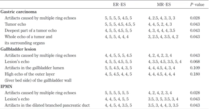
Fig. 1. Typical image of advanced gastric cancer obtained byER-ES.
Scores of six independent evaluators were : 5, 5, 4, 5, 5, and 5 for absence of artifacts caused by multiple ring echoes ; 5, 5, 5, 5, 5, and 5 for visibility of tumor echo ; 5, 5, 5, 5, 5, and 5 for visibility of deepest part of tumor ; 5, 5, 5, 4, 4, and 5 for visibility of whole echo of tumor and its surrounding organs.
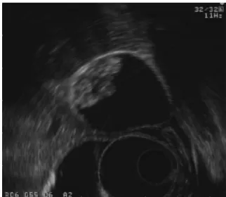
Fig. 2. Typical image of advanced gastric cancer obtained by MR-ES.
Scores of six independent evaluators were : 4, 4, 4, 4, 3, and 4 for absence of artifacts caused by multiple ring echoes ; 4, 4, 5, 4, 4, and 3 for visibility of tumor echo ; 4, 4, 4, 5, 4, and 3 for visibility of deepest part of tumor ; 4, 2, 4, 2, 3, and 1 for and visibility of whole echo of tumor and its surrounding organs.
Fig. 3. Typical image of gallbladder lesion (gallstone) obta- ined by ER-ES.
Scores of six independent evaluators were : 4, 4, 5, 5, 5, and 5 for absence of artifacts caused by multiple ring echoes ; 4, 4, 5, 5, 5, and 5 for visibility of small lesions in gallbladder ; 4, 5, 5, 5, 3, and 5 for absence of artif acts in gallbladder lumen ; 3, 3, 5, 5, 3, and 5 for visibility of high echo of outer layer (liver bed side) of gallbladder wall.
Fig. 4. Typical image of gallbladder lesion (gallstone) obta- ined by MR-ES.
Scores of six independent evaluators were : 4, 1, 2, 1, 2, and 4 for absence of artifacts caused by multiple ring echoes ; 5, 4, 4, 4, 4, and 5 for visibility of small lesions in gallbladder ; 4, 5, 4, 4, 4, and 4 for absence of artifacts in gallbladder lumen ; 4, 5, 5, 5, 5, and 4 for visibility of high echo of outer layer (liver bed side) of gallbladder wall.
the depth of invasion and lymph node metastasis.
As for gallbladder lesions, we decided to use small lesions, such as stones, polyps, and debris, because in chronic cholecystitis and advanced gallbladder carcinoma, the gallbladder lumen and wall structure are difficult to evaluate. Branch
-duct type IPMN was chosen because EUS is useful for its qualitative diagnosis. In the United States and Europe, EUS
-FNA is performed to obtain specimens of cystic pan- creatic lesions such as IPMN for cytopathologic analysis and determination of carcinoembryonic antigen. Sedlack
et al.25), however, reported that EUS was sensitive and accurate enough to identify malignant or potentially malignant pancreatic cystic lesions, and that EUS
-FNA did not enhance diag- nostic yield.
In all three groups of patients, the median scores for the ER
-ES were significantly higher than those for the MR
-ES with respect to absence of arti- facts caused by multiple ring echoes. This means that with the ER
-ES, diagnosis is hardly affected by these artifacts. In patients with gastric cancer, the median scores for the ER
-ES were significantly higher than those for the MR
-ES with respect to visibility of a tumor echo, visibility of the deepest part of a tumor, and visibility of the whole echo of a tumor and its surrounding organs, showing the superiority of the ER
-ES in staging gastric cancer.
These findings clearly indicate that with the ER
-ES, the EUS images obtained using standard B
-mode imaging are of excellent quality, that there is very little echo decrement, and that when combined with THI, the ER
-ES can visualize both a lesion and the
borders with its surrounding organs more clearly.
However, there were no statistically significant dif- ferences between the ER
-ES and MR
-ES with respect to visibility of small lesions in the gallblad- der, absence of artifacts in the gallbladder lumen, and visibility of a high echo of the outer layer (liver bed side) of the gallbladder wall. The reason for this may be that the same patients were not sub- jected to EUS using both the ER
-ES and MR
-ES.
The results may have been different in these items if the sample size was larger, or if the number of evaluators was greater. Nevertheless, when pati- ents with gallbladder lesions and those with IPMN were taken together, the median scores for the ER
-ES were significantly higher in four of the seven items, showing the definite superiority of the ER
-ES in patients with pancreaticobiliary lesions.
In conclusion, the ER
-ES is certainly superior to the MR
-ES in the quality of EUS images of GI tract and pancreaticobiliary lesions. We hope that electronic scanning EUS combining the functions of the ER
-ES and EUS
-FNA will be developed in the near future.
REFERENCES
1. DiMagno EP, Buxton JL, Regan PT, Hattery RR, Wilson DA, Suarez JR, Green PS. Ultrasonic endoscope. Lancet, 22: 629-631, 1980.
2. Strohm WD, Phillip J, Hagenmuller F, Classen M.
Ultrasonic tomography by means of an ultrasonic Fig. 5. Typical image of IPMN obtained by ER-ES.
Scores of six independent evaluators were : 4, 4, 5, 5, 4, and 5 for absence of artifacts caused by multiple ring echoes ; 4, 5, 5, 5, 5, and 5 for visibility of all lesions ; 4, 5, 5, 4, 4, and 5 for absence of artifacts in dilated branched pancreatic duct.
Fig. 6. Typical image of IPMN obtained by MR-ES.
Scores of six independent evaluators were : 4, 4, 4, 2, 3, and 4 for absence of artifacts caused by multiple ring echoes ; 5, 5, 5, 4, 5, and 4 for visibility of all lesions ; 5, 5, 5, 4, 5, and 4 for absence of artifacts in dilated branched pancreatic duct.
106 T. HIKICHI et al.
fiberendoscope. Endoscopy, 12: 241-244, 1980.
3. DiMagno EP, Regan RT, Clain JE, Buxton JL.
Human endoscopic ultrasonography. Gastroent- er ology, 83: 824-829, 1982.
4. Rosch T. Endoscopic ultrasonography. Endo- scopy, 24: 144-153, 1992.
5. Chak A. Endoscopic ultrasonography. Endo- scopy, 32: 146-152, 2000.
6. Caletti G, Fusaroli P. Endoscopic ultrasonography.
Endoscopy, 33: 158-166, 2001.
7. Fusaroli P, Vallar R, Togliani T, Khodadadian E, Caletti G. Scientific publications in endoscopic ultrasonography : a 20-year global survey of the literature. Endoscopy, 34: 451-456, 2002.
8. Fusaroli P, Caletti G. Endoscopic ultrasonography.
Endoscopy, 39: 17-20, 2007.
9. Noh KW, Woodward TA, Raimondo M, Savoy AD, Pungpapong S, Hardee JD, Wallace MB. Chan- ging trends in endosonography : linear imaging and tissue are increasingly the issue. Dig Dis Sci, 52: 1014-1018, 2007.
10. Vilmann P, Jacobsen GK, Henriksen FW, Hancke S.
Endoscopic ultrasonography with guided fine nee- dle aspiration biopsy in pancreatic diseases. Gas- trointest Endosc, 38: 172-173, 1992.
11. Hikichi T, Irisawa A, Bhutani MS, Takagi T, Shibukawa G, Yamamoto G, Wakatsuki T, Imamura H, Takahashi Y, Sato A, Sato M, Ikeda T, Hashimoto Y, Tasaki K, Watanabe K, Ohira H, Obara K. Endoscopic ultrasound-guided fine- needle aspiration of solid pancreatic masses with rapid on-site cytological evaluation by endosonog- raphers without attendance of cytopathologists. J Gastroenterol, 44: 322-328, 2009.
12. Wakatsuki T, Irisawa A, Bhutani MS, Hikichi T, Shibukawa G, Takagi T, Yamamoto G, Takahashi Y, Yamada Y, Watanabe K, Obara K, Suzuki T, Sato Y.
Comparative study of diagnostic value of cytologic sampling by endoscopic ultrasound-guided fine- needle aspiration and that by endoscopic retro- grade pancreatography for the management of pan- creatic masses without biliary stricture. J Gas t ro enterol Hepatol, 20: 1707-1711, 2005.
13. Bhutani MS, Hawes RH, Baron PL, Sanders- Cliette A, van Velse A, Osborne JF, Hoffman BJ.
Endoscopic ultrasound guided fine needle aspira- tion of malignant pancreatic lesions. Endoscopy, 29: 854-858, 1997.
14. Chang KJ, Nguyen P, Erickson RA, Durbin TE, Katz KD. The clinical utility of endoscopic ultra- sound-guided fine-needle aspiration in the diagno- sis and staging of pancreatic carcinoma.
Gastrointest Endosc, 45: 387-393, 1997.
15. Harewood GC, Wiersema MJ. Endosonography- guided fine needle aspiration biopsy in the evalua-
tion of pancreatic masses. Am J Gastroenterol, 97: 1386-1391, 2002.
16. Ando N, Goto H, Niwa Y, Hirooka Y, Ohmiya N, Nagasaka T, Hayakawa T. The diagnosis of GI stromal tumors with EUS-guided fine needle aspi- ration with immunohistochemical analysis.
Gastrointest Endosc, 55: 37-43, 2002.
17. Vazquez-Sequeiros E, Norton ID, Clain JE, Wang KK, Affi A, Allen M, Deschamps C, Miller D, Salmao D, Wiersema MJ. Impact of EUS-guided fine-needle aspiration on lymph node staging in patients with esophageal carcinoma. Gastrointest Endosc, 53: 751-757, 2001.
18. Annema JT, Versteegh MI, Veselic M, Voigt P, Rabe KF. Endoscopic ultrasound-guided fine-needle aspiration in the diagnosis and staging of lung can- cer and its impact on surgical staging. J Clin Oncol, 23: 8357-8361, 2005.
19. Catalano MF, Rosenblatt ML, Chak A, Sivak MV Jr, Scheiman J, Gress F. Endoscopic ultrasound- guided fine-needle aspiration in the diagnosis of mediastinal masses of unknown origin. Am J Gastroenterol, 97: 2559-2565, 2002.
20. Matthes K, Bounds BC, Collier K, Gutierrez A, Brugge W. EUS staging of upper GI mali- gnancies : results of a prospective randomized trial. Gastrointest Endosc, 64: 496-502, 2006.
21. Papanikolaou IS, Delicha EM, Adler A, Wegener K, Pohl H, Wiedenmann B, Rosch T. Prospective, ran d o mized comparison of mechanical and elec- tronic radial endoscopic ultrasound sys- tems : assessment of performance parameters and image quality. Scand J Gastroenterol, 44: 93-99, 2009.
22. Anderson MA, Scheiman JM. Initial experience with an electronic radial array echoendo- scope : randomized comparison with a mechanical sector scanning echoendoscope in humans. Gast- rointest Endosc, 56: 573-577, 2002.
23. Niwa K, Hirooka Y, Itoh A, Hashimoto S, Hirai T, Takeda K, Goto H. Preclinical study of endo- scopic ultrasound with electronic radial scanning echoendoscope. J Gastroenterol Hepatol, 18: 828-835, 2003.
24. Niwa K, Hirooka Y, Niwa Y, Itoh A, Ohmiya N, Hashimoto S, Ishikawa H, Okada N, Itoh T, Goto H.
Comparison of image quality between electronic and mechanical radial scanning echoendoscope in pancreatic diseases. J Gastroenterol Hepatol, 19: 454-459, 2004.
25. Sedlack R, Affi A, Vazquiz-Sequeiros E, Norton ID, Clain JE, Wiersema MJ. Utility of EUS in the evaluation of cystic pancreatic lesions. Gastro- intest Endosc, 56: 543-547, 2002.