electrochemically active bacteria, EAB
bioelectrochemical system BES EAB
EAB microbial fuel cell, MFC
EAB microbial electrosynthesis, MES EAB
Shewanella oneidensis Geobacter sulfurreducens EAB
I Acidithiobacillus EAB Acidithiobacillus BES ( ) CO2 MES Acidithiobacillus Acidithiobacillus ferrooxidans ATCC23270 16S rRNA ATCC23270 Acidithiobacillus sp. NU-1 16S rRNA NU-1 A. ferriphilus A. ferriphilus Acidithiobacillus NU-1 ATCC23270 2 (pH 2.0) NU-1 NU-1 MES Acidithiobacillus
EAB NU-1 MES
II EAB MFC MFC CT AL CL MFC AL 140 mW m-2 EAB AL MFC Geobacter G. pelophilus G. psychrophilus Geobacter III EAB EAB EAB EAB EPC
electron-acceptor EPC EA-EPC G.
sulfurreducens
Ag/AgCl 0.2 V EA-EPC 4 BES EPC FTO Citrobacter EPC Sulfurospirillum Geobacter EA-EPC Geobacter EA-EPC EAB EAB
1. Ueoka, N., Kouzuma, A., & Watanabe, K. (2016). Missing Iron-Oxidizing Acidophiles Highly Sensitive to Organic Compounds. Microbes and environments, 31(3), 244-248. 2. Ueoka, N., Sese, N., Sue, M., Kouzuma, A., & Watanabe, K. (2016). Sizes of anode and
cathode affect electricity generation in rice paddy-field microbial fuel cells. Journal of
Sustainable Bioenergy Systems, 6(01), 10.
1.1
Electrochemically active bacteria; ×EAB
EAB Hirose et al., 2018)
EAB Exoelectrogens (Logan, 2009)
EAB Electrotrophs
(Lovley, 2011) EAB
(Bioelectrochemical system; × BES; Fig. 1) (Rabaey et al., 2009) EAB BES
Fig. 2 Geobacter
Thiobacillus
(Interspecies electron transfer, IET) (Kato et al., 2012) EAB EAB (Summers et al., 2010) 1.2 Geobacter sulfurreducens (Caccavo et al., 1994) Shewanella oneidensis (Venkateswaran et al., 1999)
(Zuo et al., 2008)
Geobacter G. sulfurreducens
Shewanella S. oneidensis
(Kouzuma et al., 2015)
Citrobacter
(Huang et al., 2014)
Pseudomonas aeruginosa (Rabaey et al., 2005) Aeromonas hydrophila (Pham
et al., 2003) Clostridium butyricum (Park et al., 2001) Klebsiella pneumoniae (Rhoads et al., 2005) Thermincola ferriacetica (Wrighton et al., 2011) Lysinibacillus sphaericus (He et al., 2014)
Staphylococcus aureus (Bhuvaneswari et al., 2013)
Listeria monocytogenes (Light et al., 2018) 1.3 Sporomusa ovata CO2 (Tremblay et al., 2015)
Thiobacillus denitrificans (D P Kelly & Wood, 2000)
(Kato et al., 2012)
Acidithiobacillus ferrooxidans
BES
(Ishii et al., 2015)
Acidithiobacillus
(D P Kelly & Wood, 2000)
( )
(Matsumoto et al., 1999)
( ) Fig. 5
Amouric A. ferrooxidans 4 (Amouric et al., 2011)
A. ferrivorans (Hallberg et al.,
2009) A. ferridurans (Hedrich & Johnson, 2013) A. ferriphilus (D B Johnson & Falagán, 2016)
Acidithiobacillus
(Amouric et al., 2011)
Acidithiobacillus
(Matsumoto et al., 1999) Acidithiobacillus
Acidithiobacillus
BES
1.4 EAB
EAB
microbial fuel cell; ×MFC (Lovley et al, 2008)
MFC Ω
Ω MFC
(Miyahara et al., 2013) Ω (Asai et al.,
2017) MFC Ω
80
(microbial electrosynthesis;
×MES) (Rabaey & Rozendal, 2010) MES S. ovata
CO2 (Tremblay et al., 2015) Thiobacillus (Pous et al., 2014) 1.5 EAB ( ) (Kouzuma et al., 2018) EAB
EAB Geobacter (Lovley et al., 2011)
Shewanella (Kouzuma et al., 2015)
BES
EAB MFC MES
BES EAB
U-tube
MFC Ochrobactrum (Zuo et al., 2008)
1.6 MFC MES EAB EAB Acidithiobacillus EAB
Electrode plate culture; ×EPC
Fig. 6 (FTO )
×
EPC electron-accepting EPC EA-EPC
EPC electron-donating EPC ED-EPC
Fig. 1
CO2
CO2
Fig. 3
CO2 , H+
O2
Fig. 5 Fe3+ Fe2+
CuS
DAPI DNA AT 2 mL 350 mM 450 µL 50 µL DAPI (1 mg/mL) 25 µL ( 0.2 µm ADVANTEC) (BX60 ) Fig. 2 10 × (cell/mL) 10 2.2.3 9K 9K
Solution A Solution B Solution A × 10 x
2.2.6 DNA 100 mL 9K ε DNA 50 mL (4 9000 rpm 20 ) 1.5 mL (4 12000 g 20 ) TEN buffer (pH 2.0) ε PBS buffer (pH 7) ε 2
DNA Tissue Genomic DNA Extraction Mini Sample Kit (FAVORGEN) 200 µL FATG1 Buffer
Micropestle ± 20 µL Proteinase K (10 mg/ml) 60
200 µL FATG2 Buffer
70 10 200 µL 100%
FATG Mini Column
1 500 µL W1 Buffer 750 µL Wash
Buffer ε 3 Column
Elution Tube Elution Buffer 3 2
DNA DNA RNase
DNA DNA 1/1000 RNaseA ( ,
10 mg/mL) 37 NucleoSpin gDNA
Clean-up (MACHEREY-NAGEL) DNA DNA 150 µL
Binding Buffer DB 450 µL 5 Column
2.2.8
MEGA6.06-Mac (Tamura et al., 2013)
fasta MEGA ClustalW
mas mas
neighbor-joining National
Center for Biotechnology Information (NCBI; http://www.ncbi.nlm.nih.gov/)
2.2.9
NU-1 DNA 3 µg illumina Miseq (150 bp x
2) DNA CLC Genomics Workbench (CLC
bio) MetaGeneAnnotator
(CDS) JCM18981 JCM7812 JCM3865
(300 bp x2)
2.2.10 DNA-DNA
DSMZ in silico DNA-DNA Hybridization (DDH) GGDC
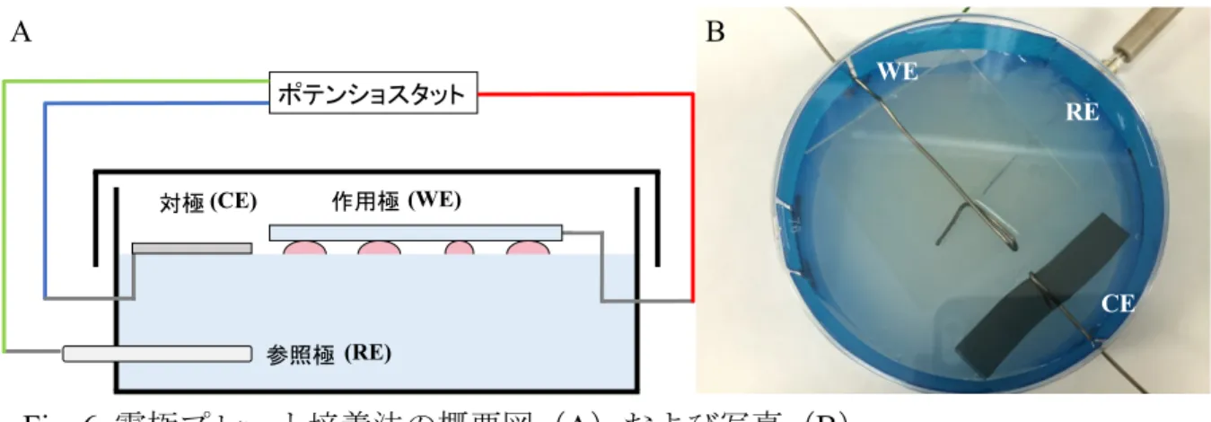
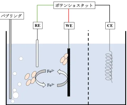
(http://ggdc.dsmz.de/) GGDC DDH 2.2.11 (Fig. 3) (3 x 4 cm) ( 0.30 mm 20 cm; ) / (Ag/AgCl) (HX-R5; )
Nafion perfluorinated membrane (SIGMA-ALDRICH) 100
mM (FeSO4) 9K 9K
150 mL 1 mL
(VMP-3 ) 0 V (vs. Ag/AgCl)
30
2.2.12 ( ) NU-1 9K 3 NU-1 1 : 1 (SEM, JSM-7500F ) 2.3 2.3.1 NU-1 Acidithiobacillus
A. ferrooxidans ATCC23270 ATCC 9K (pH 2.0) (Fig. 4) 50 61 ATCC23270 ATCC23270 Acidithiobacillus 9K (Johnson, 1995) 2 9K 5~7 (Fig. 5) 9K 2 DNA 16S rRNA PCR PCR 3 PCR (Fig. 6) ATCC23270
A. ferriphilus (Falagán & Johnson, 2016) Acidithiobacillus sp. NU-1
2.3.2
NU-1 Acidithiobacillus DSM14882
9K (Fig. 7) NU-1 (Table 2) NU-1 2.1 x 10⁸ cell/mL DSM14882 (5.6 x 10⁷ cell/mL) 3.6 2.3.3 NU-1 DSM14882 (0~20 mM) (0~10 mM) 9K (Fig. 8a 8b) NU-1 10 mM 5 mM × Acidithiobacillus DSM14882 NU-1 2.3.4 NU-1 Table 5 34,672,023bp 69 2,536,979 bp 2,521 CDS JCM18981 JCM7812 JCM3865 (Table 3) DNA-DNA 2.3.5 16S rRNA (HiPIP)
(Fig. 9-11) 16S rRNA (Fig. 9) NU-1
A. ferriphilus A .ferriphilus
rusB HiPIP iro
(Fig. 10) HiPIP (Fig. 11)
in silico DDH NU-1 DDH JCM18981 JCM7812 JCM3865 NCBI ATCC23270 SS3 NU-1 ATCC23270 JCM18981 SS3 DDH 70% × NU-1 JCM7812 JCM3865 70% NU-1 A. ferriphilus 2.3.7 NU-1 NU-1 Calvin-Benson-Bassham nifA nifH A. ferrivorans SS3
Hallberg et al., 2010; Liljeqvist et al., 2011 NU-1
IV JCM7812 nifH
NU-1
Rus I
rusA (Valdés et al., 2008) rusB
IV Rus (Amouric et al., 2011) 2.3.8 NU-1 JCM18981 7 (Fig. 12) NU-1 15 JCM18981 2.3.9
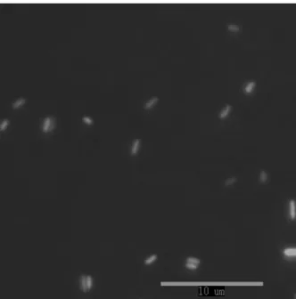
NU-1 (SEM) (Fig. 13) NU-1
1.7 x 0.6 ( 0.23 x 0.04) m
2.4
NU-1 Acidithiobacillus (Amouric et al., 2011) A. ferrooxidans JCM7811 (JCM7811-P1 JCM7811-P4) NU-1 ATCC ATCC23270 Acidithiobacillus ATCC23270 NU-1 ATCC23270 ATCC Acidithiobacillus Acidithiobacillus NU-1
(Fig. 5) NU-1 Acidithiobacillus
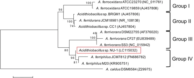
Fig. 8 NU-1 DSM14882 NU-1 Acidithiobacillus NU-1 9K (Fig. 5) Acidithiobacillus Acidithiobacillus 16S rRNA
I~IV 4 (Amouric et al., 2011) A.
ferrooxidans I A. ferridurans II A. ferrivorans III
A. ferriphilus IV Amouric (2011)
Acidithiobacillus
rusA rusB 2 (Sasaki et al., 2003;
Amouric et al., 2011) A. ferriphilus IV rusB
HiPIP iro rusB
Acidithiobacillus IV A. ferriphilus (Fig. 9) NU-1 pH 2.0 9K (Fig. 7) DSM14882 (=ATCC23270 ) 3.6 IV JCM7812 4.5 NU-1 rusA Rus rusA
NU-1 HiPIP rusA hip A. ferriphilus
(Fig. 10 Fig. 11)
Table 1.
Strain Origin Source or reference
Acidithiobacillus sp.
NU-1 ATCC23270Tculture This study
BRGM1 Sulfide ore mine, South Africa Liu et al. (2000)
CC1 Acid mine drainage, Carnoule`s, France Duquesne et al. (2003)
JCM3865 Acid mine water, Shimokawa, Japan Japan Collection of Microorganisms
(Takamori et al., 1983)
A. ferrooxidans
ATCC23270T Acid bituminous coal mine effluent, USA American Type Culture Collection
DSM14882T DSMZ
(Leathen & Braley, 1954)
ATCC19859 Acid copper mine leaching water, Canada Razzell & Trusell, 1963
A. ferridurans
ATCC33020T(= JCM18981T) Uranium mine, Japan Japan Collection of Microorganisms
(Tomizuka et al., 1976)
A.ferrivorans
NO-37T(= DSM22755T) Copper mine spoil drainage, Norway DSMZ
(Hallberg et al. 2010)
SS3 Norilsk mining area, Russia Kupka et al. (2007)
CF27 Abandoned copper/cobalt mine drainage, ID,
USA
Hallberg et al. (2010)
A. ferriphilus
M20T Galway's soufriere, Montserrat Falagán and Johnson (2015)
JCM7812 Matsuo sulfur and iron sulfide mine, Japan Japan Collection of Microorganisms
(Wakao et al., 1991)
A. caldus
Table 4. NU-1
1 Tuovinen and Kelly, 1974 2 Hedrich and Johnson, 2013 3 Hallberg et al., 2010 4 Falagan and Johnson, 2015
*DDH SS3 (4.3.4)
Strain Acidithiobacillus sp.
NU-1 A. ferrooxidansDSM14882T A. ferriduransJCM18981T A. ferrivoransDSM22755T A. ferriphilusJCM7812
Group IV I II III IV
Optimum pH 2.0 2.01 2.02 2.53 2.04
Optimum temp 30 30 1 30 2 30 3 30 4
Maximum cell density
(pH2.0, Fe9K) 2.1 x 10⁸cell/mL 5.6 x 10⁷cell/mL 1.0 x 10⁸cell/mL 3.1 x 10⁶cell/mL 4.7 x 10⁷cell/mL
Colony formation
on agarose plate - + +
Colony formation
on silica gel plate + + +
Rusticyanin rusA ? rusA rusA rusB rusB
HiPIP hip ? hip hip iro iro
Fig. 1 9K 9K 100 mM
Fig. 5 NU-1 NU-1 9K (a)
9K (b) 2
Fig. 6 Acidithiobacillus NU-1 16S
rRNA PCR
neighbor-joining
A.ferrooxidans ATCC23270 (218665024) A.ferridurans ATCC33020 (NR_108138)
Fig. 9 16S rRNA NU-1
16S rRNA
neighbor-joining Acidithiobacillus
NU-1 IV
A.ferrooxidans ATCC23270 (218665024) A.ferrooxidans ATCC19859 (AJ457808) A.ferrooxidans BRGM1 (AJ457806)
A.ferridurans ATCC33020 (NR_108138) A.ferrooxidans CC1 (AJ457804)
A.ferrivorans NO-37 (AF376020) A.ferrivorans CF27 (EU839489) A.ferrivorans SS3 (344198243) Acidithiobacillus sp. NU-1 A.ferrooxidans JCM7812 (FN686782) A.ferriphilus M20 (KR905751) A.caldus DSM8584 (Z29975) 1 0 0 4 6 1 0 0 7 4 1 0 0 5 6 8 0 8 1 9 6 0.005 Group IV Group III Group II Group I A. ferrivorans DSM22755 (AF376020) A. ferrivorans CF27 (EU839489) A. ferrivorans SS3 (NC_015942) A. ferriphilus JCM7812 (FN686782) A. ferriphilus M20 (KR905751) Acidithiobacillus sp. NU-1 (LC115032) A. ferrooxidans ATCC23270 (NC_011761) A. ferrooxidans ATCC19859 (AJ457808) Acidithiobacillus sp. BRGM1 (AJ457806)
A. ferridurans JCM18981 (NR_108138) Acidithiobacillus sp. CC1 (AJ457804)
Fig. 10 NU-1 neighbor-joining A.ferrooxidans BRGM1 (FN688760) A.ferridurans ATCC33020 A.ferrooxidans CC1 (FN688759) A.ferrooxidans ATCC19859 (FN688754) A.ferrooxidans ATCC23270 (NC_011761) Acidithiobacillus sp. NU-1
Fig. 11 HiPIP NU-1 HiPIP neighbor-joining ATCC23270T (AFE_2732) ATCC19859 (AJ621387) ATCC33020T (AJ320262) BRGM1 (AJ621388) NU-1 DSM22755T (FN688778) CF27 (FN688779) JCM3865 (FN688775) JCM7812 (FN688774) 1 0 0 1 0 0 1 0 0 9 9 9 6 9 9 0.05 hip iro A. ferrivorans DSM22755 (FN688778) A. ferrivorans CF27 (FN688779) A. ferriphilus JCM7812 (FN688774) Acidithiobacillus sp. JCM3865 (FN688775) Acidithiobacillus sp. NU-1 (LC115034) A. ferrooxidans ATCC23270 (NC_011761)
A. ferrooxidans ATCC19859 (AJ621387)
Fig. 13 NU-1 NU-1 SEM
3.1
rice paddy-field MFC, RPF-MFC
EAB
3.2 3.2.1
(Kouzuma et al., 2013) Fig.
4 GF-80-5F
Table 1 SE, small electrode; LE, large electrode; AL. anode limited; CL, cathode limited
10 cm nafion
0.1 mg/cm2 TEC10E20A
1000
HA-1510, Graphtec PC Oryza sativa L. cv.
Koshihikari Fig. 1
3.2.2
HSV-100
current vs. voltage current vs. power
Pmax [mW] (Logan et al., 2006)
Qmax [mW m-2] t
P < 0.05 3.2.3 16S rRNA
93 0.5 g 0.5 x 0.5 cm
DNA
DNA Fast DNA SPIN Kit for Soil Funakoshi
DNA 50 µL DES 16S
2013) PCR QIA quick PCR purification kit Qiagen
PCR DNA 1 ng/µL Genome
Sequencer FLX system Roche Applied Science
Silva rRNA database http://www.arb-silva.de/
2.2.8 DNA DDBJ Sequence Read Archive
Database accession number: DRA004371
3.2.4 Fe2O3 Table 2 10 mL 5 30 150 mL (Agarose L03 ) 2.25 g (121 20 ) 60 10 100 µL 30 3.3 3.3.1 Table 1 RPF-MFC
20 93 Pmax Qmax Fig. 2 LE SE
EAB 3.3.2 Geobacter (Kouzuma et al., 2013) Geobacter PCR 16S rRNA Fig. 3 AL CL Family Geobacter Geobacteraceae 0.1% CL 1.8 AL 2.4 Geobacteraceae AL Geobacter AL
CL G. pelophilus (Straub & Buchholz-cleven, 2016) G.
psychrophilus (Nevin et al., 2005) Fig. 4
Table 1 LE SE Fig. 2a SE LE Fig. 2b 2c LE SE LE Pmax Fig. 2a AL CL Pmax SE LE Pmax AL Pmax CL × 93 AL Qmax 140mW m-2 Fig. 2b Geobacter DNA 16S rRNA
Fig. 3 Geobacter Geobacteraceae 0.1%
CL 1.8 AL 2.4 Geobacteraceae
AL
Geobacter
AL CL G. pelophilus (Straub &
Buchholz-cleven, 2016) G. psychrophilus (Nevin et al., 2005)
Fig. 4 G. pelophilus G. psychrophilus
(Kouzuma et al., 2013)
Fig. 1 a 70 b
Fig. 4 AL CL Geobacter
NCBI
± 100
EA-EPC
4.1 EA-EPC Geobacter sulfurreducens 4.2 4.2.1 Geobacter sulfurreducens DSM 12127T DSMZ DSM826 Table 1 100 mL 50 mL 100 mM L- 500 µL 15 N2 H2 CO2 500 µL 30 4.2.2 EPCEPC Fig. 1 EPC
6.5 cm 3.0 cm 100 mL UV FTO 5 × 5 cm Sigma-Aldrich DSM826 2.0 g/L NaCl 100 mM L- EA-free DSM826 6.0 g/L LO3 60 Ag/AgCl HX-R5; EPC
CBB Coomassie Brilliant Blue 10 µg/mL 1 x 5 cm
EPC
Fig. 1. EPC WE FTO CE
Fig. 2. WE
EA-EPC
5.1 EA-EPC EA-EPC 5.2 5.2.1 3 AL 5.2.2 2 × 5 × 1 cm 120 mL Fig. 1 2 × 5 cm Ag/AgCl 4.2.2 EA-free DSM826 15 -0.2 V vs Ag/AgCl 30 5.2.3 EPC EPC 4.2.2 EA-free DSM826 EPC EPC -0.2 V vs Ag/AgCl 30 5.2.4 PCR EPC 16S rRNAPCR DNA Ex Taq Total DNA
16S rRNA
TACGGYTACCTTGTTACGACTT 96 30 (96 30
60 30 72 1 30 ) 30 72 7 PCR
EPC
5.2.5
PCR QIAquick PCR Purification Kit QIAGEN
PCR NCBI Nucleotide BLAST
2.2.8 MEGA6.06-Mac NCBI 5.2.6 4.2.4 5.2.7 DAPI 2.2.2 5.3 5.3.1 4 × 5.3.2 EA-EPC 1 1 cm EA-free DSM826 EPC 12 Fig. 2 12 FTO × CBB 50 Fig. 3 5.3.3 16S rRNA 45 Table 1 × 1st trial
EA-EPC Geobacter Citrobacter Macellibacteroides Parabacteroides
Eubacterium Table 1, 1st trial Citrobacter
ND-3 × Citrobacter ND-2 -0.2 V vs Ag/AgCl Fig. 4 16S rRNA Fig. 5 Geobacter Citrobacter 5.3.4 × 1st trial Citrobacter
EA-EPC × 2nd trial EA-EPC
1st trial 2nd trial 2nd trial EA-EPC
Fig. 6 Fig. 7 PCR 16S
rRNA
Sulfurospirillum Table 2, 2nd trial
Sulfurospirillum BES
(Marshall et al., 2017)
Fig. 8
3 ~ 4 µm 1 ~ 2 µm
Fig. 9
EA-EPC 3rd trial EA-EPC
Fig. 10 Fig. 11 PCR
Geobacter
Sulfurospirillum Table 1, 3rd trial
RPFA-12G-1
Fig. 12
1 ~ 2µm 16S rRNA
Fig. 13 EA-EPC Geobacter 5.4 EA-EPC Geobacter (Kouzuma et al., 2013) Geobacter
EA-EPC 1st trial EA-EPC
Fig. 3 ×
Citrobacter Table 1, 1st trial
Citrobacter (Xu & Liu,
2011)
(Huang et al., 2014) Geobacter
Fig. 4, Fig. 12 Citrobacter
Enterobacteriaceae
EA-EPC
2nd trial EA-EPC ×
Sulfurospirillum
Table 1, 2nd trial Sulfurospirillum
(Kodama et al., 2007) Sulfurospirillum BES
(Marshall et al., 2017)
Fig. 9 3rd trial EA-EPC Geobacter
Sulfurospirillum 2nd trial
EA-EPC Geobacter Sulfurospirillum
Sulfurospirillum
Geobacter Fig. 12 G.
Fig. 1 EAB WE CE
Fig. 5 Citrobacter 16S rRNA 1st trial EA-EPC Citrobacter sp. ND-3 Citrobacter sp. ND-2 Citrobacter sp. ND-1 C. amalonaticus ATCC25405 (KX450351) C. farmeri GTC01319 (AB741662) C. farmeri 2991-81 (AF025371) C. sedlakii CDC4696-86T (AF025364) C. koseri ATCC27028 (KX450352) Citrobacter sp. XS-1 (HQ845373) Citrobacter sp. sdy-48 (FJ463782) Citrobacter sp. yy-21 (FJ463779) C. rodentinum CDC1843-73T (AF025363) C. bitternis SKKU-TP7 (KJ817168) C. freundii ATCC8090 (KM515969) Citrobacter sp. Z7 (JX185134) Citrobacter sp. ND-3 Citrobacter sp. ND-2 Citrobacter sp. ND-1 Citrobacter amalonaticus ATCC25405 (KX450351) Citrobacter farmeri GTC01319 (AB741662) Citrobacter farmeri 2991-81 (AF025371)
Citrobacter sedlakii CDC4696-86T (AF025364)
Citrobacter koseri ATCC27028 (KX450352) Citrobacter sp. XS-1 (HQ845373)
Citrobacter sp. sdy-48 (FJ463782) Citrobacter sp. yy-21 (FJ463779) Citrobacter rodentium CDC1843-73T (AF025363) Citrobacter bitternis SKKU-TP7 (KJ817168)
Fig. 9 Fig. 8
Fig. 13 Geobacter 16S rRNA 3rd trial EA-EPC G. sulfurreducens G. bemidjiensis Bem (NR_075007) G. bremensis Dfr1 (NR_026076) G. humireducens (AY187306) G. uraniireducens Rf4 (NR_074940) G. lovleyi SZ (NR_115337) G. argillaceus G12 (NR_043575) G. pelophilus Dfr2 (NR_026077) G. chapellei 172 (U41561) G. psychrophilus P35 (NR_043075) G. hydrogenophilus (U46860) G. metallireducens GS-15 (NC_007517) G. anodireducens SD-1 (NR_126282) G. sulfurreducens PCA (NC_002939)
G. sulfurreducens subsp. ethanolicus (AB762695) RPFA-12G-1
RPFA-12G-4
Desulfuromonas acetoxidans (AY187305) 1 0 0 6 5 1 0 0 1 0 0 1 0 0 8 8 8 3 5 7 4 7 9 9 1 0 0 9 7 6 9 9 2 0.01 G. bemidjiensis Bem (NR_075007) G. bremensis Dfr1 (NR_026076) G. humireducens (AY187306) G. uraniireducens Rf4 (NR_074940) G. lovleyi SZ (NR_115337) G. argillaceus G12 (NR_043575) G. pelophilus Dfr2 (NR_026077) G. chapellei 172 (U41561) G. psychrophilus P35 (NR_043075) G. hydrogenophilus (U46860) G. metallireducens GS-15 (NC_007517) G. anodireducens SD-1 (NR_126282)
G. sulfurreducens subsp. ethanolicus (AB762695)
Desulfuromonas acetoxidans (AY187305)
← (3rdtrial)
←
G. sulfurreducens DSM12127 (NC_002939)
6.1 ( ) EAB MFC MES EAB EAB EAB BES EAB MFC MES EAB EAB EAB Acidithiobacillus EAB
Acidithiobacillus EAB NU-1 NU-1
EAB NU-1
Acidithiobacillus
6.2 6.2.1 EA-EPC × EA-EPC ± 16S rRNA 6.2.2 EPC
EPC electron donating EPC, ED-EPC
T. denitrificans S. ovata EA-EPC 16S rRNA 6.2.3 Acidithiobacillus EPC Acidithiobacillus Acidithiobacillus
ED-EPC At. ferrooxidans ED-EPC
ED-EPC
pH
ED-EPC ED-EPC
Acidithiobacillus NU-1
μ
EAB
±
(1)
1. Ueoka, N., Kouzuma, A., & Watanabe, K. (2016). Missing Iron-Oxidizing Acidophiles Highly Sensitive to Organic Compounds. Microbes and environments, 31, 244-248.
2. Ueoka, N., Sese, N., Sue, M., Kouzuma, A., & Watanabe, K. (2016). Sizes of anode and cathode affect electricity generation in rice paddy-field microbial fuel cells. Journal of
Sustainable Bioenergy Systems, 6, 10.
3. Ueoka, N., Kouzuma, A., & Watanabe, K. (2018). Electrode plate-culture methods for colony isolation of exoelectrogens from anode microbiomes. Bioelectrochemistry, 124, 1-6.
(2) 1. , μ . . Electrochemistry, 84, 104-106 (2016). 2. μ . . 10:19-22. (3) 1.
N. UEOKA, A. KOUZUMA, K. WATANABE. Isolation and electrochemical cultivation of a novel iron-oxidizing bacterium NU-1 affiliated with the genus Acidithiobacillus. The 5th international meeting on microbial electrochemistry and technologies, Arizona, USA, October, 2015.
2.
Amouric, A., Brochier-Armanet, C., Johnson, D. B., Bonnefoy, V., & Hallberg, K. B. (2011). Phylogenetic and genetic variation among Fe(II)-oxidizing acidithiobacilli supports the view that these comprise multiple species with different ferrous iron oxidation pathways.
Microbiology, 157(1), 111–122. https://doi.org/10.1099/mic.0.044537-0
Asai, Y., Miyahara, M., Kouzuma, A., & Watanabe, K. (2017). Comparative evaluation of wastewater-treatment microbial fuel cells in terms of organics removal, waste-sludge production, and electricity generation. Bioresources and Bioprocessing, 4(1), 30. https://doi.org/10.1186/s40643-017-0163-7
Bhuvaneswari, A., Navanietha, K. R., & Berchmans, S. (2013). Metamorphosis of pathogen to electrigen at the electrode/electrolyte interface: Direct electron transfer of Staphylococcus
aureus leading to superior electrocatalytic activity. Electrochemistry Communications, 34,
25–28. https://doi.org/10.1016/j.elecom.2013.05.013
Caccavo, F., Lonergan, D. J., Lovley, D. R., Davis, M., Stolz, J. F., & McInerney, M. J. (1994).
Geobacter sulfurreducens sp. nov., a hydrogen- and acetate-oxidizing dissimilatory
metal-reducing microorganism. Applied and Environmental Microbiology, 60(10), 3752–3759. https://doi.org/0099-2240/$04.00+0
Falagán, C., & Barrie Johnson, D. (2016). Acidithiobacillus ferriphilus sp. nov., a facultatively anaerobic iron- and sulfur-metabolizing extreme acidophile. International Journal of
Systematic and Evolutionary Microbiology, 66(1), 206–211.
https://doi.org/10.1099/ijsem.0.000698
Hallberg, K. B., González-Toril, E., & Johnson, D. B. (2009). Acidithiobacillus ferrivorans, sp. nov.; facultatively anaerobic, psychrotolerant iron-, and sulfur-oxidizing acidophiles isolated from metal mine-impacted environments. Extremophiles, 14(1), 9–19. https://doi.org/10.1007/s00792-009-0282-y
Hedrich, S., & Johnson, D. B. (2013). Acidithiobacillus ferridurans sp. nov., an acidophilic iron-, sulfur- and hydrogen-metabolizing chemolithotrophic gammaproteobacterium.
International Journal of Systematic and Evolutionary Microbiology, 63(PART 11), 4018–
4025. https://doi.org/10.1099/ijs.0.049759-0
Hirose, A., Kasai, T., Aoki, M., Umemura, T., Watanabe, K., & Kouzuma, A. (2018). Electrochemically active bacteria sense electrode potentials for regulating catabolic pathways. Nature Communications, 9(1), 1083. https://doi.org/10.1038/s41467-018-03416-4
Huang, J., Zhu, N., Cao, Y., Peng, Y., Wu, P., & Dong, W. (2014). Exoelectrogenic Bacterium Phylogenetically Related to Citrobacter freundii, Isolated from Anodic Biofilm of a Microbial Fuel Cell. Applied Biochemistry and Biotechnology, 175(4), 1879–1891. https://doi.org/10.1007/s12010-014-1418-9
Ishii, T., Kawaichi, S., Nakagawa, H., Hashimoto, K., & Nakamura, R. (2015). From chemolithoautotrophs to electrolithoautotrophs: CO2 fixation by Fe(II)-oxidizing bacteria coupled with direct uptake of electrons from solid electron sources. Frontiers in
Microbiology, 6(SEP), 1–9. https://doi.org/10.3389/fmicb.2015.00994
Johnson, D. B. (1995). Selective solid media for isolating and enumerating acidophilic bacteria.
Journal of Microbiological Methods, 23(2), 205–218.
https://doi.org/10.1016/0167-7012(95)00015-D
Johnson, D. B., & Falagán, C. (2016). Acidithiobacillus ferriphilus sp. nov., a facultatively anaerobic iron- and sulfur-metabolizing extreme acidophile. International Journal of
Systematic and Evolutionary Microbiology, 66(1), 206–211.
https://doi.org/10.1099/ijsem.0.000698
Kaku, N., Yonezawa, N., Kodama, Y., & Watanabe, K. (2008). Plant/microbe cooperation for electricity generation in a rice paddy field. Applied Microbiology and Biotechnology, 79(1), 43–49. https://doi.org/10.1007/s00253-008-1410-9
Kato, S., Hashimoto, K., & Watanabe, K. (2012). Microbial interspecies electron transfer via electric currents through conductive minerals. Proceedings of the National Academy of
Kelly, D. P., & Wood, A. P. (2000). Confirmation of Thiobacillus denitrificans as a species of the genus Thiobacillus, in the beta-subclass of the Proteobacteria, with strain NCIMB 9548 as the type strain. International Journal of Systematic and Evolutionary Microbiology, 50(2), 547–550. https://doi.org/10.1099/00207713-50-2-547
Kelly, D. P., & Wood, A. P. (2000). Reclassification of some species of Thiobacillus
Acidithiobacillus gen . nov ., Halothiobacillus. International Journal of Systematic and Evolutionary Microbiology, (2000), 511–516.
Kodama, Y., Ha, L. T., & Watanabe, K. (2007). Sulfurospirillum cavolei sp. nov., a facultatively anaerobic sulfur-reducing bacterium isolated from an underground crude oil storage cavity.
International Journal of Systematic and Evolutionary Microbiology, 57(4), 827–831.
https://doi.org/10.1099/ijs.0.64823-0
Kouzuma, A., Ishii, S., & Watanabe, K. (2018). Metagenomic insights into the ecology and physiology of microbes in bioelectrochemical systems. Bioresource Technology, 255, 302– 307. https://doi.org/10.1016/j.biortech.2018.01.125
Kouzuma, A., Kaku, N., & Watanabe, K. (2014). Microbial electricity generation in rice paddy fields: recent advances and perspectives in rhizosphere microbial fuel cells. Applied
Microbiology and Biotechnology, 98(23), 9521–9526.
https://doi.org/10.1007/s00253-014-6138-0
Kouzuma, A., Kasai, T., Hirose, A., & Watanabe, K. (2015). Catabolic and regulatory systems in shewanella oneidensis MR-1 involved in electricity generation in microbial fuel cells.
Frontiers in Microbiology, 6(JUN), 1–11. https://doi.org/10.3389/fmicb.2015.00609
Kouzuma, A., Kasai, T., Nakagawa, G., Yamamuro, A., Abe, T., & Watanabe, K. (2013). Comparative metagenomics of anode-associated microbiomes developed in rice paddy-field microbial fuel cells. PLoS ONE, 8(11), 2–11. https://doi.org/10.1371/journal.pone.0077443
Light, S. H., Su, L., Rivera-Lugo, R., Cornejo, J. A., Louie, A., Iavarone, A. T., … Portnoy, D. A. (2018). A flavin-based extracellular electron transfer mechanism in diverse Gram-positive bacteria. Nature, 562(7725), 140–144. https://doi.org/10.1038/s41586-018-0498-z
Logan, B. E. (2009). Exoelectrogenic bacteria that power microbial fuel cells. Nature Reviews
Microbiology, 7, 375–381. https://doi.org/10.1038/nrmicro2113
Logan, B. E., Hamelers, B., Rozendal, R., Schröder, U., Keller, J., Freguia, S., … Rabaey, K. (2006). Microbial fuel cells: Methodology and technology. Environmental Science and
Technology, 40(17), 5181–5192. https://doi.org/10.1021/es0605016
Lovley, D. R. (2008). The microbe electric: conversion of organic matter to electricity. Current
Opinion in Biotechnology, 19(6), 564–571. https://doi.org/10.1016/j.copbio.2008.10.005
Lovley, D. R. (2011). Powering microbes with electricity: Direct electron transfer from electrodes to microbes. Environmental Microbiology Reports, 3(1), 27–35. https://doi.org/10.1111/j.1758-2229.2010.00211.x
Lovley, D. R., Ueki, T., Zhang, T., Malvankar, N. S., Shrestha, P. M., Flanagan, K. A., … Nevin, K. P. (2011). Geobacter. The Microbe Electric’s Physiology, Ecology, and Practical Applications. Advances in Microbial Physiology (Vol. 59). https://doi.org/10.1016/B978-0-12-387661-4.00004-5
Marshall, C. W., Ross, D. E., Handley, K. M., Weisenhorn, P. B., Edirisinghe, J. N., Henry, C. S., … Norman, R. S. (2017). Metabolic reconstruction and modeling microbial electrosynthesis. Scientific Reports, 7(1), 1–12. https://doi.org/10.1038/s41598-017-08877-z
Matsumoto, N., Yoshinaga, H., Ohmura, N., Ando, A., & Saiki, H. (1999). Extension of logarithmic growth of Thiobacillus ferrooxidans using potential controlled electrochemical cultivation system. Process Metallurgy, 9(C), 757–766. https://doi.org/10.1016/S1572-4409(99)80078-9
cell for wastewater treatment. Journal of Bioscience and Bioengineering, 115(2), 176–181. https://doi.org/10.1016/j.jbiosc.2012.09.003
Nevin, K. P., Holmes, D. E., Woodard, T. L., Hinlein, E. S., Ostendorf, D. W., & Lovley, D. R. (2005). Geobacter bemidjiensis sp. nov. and Geobacter psychrophilus sp. nov., two novel Fe(III)-reducing subsurface isolates. International Journal of Systematic and Evolutionary
Microbiology, 55(4), 1667–1674. https://doi.org/10.1099/ijs.0.63417-0
Park, H. S.; Kim, B. H.; Kim, H. S.; Kim, H. J.; Kim, G. T.; Kim, M.;Chang, I. S.; Park, Y. K.; Chang,H. I. (2001). Anovel electrochemically active and Fe(III)-reducing bacterium phylogenetically related to Clostridium butyricum isolated from a microbial fuel cell.
Anaerobe, 7, 297-306.
Pham, C. A., Jung, S. J., Phung, N. T., Lee, J., Chang, I. S., Kim, B. H., … Chun, J. (2003). A novel electrochemically active and Fe(III)-reducing bacterium phylogenetically related to
Aeromonas hydrophila, isolated from a microbial fuel cell. FEMS Microbiology Letters,
223(1), 129–134. https://doi.org/10.1016/S0378-1097(03)00354-9
Pous, N., Koch, C., Colprim, J., Puig, S., & Harnisch, F. (2014). Extracellular electron transfer of biocathodes: Revealing the potentials for nitrate and nitrite reduction of denitrifying microbiomes dominated by Thiobacillus sp. Electrochemistry Communications, 49, 93– 97. https://doi.org/10.1016/j.elecom.2014.10.011
Rabaey, K., & Rozendal, R. A. (2010). Microbial electrosynthesis - revisiting the electrical route for microbial production. Nature Reviews. Microbiology, 8(10), 706–16. https://doi.org/10.1038/nrmicro2422
Rabaey, K., Angenent, L., Schroder, U., & Keller, J. (Eds.). (2009). Bioelectrochemical systems. IWA publishing.
Rabaey, K., Boon, N., Höfte, M., & Verstraete, W. (2005). Microbial phenazine production enhances electron transfer in biofuel cells. Environmental Science and Technology, 39(9), 3401–3408. https://doi.org/10.1021/es048563o
respiration as an anodic reaction and biomineralized manganese as a cathodic reactant.
Environmental Science and Technology, 39(12), 4666–4671.
https://doi.org/10.1021/es048386r
Sasaki, K., Ida, C., Ando, A., Matsumoto, N., Saiki, H., & Ohmura, N. (2003). Respiratory isozyme, two types of rusticyanin of Acidithiobacillus ferrooxidans. Bioscience,
Biotechnology, and Biochemistry, 67(5), 1039–1047. https://doi.org/10.1271/bbb.67.1039
Straub, K. L., & Buchholz-cleven, B. E. E. (2016). Geobacter bremensis sp. nov. and Geobacter
pelophilus sp. nov., two dissimilatory ferric-iron-reducing bacteria. International Journal of Systematic and Evolutionary Microbiology, 51(2001), 1805–1808.
Summers, Z. M., Fogarty, H. E., Leang, C., Franks, A. E., Malvankar, N. S., & Lovley, D. R. (2010). Direct Exchange of Electrons Within Aggregates of an Evolved Syntrophic Coculture of Anaerobic Bacteria. Science, 330(6009), 1413–1415. https://doi.org/10.1126/science.1196526
Tamura, K., Stecher, G., Peterson, D., Filipski, A., & Kumar, S. (2013). MEGA6: Molecular Evolutionary Genetics Analysis version 6.0. Molecular biology and evolution, 30(12), 2725-9.
Tremblay, P. L., Höglund, D., Koza, A., Bonde, I., & Zhang, T. (2015). Adaptation of the autotrophic acetogen Sporomusa ovata to methanol accelerates the conversion of CO2 to organic products. Scientific Reports, 5(October), 1–11. https://doi.org/10.1038/srep16168
Valdés, J., Pedroso, I., Quatrini, R., Dodson, R. J., Tettelin, H., Blake, R., … Holmes, D. S. (2008). Acidithiobacillus ferrooxidans metabolism: from genome sequence to industrial applications. BMC Genomics, 9, 597. https://doi.org/10.1186/1471-2164-9-597
Venkateswaran, K., Moser, D. P., Dollhopf, M. E., Lies, D. P., Saffarini, D. A., MacGregor, B. J., … Nealson, K. H. (1999). Polyphasic taxonomy of the genus Shewanella and description of Shewanella oneidensis sp. nov. International Journal of Systematic
Bacteriology, 49(2), 705–724. https://doi.org/10.1099/00207713-49-2-705
7633–7639. https://doi.org/10.1128/AEM.05365-11
Xu, S., & Liu, H. (2011). New exoelectrogen Citrobacter sp. SX-1 isolated from a microbial fuel cell. Journal of Applied Microbiology, 111(5), 1108–1115. https://doi.org/10.1111/j.1365-2672.2011.05129.x
Zuo, Y., Xing, D., Regan, J. M., & Logan, B. E. (2008). Isolation of the exoelectrogenic bacterium Ochrobactrum anthropi YZ-1 by using a U-tube microbial fuel cell. Applied
and Environmental Microbiology, 74(10), 3130–3137.