birpublications.org/dmfr
ReseaRch aRticle
Prevalence and imaging characteristics of nasopharyngeal and
eustachian tube tonsilloliths in 2244 patients determined using
computed tomography
1Akira Takahashi, 2Chieko Sugawara, 1Kazuya Akita, 1Kumiko Kamada, 1Keiko Kudoh, 1Tetsuya
Tama-tani and 1Youji Miyamoto
1Department of Oral Surgery, Oral Sciences, Clinical Dentistry, Institute of Biomedical Sciences, Tokushima University Graduate School, Tokushima, Japan; 2Department of Oral Surgery, Tokushima Prefectural Central Hospital, Tokushima, Japan
Objectives: Nasopharyngeal tonsilloliths (NT) and eustachian tube tonsilloliths (ET) are not
as well-known to radiologists as palatine and lingual tonsilloliths. The aim of this investiga-tion was to determine the prevalence and imaging characteristics of NT and ET using CT and panoramic radiographs.
Methods: We retrospectively assessed the scans of 2244 patients who underwent consecutive
CT and panoramic radiographs of the maxillofacial region. The prevalence, size, number, and position of NT and ET were analysed.
Results: NT and ET were detected in 14 (0.6%) and 6 (0.3%) of 2244 patients on CT,
respec-tively, but they were undetectable on panoramic radiographs. No significant difference was found in the prevalence with respect to sex. Although there was also no significant difference in the prevalence among age groups, tonsilloliths were most commonly noted in patients over 40 years old; they appeared as small and round calcified bodies, ranging from 1 to 3 mm in diameter. All NT were found 0 to 3 mm beneath the nasopharyngeal mucosal surface.
conclusions: The prevalence of NT and ET on CT was lower than that of palatine and
lingual tonsilloliths. However, since they are encountered more frequently than clinically significant calcifying diseases such as retropharyngeal calcific tendinitis, clinicians should be able to correctly diagnose NT and ET based on their anatomical features.
Dentomaxillofacial Radiology (2018) 47, 20180052. doi: 10.1259/dmfr.20180052
cite this article as: Takahashi A, Sugawara C, Akita K, Kamada K, Kudoh K, Tamatani T,
et al. Prevalence and imaging characteristics of nasopharyngeal and eustachian tube tonsillo-liths in 2244 patients determined using computed tomography. Dentomaxillofac Radiol 2018;
47: 20180052.
Keywords: nasopharynx; eustachian tube; tonsil; calculi; computed tomography; X-Ray introduction
Tonsilloliths are concretions within enlarged tonsillar crypts. Most tonsilloliths are asymptomatic and no treatment is required.1 However, recent studies suggest
that tonsilloliths are clinically related to halitosis, throat irritation, foreign body sensation upon swallowing, and dysphagia.2,3 In addition, tonsilloliths may cause
glosso-pharyngeal neuralgia or orofacial pain,4 upper airway
obstruction,5 and sleep-disordered breathing.6
Further-more, large tonsilloliths may cause life-threatening conditions including dyspnoea, oesophageal perfora-tion, and mediastinitis.7
Waldeyer’s tonsillar ring is composed of pala-tine, lingual, pharyngeal, and eustachian tube tonsils. Previous studies reported that palatine tonsilloliths are commonly detected on CT and panoramic radi-ography at an incidence of 16–46.6 and 7.3–13.4%, respectively,8–13 while lingual tonsilloliths are detected at
Correspondence to: Dr Akira Takahashi, E-mail: atakahashi- dent@ tokushima- u. ac. jp
an incidence of 4.8% on CT and 1.5% on panoramic radiography.14 Studies have shown the clinical
impor-tance of differentiating palatine and lingual tonsilloliths from pathological calcifications including submandib-ular sialoliths and phleboliths. However, there is limited data on the prevalence and imaging characteristics of nasopharyngeal tonsilloliths (NT) and eustachian tube tonsilloliths (ET). Thus, we conducted this retrospective survey in order to describe the radiological features and to determine the prevalence of tonsilloliths on CT and panoramic radiography.
Methods and materials
This study included 2244 pairs of CT images and panoramic radiographs that were obtained from patients with oral and maxillofacial diseases unre-lated to tonsillar conditions between 2004 and 2012 at Tokushima University Hospital. This clinical study was approved by the Ethics Committee of Tokushima University Hospital. Informed consent was obtained from all patients included in this study.
Panoramic radiographs were acquired using Vera-viewepocs (Morita, Osaka, Japan) with standard locus and image processing. CT images were obtained using Somatom (Siemens, Erlangen, Germany) with a single-row detector or Aquilion (Toshiba, Tokyo, Japan) with a 16-row multidetector. CT scan param-eters included a tube voltage of 140 kVp, with both the scan and reconstruction thickness set at 1 mm in a plane parallel to the occlusal plane and/or inferior border of the mandible. Images were obtained using standard soft tissue and bone algorithms. All images were observed on display monitors used for medical purposes.
Prior to the study, interobserver agreement was calculated by κ statistics. We randomly selected CT images from 200 patients. Two radiologists (AT and CS) independently assessed the presence of NT and ET. The interobserver κ was 1.00 (perfect) for NT and 0.91 (almost perfect) for ET, suggesting that the interobserver difference might be negligible in detecting tonsilloliths. Therefore, the evaluation of all 2244 patients was performed by a single radiolo-gist (A.T.). The number, size, and position in relation to the retropharyngeal mucosal surface, longus colli muscle, and pharyngobasilar fascia of the tonsilloliths were examined. In patients with multiple tonsilloliths, we only included the largest calculus in the analysis. The prevalence of coexisting NT, ET, palatine tonsil-loliths, and lingual tonsilloliths was recorded. Patients with NT and/or ET detected on CT were assessed using panoramic radiographs. Commercially available software (SPSS Statics v. 23.0.0; IBM, Chicago, IL) was used for data evaluation. The Χ2 test was used to
compare categorical values. The results were consid-ered significant when p < 0.05.
Results
Of the 2244 patients evaluated using CT, 14 (0.6%) had NT (Table 1 and Figure 1), comprising 6 male and 8 female patients. There was no significant difference in the prevalence of tonsilloliths with respect to sex. The age of patients ranged between 49 and 79 years with a mean of 61.0 ± 8.2 years (61.4 ± 9.4 in males, 60.8 ± 7.4 in females) (Table 2). There was no signifi-cant difference in the prevalence among any age group. Out of the 14 patients with NT, 13 (92.9 %) had 1 and 1 (7.1 %) had 4 tonsilloliths. The sizes of NT ranged from 1 to 3 mm, 10 (71.4%) were 1 mm, 2 (14.2%) were 2 mm, and 2 (14.2%) were 3 mm in diameter (Table 3). Their shapes varied between round and ovoid. On CT, the distance from NT to the mucosal surface was <4 mm in all patients (Table 4). All detected NT were located anterior to the longus colli muscle and pharyngobasilar fascia (Figure 1).
ET were found on CT in six (0.3%) patients (Figure 2). Among those, two were male and four were female patients. There was no significant difference in the prevalence of tonsilloliths with respect to sex. The age of the patients ranged between 21 and 62 years with a mean of 50.3 ± 14.8 years (53.5 ± 3.5 in males, 48.8 ± 18.8 in females) (Table 2). There was no signifi-cant difference in the prevalence among any age group. Out of the six patients with ET, five had one and one had three tonsilloliths. All detected ET were round and 1 mm in size (Table 3).
Among the 14 patients with NT, palatine tonsilloliths were detected in 6 (42.3%) (Figure 3). Neither lingual tonsilloliths nor ET were found in patients with NT. In the six patients with ET, neither palatine nor lingual tonsilloliths were detected.
table 1 Detection rates of NT and ET on CT
Sex NT ET
Total Cases (%) Cases (%)
Male 1060 6 (0.6%) 2 (0.2%)
Female 1184 8 (0.7%) 4 (0.3%)
Total 2244 14 (0.6%) 6 (0.3%)
CT, computed tomography; ET, eustachian tube tonsilloliths; NT, nasopharyngeal tonsilloliths.
Figure 1 CT images of a nasopharyngeal tonsillolith (a): axial
image, (b): sagittal reconstructed image A small round calcified body (arrows) was detected beneath the nasopharyngeal mucosal surface.
Panoramic radiographs were unable to detect both NT and ET.
Discussion
To our knowledge, this is the first study to determine the prevalence and imaging characteristics of ET. There are only two reports on NT. Bonneville et al15 reported
the prevalence to be 0.1%, with patient ages ranging between 30 and 60 years. This finding did not agree with our prevalence results, which were higher. The difference in prevalence may be explained by the difference in the imaging modality used as the investigators mainly used conventional tomography, which has lower sensitivity to small calcifications compared to CT. In the other report, NT were detected in 31 (6%) out of 515 French patients.16 Although the imaging modality used in that
report was similar to ours, the prevalence of NT was almost 10 times higher than our results. Furthermore, palatine tonsilloliths were found in 37 (24.6%) out of 150 French patients,9 whereas they were found in 40.7%
of patients in our previous Japanese study.13 Therefore,
their results show a relative prevalence pattern of pala-tine tonsilloliths and NT inverse to ours. Although the reason is unclear, it is possible that interracial differences or lifestyle habits affect the development of tonsilloliths.
Bonneville et al15 discussed the possible
relation-ship between NT and the notochord. The notochord is located just behind the posterior wall of the pharynx,
which normally regresses by the fourth gestational month. However, remnants were found in 3% of adult autopsy specimens,15 which had developed into NT.
According to their hypothesis, NT should be found at a younger age since they are congenital condi-tions. However, in our study, NT were observed at ages ranging between 49 and 79 years. This distribu-tion suggests that NT may be an acquired condidistribu-tion rather than congenital. The mechanism of NT devel-opment is still unclear. However, previous investiga-tions suggested that palatine tonsilloliths are thought to result from unresolved tonsillitis; with infectious agents such as fungi and bacteria combining with pus cells to create an ideal environment for stone formation.2,17 Considering the similar age distribution
among patients with palatine tonsilloliths, NT, and ET, we assume the same mechanism of development between these tonsilloliths exists.
Palatine tonsilloliths are commonly encountered in clinical practice. Previous reports showed that palatine tonsilloliths were one of the most common findings among pathological and physiological calci-fications in the head and neck region, ranging from 16 to 46.1%.8–13 In addition, lingual tonsilloliths are
commonly encountered with a prevalence of 4.8%.14
Compared to those, NT and ET are much less preva-lent and smaller among the same population.11,13,14 This
may be explained by the sizes of the nasopharyngeal and eustachian tube tonsils, which become smaller than the palatine tonsils following the preadolescence period, making it difficult for NT and ET to grow larger in size. Furthermore, in this study, we occasion-ally encountered a pocket-like depression or pouch on the mucosal surface of the nasopharynx (Figure 4), which may have been caused by the dislodgement of
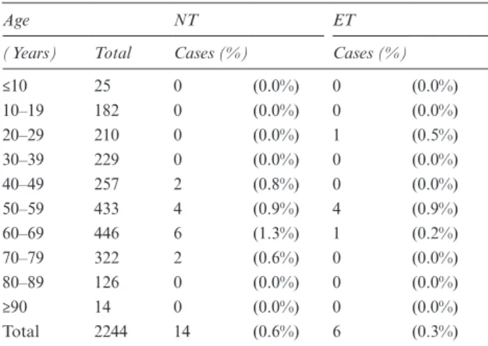
table 2 Prevalence of NT and ET by age group
Age NT ET
(Years) Total Cases (%) Cases (%)
≤10 25 0 (0.0%) 0 (0.0%) 10–19 182 0 (0.0%) 0 (0.0%) 20–29 210 0 (0.0%) 1 (0.5%) 30–39 229 0 (0.0%) 0 (0.0%) 40–49 257 2 (0.8%) 0 (0.0%) 50–59 433 4 (0.9%) 4 (0.9%) 60–69 446 6 (1.3%) 1 (0.2%) 70–79 322 2 (0.6%) 0 (0.0%) 80–89 126 0 (0.0%) 0 (0.0%) ≥90 14 0 (0.0%) 0 (0.0%) Total 2244 14 (0.6%) 6 (0.3%)
ET, eustachian tube tonsilloliths; NT, nasopharyngeal tonsilloliths. No significant differences were found among any age groups.
table 3 Size distribution of NT and ET on CT
Size (mm) on CT NT ET
1 10 6
2 2 0
3 2 0
Total 14 6
ET, eustachian tube tonsilloliths; NT, nasopharyngeal tonsilloliths.
table 4 Distance from NT to the mucosal surface on CT
Distance from the mucosal surface (mm) Number of NT
<1 3 (21.4%) 1 3 (21.4%) 2 7 (50.0%) 3 1 (7.1%) Total 14 (100.0%) NT, nasopharyngeal tonsilloliths.
Figure 2 CT images of a case with three eustachian tube tonsilloliths
(a): axial image, (b): another axial image in the caudal position. Two small tonsilloliths (arrows) were detected near the eustachian tube.
References
1. de Moura MD, Madureira DF, Noman-Ferreira LC, Abdo EN, de Aguiar EG, Freire AR. Tonsillolith: a report of three clinical cases. Med Oral Patol Oral Cir Bucal 2007; 12: E130–3.
2. Pruet CW, Duplan DA. Tonsil concretions and tonsilloliths.
Otolaryngol Clin North Am 1987; 20: 305–9.
3. Mandel L. Multiple bilateral tonsilloliths: case report. J Oral
Maxillofac Surg 2008; 66: 148–50. doi: https:// doi. org/ 10. 1016/ j. joms. 2006. 05. 047
4. Siber S, Hat J, Brakus I, Biočić J, Brajdić D, Zajc I, et al. Tonsillo-lithiasis and orofacial pain. Gerodontology 2012; 29: e1157–e1160. doi: https:// doi. org/ 10. 1111/ j. 1741- 2358. 2011. 00456.x
5. Lo RH, Chang KP, Chu ST. Upper airway obstruction caused by bilateral giant tonsilloliths. J Chin Med Assoc 2011; 74: 329–31. doi: https:// doi. org/ 10. 1016/ j. jcma. 2011. 05. 011
6. Chang YL, Chen PY, Hung SH. Large tonsillolith: Possible cause of sleep-disordered breathing. J Formos Med Assoc 2015; 114: 1293–4. doi: https:// doi. org/ 10. 1016/ j. jfma. 2014. 11. 005
7. Navas Cuéllar JA, López Bernal F, Ibáñez Delgado F. Giant tonsillolith causing dyspnea, esophageal perforation, and medias-tinitis. Emergencias 2015; 27: 280.
8. Aspestrand F, Kolbenstvedt A. Calcifications of the palatine tonsillary region: CT demonstration. Radiology 1987; 165: 479– 80. doi: https:// doi. org/ 10. 1148/ radiology. 165. 2. 3659369
9. Fauroux MA, Mas C, Tramini P, Torres JH. Prevalence of pala-tine tonsilloliths: a retrospective study on 150 consecutive CT examinations. Dentomaxillofac Radiol 2013; 42: 20120429. doi:
https:// doi. org/ 10. 1259/ dmfr. 20120429
10. Oda M, Kito S, Tanaka T, Nishida I, Awano S, Fujita Y, et al. Prevalence and imaging characteristics of detectable tonsilloliths on 482 pairs of consecutive CT and panoramic radiographs. BMC
the NT. NT are likely to become dislodged from the surface of the nasopharynx due to their small size and close proximity to the mucosal surface. Accordingly, this may lead to an underestimation of the true prev-alence of NT.
In contrast to palatine and lingual tonsilloliths, neither NT nor ET were detected on panoramic radio-graphs in our study. This may again be explained by their smaller sizes and the superimposition on the bony compartments of the temporomandibular joint and skull base, making them difficult to detect on panoramic radiographs.
Among the differential diagnoses of calcifications in the retropharyngeal region is retropharyngeal calcific tendinitis (RCT), which is a significant disease occurring in 1 per 200,000 patients. RCT is caused by an inflammatory process leading to hydroxyapatite crystal deposits in the longus colli muscle tendon. It commonly presents with neck pain and odyno-phagia18 and is diagnosed by detecting the
retropha-ryngeal calcified bodies using CT. Differentiation between RCT and NT is based on the location, with RCT normally detected dorsal to the pharyngobasilar fascia. Thus, to avoid misdiagnosis, clinicians should be able to identify the imaging characteristics of NT. From a clinical perspective, since NT and ET are small, they can be asymptomatic. However, as pala-tine tonsilloliths might cause clinical symptoms, we
recommend considering NT and ET in the differen-tial diagnosis as possible causes of similar symptoms including halitosis, throat pain, and earache.
In this investigation, we included over 2000 patients. However, most patients were of Oriental Mongolian descent. Further investigations are needed to deter-mine the prevalence and radiological features of NT and ET among patients with other racial descents and lifestyles. Furthermore, since the development and discharge of the NT and ET are unknown, a follow-up study of these concretions is necessary.
conclusions
The detection rates of NT and ET were lower than those of palatine and lingual tonsilloliths. However, as they are more frequently observed than the clinically signif-icant calcifying diseases such as RCT, clinicians should be able to correctly diagnose NT and ET based on their anatomical features.
acknowledgement
We would like to thank Editage (www. editage. jp) for English language editing.
Figure 3 CT images of a case with multiple nasopharyngeal and
palatine tonsilloliths (a): axial image of three nasopharyngeal tonsillo-liths (arrow), (b): axial image of multiple bilateral palatine tonsillotonsillo-liths (arrowheads), (c): coronal reconstructed image of the same patient nasopharyngeal (arrow) and palatine tonsilloliths (arrowheads) were observed.
Figure 4 CT images of a case with a pouch-like depression on the
mucosal surface of the nasopharynx (arrows) (a): axial image, (b): sagittal reconstructed image.
Oral Health 2013; 13: 54. doi: https:// doi. org/ 10. 1186/ 1472- 6831- 13- 54
11. Takahashi A, Sugawara C, Kudoh T, Uchida D, Tamatani T, Nagai H, et al. Prevalence and imaging characteristics of palatine tonsilloliths detected by CT in 2,873 consecutive patients.
Scien-tific World Journal 2014; 2014: 4–. doi: https:// doi. org/ 10. 1155/ 2014/ 940960
12. Ergun T, Lakadamyali H. The prevalence and clinical importance of incidental soft-tissue findings in cervical CT scans of trauma population. Dentomaxillofac Radiol 2013; 42: 20130216. doi:
https:// doi. org/ 10. 1259/ dmfr. 20130216
13. Takahashi A, Sugawara C, Kudoh T, Ohe G, Takamaru N, Tamatani T, et al. Prevalence and imaging characteristics of pala-tine tonsilloliths evaluated on 2244 pairs of panoramic radio-graphs and CT images. Clin Oral Investig 2017; 21: 85–91. doi:
https:// doi. org/ 10. 1007/ s00784- 016- 1752-0
14. Takahashi A, Sugawara C, Kudoh K, Yamamura Y, Ohe G, Tamatani T, et al. Lingual tonsillolith: prevalence and imaging
characteristics evaluated on 2244 pairs of panoramic radiographs and CT images. Dentomaxillofac Radiol 2018; 47: 20170251. doi:
https:// doi. org/ 10. 1259/ dmfr. 20170251
15. Bonneville JF, Belloir A, Mawazini H, Manzoni JM, Runge M, Monnier G, et al. Calcified remnants of the notochord in the roof of the nasopharynx. Radiology 1980; 137: 373–7. doi: https:// doi. org/ 10. 1148/ radiology. 137. 2. 7433671
16. Ben Salem D, Guiu B, Duvillard C, Couaillier J, Ricolfi F. Naso-pharyngeal tonsillolith: a report of 31 cases. J Radiol 2007; 88: 259–62.
17. Mesolella M, Cimmino M, Di Martino M, Criscuoli G, Albanese L, Galli V. Tonsillolith. Case report and review of the literature. Acta Otorhinolaryngol Ital 2004; 24: 302–7.
18. Horowitz G, Ben-Ari O, Brenner A, Fliss DM, Wasserzug O. Inci-dence of retropharyngeal calcific tendinitis (longus colli tendin-itis) in the general population. Otolaryngol Head Neck Surg 2013;