INTRODUCTION
Autism is a pervasive developmental disorder with severe disruption of cognitive and social function. The abnormality of anatomical structure in the brain with autistic patients has been published using MRI or CT study (1). We showed the metabolic difference with autism by proton magnetic resonance spectroscopy (MRS)(2), and Muller et al. reported the difference of cerebral perfusion in autism by positron emission tomography (PET) with O-15 (3). It has been speculated that developmental language disabilities of autistic
patients may have a hemispheric basis involving atypical cerebral dominance. Recently the advent of functional magnetic resonance imaging (fMRI), as a hemodynamic-based technique without ionizing radiation, has began to expand the use of functional neuroimaging even in the clinical setting. We considered that the difference of hemodynamic response with autism might be de-tected by fMRI after conduction of a language task. In this paper, cerebral function of autistic patients was evaluated by using fMRI with a language task and com-pared with normal controls.
MATERIALS AND METHODS
Ten patients were diagnosed as autism using DSM-ⅢR criteria, and the I.Q. of autistic patients was within the normal range. The clinical data of the subjects were
ORIGINAL
Difference of signal change by a language task on autistic
patients using functional MRI
Mayumi Takeuchi
1, Masafumi Harada
2, Kenji Matsuzaki
1, Hiromu Nishitani
1, and Kenji Mori
31Department of Radiology,2Department of Radiologic Technology, School of Health Sciences, and 3Department of Pediatrics, The University of Tokushima School of Medicine, Tokushima, Japan
Abstract : [Objective] Cerebral function with a language task was evaluated by functional magnetic resonance imaging (fMRI), and the differences of activated pattern and signal changes were compared between autistic patients and normal controls.
[Methods] Ten autistic and ten normal subjects were tested by fMRI with a language task requiring the attribution of complex mental states. Activation maps analyzed between two groups were generated and the asymmetry indexes calculated by the quotient of activated pixels of the right frontal lobe divided by those of the left frontal lobe were statistically com-pared by unpaired t-test.
[Results] Both the autistic and the normal subjects showed activation at the bilateral prefrontal cortical areas and the ventral occipito-temporal regions. However, the autistic patients dem-onstrated more activation at the right frontal lobe than the normal controls. Thus it was considered that in the autistic patients the right-hemisphere was more dominant for the language task than that of the normal controls. The result is consist to the theory that autism is related to early left-hemisphere dysfunction.
[Conclusions] We considered that fMRI may be a useful non-invasive method to evaluate the cerebral functional abnormality in autistic patients. J. Med. Invest. 51 : 59-62, February, 2004
Keywords : autism ; functional MRI (fMRI) ; language task
Received for publication November 7, 2003 ; accepted January 7, 2004.
Address correspondence and reprint requests to Mayumi Takeuchi, M.D., Department of Radiology, The University of Tokushima School of Medicine, Kuramoto-cho, Tokushima 770-8503, Japan and Fax : +81-88-633-7174.
The Journal of Medical Investigation Vol. 51 2004
summarized in Table1. Ten subjects in the normal group were matched for mean age, handedness, gender, and intelligence quotient (I.Q.), with ten patients of autism. Normal controls had no history of drug abuse, special education, major medical or psychiatric illness, and developmental or neurological disorder. Informed con-sent was obtained from all patients and normal controls before they were recruited into the study. We tested the autistic and the normal controls by fMRI with a language task requiring the attribution of complex mental states. The sample of sentences and questions is shown in Fig.1. Comprehension of the subjects about the story was ascertained by their answer to
question-naire immediately after completion of fMRI studies. The sentences were displayed on the mirror attached to a head coil during a task period and read by a subject after indication from the operator. At rest periods, sub-jects were not allowed to read sentences and were in-dicated to consider nothing. The head of a subject was fixed tightly on the coil to avoid gross motion of the head. The functional paradigm was conducted as rest/ task/rest each of 1-minute duration. The fMRI study was performed on a clinical MRI instrument (1.5 Tesla, Signa Horizon, GE, MI) with a standard head coil. The functional images were acquired using echo planar im-aging (TR/TE=3000ms/82ms, matrix=64×64, FOV= 24cm). Data were transferred to a workstation and analyzed using Statistical Parametric Mapping (SPM 99) installed in Medx Ver 3.4 (Sensor system, USA). The time series of imaging were realigned using the first image and spatially normalized to the stereotactic space of Tailarach and Tournoux. These data were subsequently smoothed with an isotropic Gaussian kernel of 6mm at full width half maximum. Activation maps were generated by fitting time course to a boxcar type as the reference function with a statistical sig-nificancy (p<0.01). The number of activated pixels de-picted was counted in the right and left frontal lobe separately, and the asymmetry index was calculated as following : the R/L ratios=(the number of activated pixels of the right frontal lobe) /(the number of ac-tivated pixels of the left frontal lobe). The higher ratio of this index thus indicates greater right-hemisphere activation. The statistical comparison between the autis-tic patients and the normal controls was conducted by unpaired t-test. The activation maps of group analysis
Table 1. Mean age and IQ, handedness of subjects in the experiment.
Autism Controls
Age (years) Mean(±SD) 11.2±2.6 9.4±2.3
Gender Male : Female 8 : 2 8 : 2
IQ Mean(±SD) 110.4±14.8 108±12.1
handedness Right : Left 10 : 0 10 : 0
Fig. 1. Language task
normal
autism
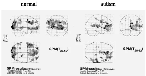
Fig. 2. A statistical parametric map (SPM) as a maximum intensity projection showing the activated areas.
M. Takeuchi et al. Cerebral function of autism evaluated by fMRI
between the autistic group (n=10) and the normal con-trols (n=10) were conducted using MEDx Ver.3.4 after the individual analysis by SPM 99, and the statistical threshold for depicting activated areas was set at p< 0.01.
RESULTS
The experimental procedures were successfully carried out in all subjects. All subjects were confirmed to conduct the language task by their answers for the questions. Only one autistic patient gave correct answers and the other patients gave wrong answers. All of the normal controls could answer correctly. Though activation at the bilateral prefrontal cortical regions and the ventral occipito-temporal regions was observed in both the autistic and the normal controls, the autistic patients demonstrated more activation on the right
fron-tal lobe than the normal controls. The typical individual results of the autistic and the normal controls were shown in Fig. 2.
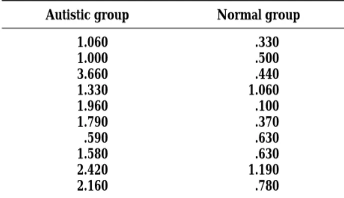
The asymmetric ratios (R/L ratio) between the autis-tic group and the normal group were shown in Table 2. The autistic subjects had higher ratios than the nor-mal controls indicating their right hemisphere domi-nancy. The difference of the averaged asymmetric ratios between two groups was demonstrated in Fig. 3 and the statistical difference was found at p=0.0035. The activation maps analyzed between two groups were shown in Fig. 4. The activated areas of the autistic group were more dominant in the right hemisphere compared with the normal group.
DISCUSSION
The fMRI studies are recently applied to various
Fig. 3. The difference of the averaged asymmetric ratios between the normal group and the autistic group.
Table 2. Mean R/L ratios for language tasks for autistic and normal controls.
Autistic group Normal group
1.060 1.000 3.660 1.330 1.960 1.790 .590 1.580 2.420 2.160 .330 .500 .440 1.060 .100 .370 .630 .630 1.190 .780
Fig. 4. Repetitive activation maps showing language related activation.
patients and there are quite a few papers reporting the usefulness of fMRI to clinical application (4). Autism is a developmental disorder defined by impairments of mutual social interaction, verbal and nonverbal com-munication, and a markedly restricted repertoire of activities and interests (5). It is expected that fMRI will evaluate cerebral function of autism in the clinical setting, and our study showed that autistic patients exhibited right-hemisphere lateralization for a language task different from normal controls. Dawson et al. re-ported an atypical pattern of cerebral lateralization in the early infantile autism based on EEG measures of hemispheric activation during cognitive processing. The autistic patients showed a pattern of hemispheric specialization, i.e.“reversal”in lateralization, which was rarely seen in the normal population, reflective of a lack of left-hemisphere specialization for linguis-tic functions (6). Muller et al. conducted an O-15PET study to examine the neurofunctional organization of audition and language (3). The result of our study was consistent with the previous results observed by PET and EEG studies, and these results indicated a sig-nificant reversal of normal left hemisphere dominance for receptive language (listening to sentences) in the patient group. Since leftward dominance has been linked to the maturation of the left hemisphere, this reversal might arise as a result of language delay. Our result and the previous data by PET and EEG may suggest that right hemisphere may compensate for linguistic disability of the left hemisphere to some extent.
A few papers of fMRI with autism have been pub-lished. Gallagher et al. studied fMRI involving a story task and a cartoon task both designed to tap‘theory of mind’-the attribution of mental states, and brain activation was shown in the medial prefrontal cortex (7). Luna et al. studied fMRI involving an oculomotor special working memory task and a visually guided saccade task, and the autistic subjects demonstrated significantly less activation in dorsolateral prefrontal cortex and posterior cingulate cortex suggesting a functional disconnectivity of the circuitry underlying working memory (8). Their results and our study showed the usefulness of fMRI to find differences of cerebral function with autistic patients and fMRI will be appli-cable to evaluate cerebral dysfunction of individual patients with autism.
CONCLUSIONS
FMRI is a suitable non-invasive clinical modality for evaluating the cerebral functional abnormality in autistic patients.
REFERENCES
1. Townsend J, Westerfield M, Leaver E, Makeig S, Jung T, Pierce K, Courchesne E : Event-related brain response abnormalities in autism : evidence for impaired cerebello-frontal spatial attention networks. Cognitive Brain Research 11 : 127-145, 2001
2. Hisaoka S, Harada M, Nishitani H, Mori K : Re-gional magnetic resonance spectroscopy of the brain in autistic individuals. Neuroradiology 43 : 496-498, 2001
3. Muller RA, Behen ME, Rothermel RD, Chugani DC, Muzik O, Mangner TJ, Chugani HT: Brain mapping of language and auditory perception in high-functioning autistic adults : A PET study. J Autism Dev Disord 29 : 19-31, 1999
4. Hennig J, Speck O, Koch MA, Weiller C : Func-tional magnetic resonance imaging : A review of methodological aspects and clinical applica-tions. J Magn Reson Imaging 18 : 1-15, 2003 5. Rumsey JM, Ernst M:Functional neuroimaging
of autistic disorders. Ment Retard Dev Disabil Res Rev 6 : 171-179, 2000
6. Dawson G, Warrenburg S, Fuller P : Cerebral lateralization in individuals diagnosed as autistic in early childhood. Brain and Language15:353-368, 1982
7. Gallagher HL, Happe F, Brunswick N, Fletcher PC, Frith U, Frith CD:Reading the mind in car-toons and stories : an fMRI study of‘theory of mind’in verbal and nonverbal tasks. Neuropsy-chologia 38 : 11-21, 2000
8. Luna B, Minshew NJ, Garver KE, Lazar NA, Thulborn KR, Eddy WF, Sweeney JA : Neocortical system abnormalities in autism : An fMRI study of spatial working memory. Neurology 59 : 834-840, 2002
M. Takeuchi et al. Cerebral function of autism evaluated by fMRI