A Case of Invasive Cortical Thymoma Involving
Unusual Multifocal B-Cell Hyperplasia with
Germinal Centers
著者
HASUI Kazuhisa, SATO Eiichi, SUEYOSHI
Kazunobu, MATSUSHITA Yoshifumi, SAKAE
Kiyohiro, MAEMURA Makoto, MAEMURA Kousei,
FUNASAKO Susumu, TACHIWADA Motomu
journal or
publication title
鹿児島大学医学雑誌=Medical journal of
Kagoshima University
volume
47
number
Suppl. 2
page range
125-128
URL
http://hdl.handle.net/10232/18329
Med. J. Kagoshima Univ., Vol. 47, Suppl. 2. 125-128, November, 1995
Case Report
A Case of Invasive Cortical Thymoma
Involving Unusual Multifocal B-Cell Hyperplasia with Germinal Centers
Kazuhisa HASUI1, Eiichi SATO1,
Kazunobu SUEYOSHI1, Yoshifumi MATSUSHITA1, Kiyohiro SAKAE2,
Makoto MAEMURA3, Kousei MAEMURA3, Susumu FUNASAKO3 and Motomu TACHIWADA3
'Second Department of Pathology, Faculty of Medicine Kagoshima University, Kagoshima, Japan
2School of Allied Medicine, Kagoshima University
3Department of Surgery, Kagoshima Prefectural Kanoya Hospital, Kanoya, Japan
Summary
An invasive cortical thymoma in the anterior left mediastinum of a 78 year-old Japanese female involved unusual multifocal B-cell hyperplasia forming germinal centers in its invasion. The invasive cortical thymoma showed a small number of keratin+epithelioid cells in lymphocytic stroma around some of blood-pooling microcysts and in the degenerative as well as invasive
areas. In the invasive areas T-cell-rich stroma was
associated with multifocal B-cell hyperplasia forming germinal centers. The Tcells included many proliferat ing cells. In the analysis of DNA extracted from paraffin sections by means of polymerase chain
reaction for T-cell receptor P and 7 and immunoglo
bulin genes, no clonal proliferation of T- and B-cells was detected. An invasive cortical thymoma was diagnosed because of no obviously detectable neoplas tic natures in the lymphocytes, although the association of unusual multifocal B-cell hyperplasia forming germinal centers suggested a possibility of a secondary T-cell low grade malignant lymphoma corresponding to the nodal T-zone lymphoma.
Key Words: Thymoma, T-cell, B-cell, paraffin-im
munohistochemistry, polymerase chain reaction, TCR
P, TCR 7, immunoglobulin heavy chain gene
Address of Correspondence: Kazuhisa HASUI,
Second Department of Pathology, Faculty of Medicine Kagoshima University, Sakuragaoka 8-35-1, Kagoshima 890 Japan
Introduction
Primary thymic tumors in aged patients in Japan are
thymomas, teratomas and lymphomas1^ There were
some reports that a long-standing thymoma had
non-Hodgkin malignant lymphoma2'3), indicating a possibil
ity of the secondary occurrence of low-grade T-cell
malignant lymphoma in cortical thymoma. But it was reported that the clonal proliferation of T-cells had not yet been recognized in lymphocytic thymomas. We
experienced a case of invasive cortical thymoma involving multifocal B-cell hyperplasia forming germin
al centers.
We report here the case of invasive cortical thymoma
with analysis of T-cell receptor P and 7 chain genes
and immunoglobulin heavy chain gene by means of
polymerase chain reaction and with discussion of its differential diagnosis from a composite case of cortical thymoma and non-Hodgkin malignant lymphoma.
Case
The patient was 78 year old Japanese woman. She
complained sometimes of pain on her neck. Thymic
mass was pointed out on her chest X-ray examination in
medical check-up. Neither swelling of lymph nodes nor
splenomegaly was noted. Number and nature of blood cells were not abnormal in her peripheral blood. The all laboratory data of her peripheral blood were within the normal range. Computed tomograph detected a thymic tumor revealing high and low density areas and clear
outline and associating no regional swollen lymph
nodes in her anterior left mediastinum. Under a clinical diagnosis of thymoma the tumor was removed. After
the operation she was free from any complains and abnormal physiological and laboratory findings.
[126] Med. J. Kagoshima Univ.. Vol. 47. Suppl. 2, November. 1995
Pathology and paraffin-immunohistochemistry
The removed thymic mass was a 11X6X6 cm large, lobulated and solid, involving ligament of thyroid. On cut surface of the tumor, dark-red tumor had capsule like tissue demarcating it from whitish invasion. There was a small amount of persistent thymic tissue around
the tumor.
Microscopically the dark-red tumor parenchyma comprised blood-pooling microcysts surrounded by
lymphocytic stroma and degenerated areas (Fig. la). In the degenerated areas spindle epithelioid cells were seen in epithelial arrangement (Fig. lb).
Paraffin-immunohistochemistry of anti-keratin antibody showed keratin + spindle cells along the degenerated cystic lesions (Fig. lc). Some of the microcysts had keratin +
factor II- epithelial lining. The cellular stroma (Fig. lc) comprised many small CD3 + UCHL-1 + M1-
T-cells, small clusters of L26 + Mx-pan B+ MB-1-
LN-1-LN-2-LN-3- cells and some histiocytes. Some his tiocytes were positive for S100 protein. Epithelioid cells among the lymphocytes were negative for EMA, LN-2 and LN-3. Only a few epithelioid cells were positive for
keratin.
The tumor parenchyma was demarcated by thick fibrosclerotic capsular tissue from the extracapsular whitish areas (Fig. 2a). The extracapsular areas were the invasion, comprising T-cell rich areas (Fig. 2b) with a few keratin + epithelioid cells, fibrosclerotic stroma and multifocal proliferation of L26 + LN-1 +LN-2 + LN-3 + pale B-cells associating a small number of CD3
+ UCHL-1 + T-cells. In some of the foci of the pale B-cells germinal center with mantle zone was formed (Fig. 2c). The lymphocytes in the T-cell rich areas and in the B-cell hyperplastic areas showed a few regular mitoses and included many proliferating cells labeled in antigen-retrieval paraffin-immunohistochenistry of PCNA and MIB1 (Ki-67).
b)
c)
Fig. 1. Parenchyma of the cortical thymoma
a) Blood-pooling microcysts and degenerated areas
Blood-pooling microcysts are surrounded by
lymphocytic stroma and their ghosts in the
degenerated areas b) Degenerated area
A small number of epithelioid cells show
epithelial arrangement along degenerated cysts
or alveoli.
c) Paraffin-immunohistochemistry of anti-keratin antibody
Keratin-positive epithelioid cells line along the degenerated microcysts. •?• • .. . ..'> •-<<.••• ;;'" .• •-.1* • -. •-• A-'; '•w. '>'?£>-?• '•'!?"•; *' -•) • .-'•1— Zi't.•$•••:
'"-."•
*•' 7*. ~ -• • v • - * • " **•y f
*•*£ ';-*» 1,.:.\. ?^g.Q-- V
'y'i
'.- "t-v^y ;•- *-~- -"-"•; '' •, • »*• h I3^
'S^i^^m
*> * ---. ;.£•:**:0^^me&
.;'<•••;'
, *• "*< • "" *. S-: f I,* f 5 ii&< '.%l
•V
y y
;• „"" e.: . . -J.'.'li'" V-i .
-v?" r°j;-''': •
-"' •
-Invasive Cotical Thymoma with B-Cell Hyperplasia (127) a)
KUl
"ste?/
* &yy • S I #
ft
«
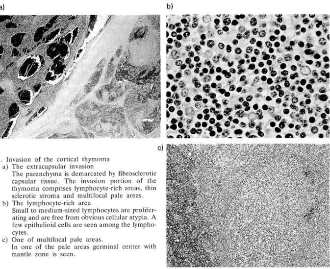
Fig. 2. Invasion of the cortical thymoma
a) The extracapsular invasion
The parenchyma is demarcated by fibrosclerotic
capsular tissue. The invasion portion of the thymoma comprises lymphocyte-rich areas, thin sclerotic stroma and multifocal pale areas. b) The lymphocyte-rich area
Small to medium-sized lymphocytes are prolifer
ating and are free from obvious cellular atypia. A
few epithelioid cells are seen among the lympho
cytes.
c) One of multifocal pale areas.
In one of the pale areas germinal center with
mantle zone is seen.
Polymerase chain reaction (PCR) of T-cell receptor (TCR) P and V chain genes and immunoglobulin
heavy chain gene
DNA extracted from one 10 micrometer thick
paraffin section of this thymic tumor was examined by
PCR to see whether any of TCR ft and 7 -chain and
immunoglobulin heavy chain genes of the lymphocytes
is rearranged or not. When an enough amount of the
extracted DNA was the template DNA of the PCR,
amplified DNA bands could not detected in the agar-gel electrophoresis of the PCR products of 6 pairs of
primers TCR/? V, D1,D2, Jl and J24), 4 pairs ofTCR
7 Vll, V101, J 7 and Jp5' and a pair of IgJHI and
IgJH26).
Discussion
Histopathological differentiation of lymphocytic
thymoma from the primary thymic lymphoma is sometimes difficult but the existence of cyst is considered to be pathognomic for lymphocytic
thymoma7'. But there were some thymoma patients
reported to suffer from secondary non-Hodgkin
J§IIP#
.:«•?;.,•.';.-:--";'••'';,: ':^y.'.- • -V.'.V.Y-"- r.v<:'v,i*.-' ::•<•;•<:-•>
MnMttitt
''.':.;V:-':•"•••• ••'*•'.malignant lymphoma such as T-cell lymphoblastic
lymphoma and chronic lymphocytic leukemia2'3', while
a composite case of thymoma and non-Hodgkin malignant lymphoma have not yet been reported. Molecular analysis of lymphocytes in lymphocytic thymomas could not show their monoclonal
proliferation8', although it is unknown whetherunusual
lymphocytic thymomas like this case were included in those cases studied. There seems to be a possibility of outcome of low grade malignant lymphoma in lym phocytic thymoma.
Lymphocytic thymoma is recently classified as
cortical type thymoma9'. Cortical type thymoma often
associates Myasthenia gravis and its stromal lympho
cytes are regarded as stimulated peripheral T cells10'.
L26 + non-lymphoid cells are reported to be found in
thymomas and normal thymus11' and clusters of the
L26+ Mx-pan B + MB- 1- LN-1- LN-2- LN-3- cells in this case may be the L26+ non-lymphoid cells. The stimulated T-cells may induce proliferation of B-cells.
Multifocal B-cell hyperplasia in the invasion areas of
this cortical thymoma might be explained by the function of the activated peripheral T-cells in a cortical thymoma. But multifocal B-cell hyperplasia forming
[128] Med. J. Kagoshima Univ., Vol. 47, Suppl. 2, November, 1995
germinal centers can not be often found in a cortical thymoma. And such hyperplastic lymph follicles are
seen in nodal T-zone lymphoma12).
It is the problem in the differential diagnosis of this invasive cortical thymoma from the secondary outcome of T-zone lymphoma in the long-standing thymoma what method can detect neoplastic features of peripher al T-cells in a cortical thymoma. The lymphocytes in this thymoma proliferated, showing many labeled cells by PCNA and MIB1 (Ki-67) and regular mitosis. No obvious presence of atypical mitoses in them suggests their reactive nature rather than neoplastic one. Recently, PCR analysis is introduced in hematopathol
ogy to detect rearrangement of TCR P and 7 -chains
and immunoglobulin heavy chain genes in malignant
lymphomas and lymphocytic leukemias4'5'6\ Rear
rangement in one or the both genes of TCR P and 7-chain, a proof of clonal proliferation of T-cells, is
reported to be detected in more than 80% cases of
T-cell malignant lymphomas5). The PCR analysis of this
cortical thymoma could not show rearrangement in any of the genes, suggesting no obvious neoplastic nature in the T- and B-cells in this cortical thymoma.
Consequently, this case should be diagnosed as invasive cortical thymoma involving unusual multifocal B-cell hyperplasia forming germinal centers, although a possibility of a secondary T-cell lymphoma correspond ing to the nodal T-zone lymphoma in this invasive cortical thymoma could not be denied completely.
Acknowledgement
This thymic tumor was consulted to Prof. K. H. Muller-Hermelink (Konsultations- und Referenzzen-trum fur Lymphknotenpathologie, Pathologisches
Institut, tJniversitat Wiirzburg, Wiirzburg, Germany)
and diagnosed as an invasive cortical thymoma according to a new classification of thymoma. Authors thank him for the diagnosis and for his lecture about
the new classification of thymoma in Fukuoka10).
References
1) Akashi A, Nakahara K, Ohno K, Fujii Y, Maeda H et al. Primary mediastinal tumors in children-comparison with mediastinal tumors in adults. Nippon-Kyobu-Geka-Gakkai-Zasshi 1993, 41:
2180-4.
2) Macon WR, Rynalski TH, Swerdlow SH and Cousar JB. T-cell lymphoblastic leukemia/lymphoma presenting in a recurrent thymoma. Mod Pathol.
1991, 4: 524-8.
3) Friedman HD, Inman DA, Hutchison RE and
Poiesz BJ. Concurrent invasive thymoma and T-cell lymphoblastic leukemia and lymphoma. A case
report with necropsy findings and literature review
of thymoma and associated hematologic neoplasm.
Am J Clin Pathol. 1994, 101: 432-7.
4) McCarthy KP, Sloane JP, Kabarowski JHS, Ma-tutes E and Wiedemann LM. The rapid detection of clonal T-cell proliferations in patients with lymphoid
disorders. Am J Pathol 1991, 138: 821-8.
5) McCarthy KP, Sloane JP, Kabarowski JHS,
Ma-tutes E and Wiedemann LM. A simplified method of detection of clonal rearrangements of the T-cell receptor-7 chain gene. Diagnostic Molecular Pathology 1992, 1(3): 173-9.
6) McCarthy KP, Sloane JP, Wiedemann LM. Rapid
method for distinguishing clonal from polyclonal B-cell populations in surgical biopsy specimens. J Clin
Pathol. 1990, 43: 429-32.
7) Henry K. The thymic gland. In: Henry K and
Symmers W StC, editors. Systemic Pathology, third edition, vol.7: Thymus, lymph nodes, spleen and lymphatics. Churchill Livingston, 1992: 27-139. 8) Katzin WE, Fishleder AJ, Linden MD and Tubbs
RR. Immunoglobulin and T-cell receptor genes in thymomas-genotypic evidence supporting the non neoplastic nature of the lymphocytic component.
Hum Pathol. 1988, 19: 323-8.
9) Kirchner T, Schalke B, Mar A and Muller-Herme link HK. Evaluation of prognostic features in thymic epithelial tumours. Thymus 1989, 14:
195-203.
10) Muller-Hermelink HK. Lecture about new classi fication of thymoma in the 108th Fukuoka-Ketueki-Konwakai in July 1994 in Fukuoka. 11) Taubenberger JK, Jaffe ES and Medeiros LJ.
Thymoma with abundant L26-positive asteroid cells. A case report with an analysis of normal thymus and thymoma specimens. Arch Pathol Lab
Med. 1991, 115: 1254-7.
12) Suchi T, Lennert K, Tu LY, Kikuchi M, Sato E et al. Histopathology and immunohistochemistry of peripheral T-cell lymphomas, a proposal for their