2020
T172A007
Yumiko Masuda Doctor Thesis
Gunma University
Study on purification method of monoclonal antibodies by ion-exchange chromatography in a flow-through mode:
evaluation of virus removal performance and application to
antibodies with asymmetric charge distribution
2
Chinese Hamster Ovary (CHO) CHO
DNA CHO
Protein A
(CEX) (AEX)
CEX AEX
CEX
3
CEX /
/ 10
CEX
/
/ CEX
/ 20
/
CEX
AEX
AEX
A AEX
A AEX
Fv
4
AEX
A
NatriFlo HD-Q
5
Abstract
Therapeutic monoclonal antibodies (mAbs) have been widely focused as molecular targeted drug for cancer and autoimmune disease, because of their higher antigen specificity and lower side effects. Their worldwide market is year-by-year expanding.
Many therapeutic mAbs are produced by mammalian cell expression system with Chinese hamster ovary (CHO) cells as a host. Culture broth of CHO cells contains not only mAbs but also product and process related impurities such as high-molecular-weight species, host cell protein, and host cell DNA. In addition, it is well-known that retrovirus- like particles which show non-infectious features are included in CHO cells and often released from them. A performance to remove most impurities including retrovirus-like particles and adventitious viruses are required to meet an acceptable level in an
appropriate purification process.
Many of the humanized mAbs, which possess a common framework, have similar physicochemical properties. Consequently, similar purification processes, so-called
“platform processes,” are often applied as general manufacturing protocols with minimal alterations. Protein A chromatography is used as the first capture step in a downstream platform process, followed by two or three polishing steps. Cation exchange (CEX) and anion exchange (AEX) chromatographies are typically used as a polishing step.
Manufactures have often tried to streamline the manufacturing process and reduce the production cost as far as possible because of higher dose of therapeutic mAbs in clinical and medical fields and higher production cost depending on a manufacturing process using mammalian expression system. In particular, some resin and/or the other media in the chromatography steps account for a large proportion in the raw material costs for mAb manufacturing. In this study, performance of purification steps by CEX and AEX
6
chromatographies under flow-through mode conditions was investigated in order to develop an efficient platform process for mAb manufacturing.
First, a virus removal performance in the overload mode, similar to a flow-through mode, on CEX chromatography was evaluated (Chapter 2). Although CEX
chromatography is commonly used in bind/elute mode, the tested overloaded mode could be done under more than 10-times loading condition compared with bind/elute mode as efficient and alternative purification method. It has been further reported that CEX chromatography in overloaded mode is effective in removing many impurities. However, the viral clearance performance in overloaded mode has not yet been reported. If the overloaded mode can remove viruses with the same efficiency as the bind/elute mode, then the overloaded mode can take the place of the bind/elute mode on CEX
chromatography step, which requires large amount of media and buffer. The present study showed successful virus removal in overloaded mode on CEX chromatography as the first example. Even though the load amount in the overloaded mode was 20-times larger than that in the bind/elute mode, the overload mode showed the almost same viral clearance performance as a bind/elute mode. This viral clearance ability was not significantly affected by mAb features or used resins. This study showed the overloaded mode is effective to remove impurities as a polishing step for therapeutic mAbs with cost effective features.
Second, we developed the efficient purification process for the mAb having asymmetric charge distribution in flow through mode (Chapter 3). The pH of the mobile phase for AEX chromatography is typically set based on the isoelectric point (pI) of each mAb.
However, we recently encountered a tight binding of mAb A to AEX resins under
conditions set by its pI value. This anomalous adsorption behavior was suspected to be an effect of the asymmetric charge distribution on the surface of the mAb A. We predicted
7
that the use of membrane adsorbers might provide effective mAb A purification, since the supporting matrix has a network structure that would be less susceptible to interactions with the asymmetric charge distribution on the protein surface. We tested membrane adsorber under standard chromatographic conditions and found that mAb A flowed through the membrane adsorber, resulting in successful separation from virus together.
In current studies on the purification of mAbs with flow-through mode, the media such as the chromatographic resin can be efficiently used and the production cost would be reduced. In addition, it was found that mAbs can be purified under similar standard chromatographic conditions regardless of their charge distributions using specific membrane adsorber, NatriFlo HD-Q. The present outstanding findings are expected to provide universal platform process for mAb manufacturing/purification, achieving the steady supply of therapeutic mAbs, and to contribute the acceleration by becoming shorten a period for process development of mAb purification.
8
CHO Chinese hamster ovary
HMWS high-molecular-weight species HCP host cell protein
CEX cation exchange AEX anion exchange
MLV murine leukemia virus PRV pseudorabies virus Reo 3 reovirus type 3 MMV murine minute virus
TCID50 50% tissue culture infectious dose LVP large volume plating
LRV log reduction value CV column volume RT residence time HSW high-salt wash
HPLC high-performance liquid chromatography LMWS low-molecular-weight species
SE-HPLC size exclusion high-performance liquid chromatography ELISA enzyme-linked immune sorbent assay
pI isoelectric point IEX ion exchange MV membrane volume
CDR complementarity determining region
9
... 10
1.1 ... 11
1.2 ... 12
1.3 ... 13
1.4 ... 16
1.5 ... 17
... 20
2.1 ... 21
2.2 ... 22
2.3 ... 25
2.4 ... 41
2.5 ... 43
... 44
3.1 ... 45
3.2 ... 46
3.3 ... 49
3.4 ... 67
3.5 ... 67
... 71
4.1 ... 72
4.2 ... 76
4.3 ... 78
10
11
1.1
1980 1990
1,2
50 570
3,4
Chinese Hamster Ovary (CHO) CHO
(HMWS) (HCP) DNA
5 CHO
6 CHO
7
8-10
11
Fig. 1
Fc Protein A Protein A
2
12
3
(AEX) (CEX)
CEX AEX
12
60%
13
1.2
CEX AEX
/
CEX
CEX /
/ 10
13
CEX /
/
AEX
1.3
CEX
CEX
/ /
10
CEX
HCP DNA HMWS Protein A
14,15
/ /
CEX
/
AEX
14
AEX
16,17 A AEX
A AEX
Fv
AEX
pH
15
Fig.1
Cell bank
Protein A
2
(Anion exchange
1
(Cation exchange pH
Protein A CEX
AEX
16
1.4
( )
/
Fig. 2
Fig. 2 /
Ø mAb, Impurities Ø mAb/
mAb Impurities
Ø Impurities/
mAb Load
Impurities
Ø mAb, Impurities
Ø / mAb
Ø Impurities mAb
17
1.5
1. Reichert JM. Antibodies to watch in 2017. MAbs. 2017;9:167-181.
2. Vázquez-Rey M, Lang DA. Aggregates in monoclonal antibody manufacturing processes. Biotechnol Bioeng. 2011;108:1494-1508.
3. Lai Y, Wang R, Chen X, Tang D, Hu Y, Cai J, Zhang Q, Hu H. Emerging trends and new developments in monoclonal antibodies : A scientometric analysis (1980–2016). Hum Vaccine Immunother. 2017;13:1388-1397.
4. Kaplon H, Reichert JM. Antibodies to watch in 2019. MAbs. 2019;11:219-238.
5. Shukla AA, Jiang C, Ma J, Rubacha M, Flansburg L, Lee SS. Demonstration of robust host cell protein clearance in biopharmaceutical downstream processes. Biotechnol Prog.
2008;24:615-622.
6. Zhang M, Lute S, Norling L, et al. A novel, Q-PCR based approach to measuring endogenous retroviral clearance by capture protein A chromatography. Biotechnol Bioeng. 2009;102:1438-1447.
7. Anderson KP, Low M-AL, Lie YS, Keller G-A, Dinowitz M. Endogenous origin of defective retroviruslike particles from a recombinant Chinese hamster ovary cell line.
Virology. 1991;181:305-311.
8. INTERNATIONAL CONFERENCE ON HARMONISATION OF TECHNICAL REQUIREMENTS FOR REGISTRATION OF PHARMACEUTICALS FOR HUMAN USE. VIRAL SAFETY EVALUATION OF BIOTECHNOLOGY PRODUCTS
DERIVED FROM CELL LINES OF HUMAN OR ANIMAL ORIGIN Q5A(R1).
Available at:
https://www.ich.org/fileadmin/Public_Web_Site/ICH_Products/Guidelines/Quality/Q5A_
R1/Step4/Q5A_R1_Guideline.pdf. September, 1999.
9. U.S. Department of Health and Human Services, Food and Drug Administration, Center for Biologics Evaluation and Research. Points to Consider in the Manufacture and
18
Testing of Monoclonal Antibody Products for Human Use. Available at:
www.fda.gov/media/76798/download. February 1997.
10. European Medicines Agency. Evaluation of Medicines for Human Use. GUIDELINE ON VIRUS SAFETY EVALUATION OF BIOTECHNOLOGYCAL INVESTIGATIONAL MEDICINAL PRODUCTS. Available at:
https://www.ema.europa.eu/documents/scientific-guideline/guideline-virus-safety- evaluation-biotechnological-investigational-medicinal-products_en.pdf. July, 2008.
11. Liu HF, Ma J, Winter C, Bayer R. Recovery and purification process development for monoclonal antibody production. MAbs. 2010;2:480-499.
12. Shukla AA, Hubbard B, Tressel T, Guhan S, Low D. Downstream processing of monoclonal antibodies—Application of platform approaches. J Chromatogr B.
2007;848:28-39.
13. Pollock J, Coffman J, Ho SV, Farid SS. Integrated continuous bioprocessing: economic, operational, and environmental feasibility for clinical and commercial antibody
manufacture. Biotechnol Prog. 2017;33:854-866.
14. Brown A, Bill J, Tully T, Radhamohan A, Dowd C. Overloading ion-exchange
membranes as a purification step for monoclonal antibodies. Biotechnol Appl Biochem.
2010;56:59-70.
15. Liu HF, McCooey B, Duarte T, et al. Exploration of overloaded cation exchange chromatography for monoclonal antibody purification. J Chromatogr A.
2011;1218:6943-6952.
16. Ishihara T, Kadoya T. Accelerated purification process development of monoclonal antibodies for shortening time to clinic: Design and case study of chromatography processes. J Chromatogr A. 2007;1176:149-156.
17. Ishihara T, Kadoya T, Yoshida H, Tamada T, Yamamoto S. Rational methods for
predicting human monoclonal antibodies retention in protein A affinity chromatography
19
and cation exchange chromatography Structure-based chromatography design for monoclonal antibodies. J Chromatogr A. 2005;1093:126-138.
20
21
2.1
CEX /
CEX /
HCP DNA HMWS Protein A 1,2
(MLV)
3
/ CEX ( )
(HMWS HCP )
CEX
4,5 CEX
/ 100 g /L
6 10
1,000 g /L 5
( )
CEX HCP DNA HMWS
Protein A 5
/
/
CEX MLV
22
POROS XS MLV
MLV CHO
(MMV) (PRV)
3 (Reo 3) /
/
(pH )
2.2 CEX
2 1 2 (
IgG1) CEX CHO
Protein A AEX
AEX pH 5.0 0.2 µm
CEX 80
CEX 5 mS/cm AKTA Explore 100 (GE
)
280 nm
BioReliance (Stirling )
23
(50% [TCID 50]) : PG-4
(MLV) Vero (PRV) 324K (MMV) L-929 (Reo 3)
Spearman-Körber 95%
1.96
/ 5% (v/v)
1% (v/v) TCID
50 Large
volume plating (LVP) ( [LRV])
CEX
HMWS HCP POROS XS
( 0.5 cm I.D. × 5 cm [CV] =1
mL Repligen) POROS XS
Eshmuno CPX Capto S Impact (Repligen)
18 30 CV ( [RT] =2 3.3 )
/
( ) pH
5.0 50 mM 2,000 g /L
1 M (HSW)
24
/ POROS XS Eshmuno CPX Capto S Impact
( 0.8cm × 20 cm CV =10 mL Repligen)
15 CV (RT = 4 ) pH 5.0 50 mM
85g /L 180
mM ( 1) 170 mM ( 2) 50 mM
/ POROS XS
( 0.8 cm × 20 cm CV =10 mL Repligen)
15 CV (RT = 4 ) pH 5.0 50 mM
( 1) 25 g /L
5 CV
MLV PRV Reo 3
180 200 250 300 350 400 500 600 1,000 mM MMV
90 180 200 250 300 350 400 500 1,000 mM
1 15 CV
5%
PA ID (2.1 mm ×30 mm Thermo Fisher
Scientific) (HPLC)
(HMWS (LMWS) )
(SE-HPLC) TSKgel G3000SW XL (7.8 mm ×300 mm 2
5 µm ) ACQUITY UPLC Protein BEH SEC (4.6 mm ×300
mm 1.7 µm ) pH 7.0 0.1 M 0.4 M
HCP CHO HCP ELISA Kit, 3rd
25
generation (Cygnus) (ELISA)
2.3
2.3.1 /
1 Table
1 1 2,000 g /L Fig. 1
2,000 g /L 1
90%
Table 1. /
Mode Buffer conditions Flow rate
(CV/hr)
Load amount (g/L resin) overloaded Equilibration and wash:
50 mM acetate buffer, pH 5.0
18
(Residence time: 3.3 min.) 2000
bind/elute
Equilibration and wash:
50 mM acetate buffer, pH 5.0
15
(Residence time: 4 min.) 80–85 Elution:
50 mM acetate buffer containing 170 mM sodium chloride, pH 5.0
Fig. 1
90%
26
2.3.2 HMWS HCP/
1 2 2,000g /L POROS XS
HMWS HMWS
(Fig. 2) HMWS
2,000 g /L HMWS
HMWS
1.60% 0.69% ( 1) 1.83% 0.37% ( 2)
/ (Table 2)
LMWS /
HCP HCP HCP
/ (Table 2)
(a) 1 (b) 2
Fig. 2 HMWS (a) 1, (b) 2
27
Table 2 /
mAb Sample Monomer
(%)
HMWS (%)
LMWS (%)
HCP (ng/mg mAb)
mAb1
Load 97.54 1.60 0.85 2.42E+02
Recovered pool in overloaded mode
(2,000 g mAb/L resin) 98.56 0.69 0.75 1. 87
Recovered pool in bind/elute mode
(80 g mAb/L resin) 98.56 0.70 0.74 <0.84 a
mAb2
Load 97.13 1.83 1.05 1.67E+02
Recovered pool in overloaded mode
(2,000 g mAb/L resin) 98.87 0.37 0.76 <1.08 a
Recovered pool in bind/elute mode
(80 g mAb/L resin) 98.79 0.44 0.76 <1.08 a
a Quantitation limit
2.3.3 MLV/
SO3 CEX
/ (pH 5.0) MLV (>4 log10)
3,7
MLV
POROS XS 1 2 MLV
1% (v/v) pH 5.0 POROS XS 18 CV
2,000 g /L 500 g /L
TCID 50 MLV
(500 g /L ) (2,000 g /L
) MLV (Fig. 3) 2,000 g /L
MLV 1 6.09 log10 2 4.51 log10 (Table
3) 2 MLV LRV 1
2
28
2,000 g /L MLV
Fig. 3 MLV
Open symbol
Table 3. MLV
2,000 g /L
mAb MLV clearance
(log reduction value) a
mAb1 6.09 ± 0.89
mAb2 >4.51 ± 0.25
a 95%
‘>’ 2,000 g mAb/L
LVP
2.3.4 MLV/ /
POROS HS ( 18 36 CV)
DNA HMWS HCP 5
1 MLV
29
18 CV (RT = 3.3 )
30 CV (RT = 2 ) 2,000 g /L
MLV (Fig. 4) 18 CV
2,000 g /L MLV
30 CV 1,500 g /L
MLV LRV 2.89 (Table 4)
MLV HMWS HCP
HMWS HCP MLV
MLV
MLV MLV
100 120 nm 8 POROS XS (100 360 nm)
MLV
POROS XS
3 3
18 CV (RT = 3.3 )
30
Fig. 4 MLV
Open symbol
Table 4. MLV
2,000 g /L LRV
Flow rate (CV/hr)
MLV clearance (log reduction value) a
18 6.09 ± 0.89
30 2.89 ± 0.33
a 95%
2.3.5 MLV/ /
2.3.5.1 / /
MLV CEX
CEX /
POROS XS Eshmuno CPX Capto S Impact SP Sepharose Fast Flow
pH 5.0 RT = 4
(5% ) Table 4
1 Fig. 5 1995
SP Sepharose Fast Flow
31
57 g /L 3
100 g /L
Eshmuno CPX Capto S ImpAct MLV POROS XS
Fig. 5
Table 4.
Resin Support matrix Surface functionality Average particle size
(µm)
Dynamic binding capacity (g mAb1/L resin)
POROS XS
Cross-linked polystyrene- divinylbenzene
Sulfopropyl 50 107
Eshmuno CPX
Surface-grafted rigid hydrophilic- polyvinylether polymer
Sulfoisobutyl 50 101
Capto S ImpAct High-flow agarose Sulfopropyl 50 112
SP Sepharose Fast Flow 6% highly cross-linked agarose Sulfopropyl 90 57
2.3.5.2 / MLV/
MLV 3
pH 5.0 / MLV CEX
1 MLV 5% (v/v) 15 CV (RT = 4 )
32
85g /L
Monomer pH (pH 5.0) (180
mM) / 4 log10
LRV POROS XS >6 log10
/ 3 MLV
(Table 5)
Table 5. MLV
Resin MLV clearance
in bind/elute mode (log reduction value) a
MLV clearance in overloaded mode (log reduction value) a
POROS XS ≥6.06 ± 0.32 2. 89 ± 0.33
Eshmuno CPX 5.80 ± 0.24 2. 93 ± 0.31
Capto S ImpAct 4.98 ± 0.42 >2. 80 ± 0.40
a 95%
‘>’ 85 g/L / 2,000 g /L
LVP
2.3.5.3 MLV/
1 MLV 1% (v/v) 30 CV (RT
= 2 ) 2,000 g /L
4 log10 LRV (Table 5) /
3 MLV
MLV
33
2.3.6 / /
( I II )
2 ( III
) (DNA RNA)
CHO
MLV MMV PRV Reo 3
18 CV (RT = 3.3 ) 2,000 g /L
MMV LRV
PRV Reo 3 MLV
(Table 6)
Table 6.
2,000 g /L
Viral clearance (log reduction value) a
MLV MMV PRV Reo 3
6.09 ± 0.89 2.62 ± 0.39 >5.71 ± 0.38 8.03 ± 0.93
a 95%
‘>’ 2,000 g /L
LVP
2.3.7
2.3.7.1 / ( pH 5.0 POROS XS)
POROS XS pH 5.0 / MLV MMV PRV
Reo 3 (Fig. 6) MLV PRV Reo 3
34
1 160 mM
250 mM (MLV) 400 mM (PRV)
250 mM (Reo 3) 1 1
( )
MLV PRV Reo 3 /
1
MMV /
MMV MMV
(Table 6) MMV
MMV 6.2 MLV 5.8
9 pH 5.0 MLV MMV CEX
MLV MMV pH 5.0 CEX
MLV pH 5.0 CEX
3 CEX
/
/ MLV
35
pH
3
/ /
36
(a) MLV (b) MMV
(c) PRV (d) Reo 3
Fig. 6 / pH 5.0, POROS XS
Open symbol
37
2.3.7.2 / MLV MLV
pH ( pH 4.5, 5.0, 5.4)
2.3.7.1 /
pH
pH
/ MLV MLV
(Fig. 7, Table 7) CEX MLV
(>4 log10) pH pH 5.5
3 pH 4.5, 5.0 5.4
/ 1 280
mM (pH 4.5) 160 mM (pH 5.0) 100 mM (pH 5.4) pH
MLV pH 1
1 MLV pH 4.5
pH 4.5 LVP
pH 4.5 5.0 5.4 MLV 6
log10 pH MLV
pH / MLV MLV
MLV pH
38
(a) pH 4.5
(b) pH 5.0
(c) pH 5.4
Fig. 7 / MLV pH
Open symbol
39
Table 7. / MLV pH
85 g /L
pH MLV clearance
(log reduction value) a 4.5 >6.05 ± 0.28
5.0 6.19 ± 0.47
5.4 6.28 ± 0.34
a 95%
‘>’ 85 g /L
LVP
2.3.7.3 /
( pH 5.0)
2.3.7.2 pH 4.5 5.4 pH MLV
2.3.7.2
pH (50
mM pH 5.0) MLV MLV
(Fig. 8, Table 8) / 1
160 mM (pH 5.0) 120 mM
MLV pH 1
/ MLV
6 log10 MLV
/ MLV
MLV
40
(a) Acetate buffer (50 mM, pH 5.0)
(b) Citrate buffer (50 mM, pH 5.0)
Fig. 8 / MLV
Open symbol
Table 8. / MLV
85 g /L
Buffer MLV clearance (log reduction value) a Acetate 6.19 ± 0.47
Citrate 5.90 ± 0.55
a 95%
41
2.3.8 /
1 MLV MMV PRV Reo 3
/ (Table 9)
/ 20
Table 9. /
Mode Load amount
(g mAb1/L POROS XS resin)
Viral clearance (log reduction value) a
MLV MMV PRV Reo 3
Overloaded 2,000 6.09 ± 0.89 2.62 ± 0.39 >5.71 ± 0.38 8.03 ± 0.93
Bind/elute 85 6.19 ± 0.47 2.17 ± 0.51 7.43 ± 0.42 6.03 ± 0.29
a 95%
‘>’ 2,000 g /L
LVP
2.4
CEX /
CEX HMWS HCP
CEX
2,000 g /L /
20 MLV PRV
Reo 3
/
42
/
pH
/ 20
/ CEX
HMWS HCP CEX
CEX
43
2.5
1. Zhou JX, Tressel T, Yang X, Seewoester T. Implementation of advanced technologies in commercial monoclonal antibody production. Biotechnol J. 2008;3:1185-1200.
2. Shukla AA, Hubbard B, Tressel T, Guhan S, Low D. Downstream processing of monoclonal antibodies—Application of platform approaches. J Chromatogr B.
2007;848:28-39.
3. Connell-Crowley L, Nguyen T, Bach J, et al. Cation exchange chromatography provides effective retrovirus clearance for antibody purification processes. Biotechnol Bioeng.
2012;109:157-165.
4. Brown A, Bill J, Tully T, Radhamohan A, Dowd C. Overloading ion-exchange
membranes as a purification step for monoclonal antibodies. Biotechnol Appl Biochem.
2010;56:59-70.
5. Liu HF, McCooey B, Duarte T, et al. Exploration of overloaded cation exchange chromatography for monoclonal antibody purification. J Chromatogr A.
2011;1218:6943-6952.
6. Pabst TM, Suda EJ, Thomas KE, et al. Binding and elution behavior of proteins on strong cation exchangers. J Chromatogr A. 2009;1216:7950-7956.
7. Miesegaes GR, Lute S, Strauss DM, et al. Monoclonal antibody capture and viral
clearance by cation exchange chromatography. Biotechnol Bioeng. 2012;109:2048-2058.
8. Asper M, Hanrieder T, Quellmalz A, Mihranyan A. Removal of xenotropic murine leukemia virus by nanocellulose based filter paper. Biologicals. 2015;43:452-456.
9. Strauss DM, Lute S, Tebaykina Z, et al. Understanding the mechanism of virus removal by Q sepharose fast flow chromatography during the purification of CHO-cell derived biotherapeutics. Biotechnol Bioeng. 2009;104:371-380.
44
45
3.1
1 AEX
DNA
HCP HMWS / 2-
6
30 (pI) 6.4 9.1 7
IgG 1 IgG 2 pI 7.5 1
AEX AEX
1,8
AEX ( 300g /L )
3 / 1/10
×
( )
9-11 AEX
/ 12
AEX /
AEX
AEX pI
13,14 AEX
46
pH pI (pI – 0.5)
AEX pI
( A) A
AEX AEX
Fv
(IEX) IEX
15-20
AEX
AEX NatriFlo HD-Q
A A
MLV ×
3.2 AEX
3 A B
C ( IgG 1) (imaged capillary
isoelectric focusing) pI A 8.1, B 8.2, C 8.8
47
CHO Protein A
/ pH 0.2 µm
AEX 80
AEX 2 4mS/cm AKTA Explorer 100
AKTA avant 25 (GE Healthcare)
280 nm
ViSpot Inc. ( ) PG-4
(MLV) A9 (MMV) TCID 50 Spearman-
Körber 95% 1.96
1% (v/w) TCID 50
LVP (LRV)
AEX
AEX Q Sepharose Fast Flow (GE Healthcare) POROS 50 HQ (Thermo Scientific) DEAE Sepharose Fast Flow (GE Healthcare) ×
48
Q Sepharose Fast Flow 0.77
cm I.D. 10 cm CV = 4.7 mL POROS 50 HQ 0.5 cm 5
cm CV = 1.0 mL DEAE Sepharose Fast Flow 0.77 cm 10cm CV
= 4.7 mL
AEX 25 mM Tris-HCl pH 7.5
200 g /L
1 M HSW
Q Sepharose Fast Flow × (
0.77 cm I.D. 10 cm 2 ) AEX NatriFlo HD-
Q Recon Mini (Merck Millipore [MV] =0.2 mL)
AEX Q Sepharose Fast Flow
200 cm (RT = 3 ) NatriFlo HD-Q 15 MV (RT = 4 ) AEX 25 mM Tris-HCl (pH 7.5)
10 g /L (Q Sepharose Fast Flow) 8,000 g /L (Natriflo HD-Q) AEX
Q Sepharose Fast Flow 5 CV NatriFlo HD-Q
150 MV
25 50 100 200 300 400 500 1,000 mM
Q Sepharose Fast Flow POROS 50 HQ
A RT = 3 5%
49
Fv Discovery Studio version 2017R2
(Dassault Systems Biovia K.K. Japan) Fv
pH 7.5 +1 –1 kBT/e isovalue surface k B
3.3
3.3.1 AEX A
3.3.1.1 AEX 3
3 pI A 8.1, B 8.2, C 8.8
pH 7.5 AEX
pH 7.5 Q Sepharose Fast Flow 3
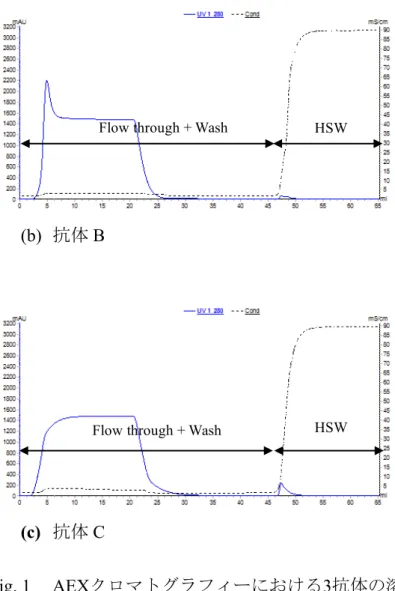
Fig. 1 B C
A HSW
(a) A
Flow through + Wash HSW
50
(b) B
(c) C
Fig. 1 AEX 3
(Q Sepharose Fast Flow)
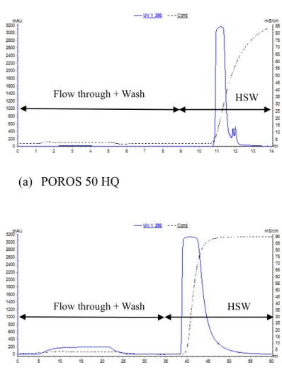
3.3.1.2 AEX A
pH 7.5 AEX A Q Sepharose Fast Flow
( ) POROS 50
HQ ( ) DEAE Sepharose Fast Flow (
) (Fig. 2) DEAE Sepharose Fast Flow
A A 95% HSW
DEAE Sepharose Fast Flow
21
POROS 50 HQ A HSW
Flow through + Wash HSW
Flow through + Wash HSW
51
A pH 7.5 AEX
(a) POROS 50 HQ
(b) DEAE Sepharose Fast Flow
Fig. 2 AEX A
(POROS 50 HQ, DEAE Sepharose Fast Flow)
3.3.1.3
pH 7.5 A AEX A AEX
/ /
Q Sepharose Fast Flow POROS 50 HQ A
Flow through + Wash HSW
Flow through + Wash HSW
52
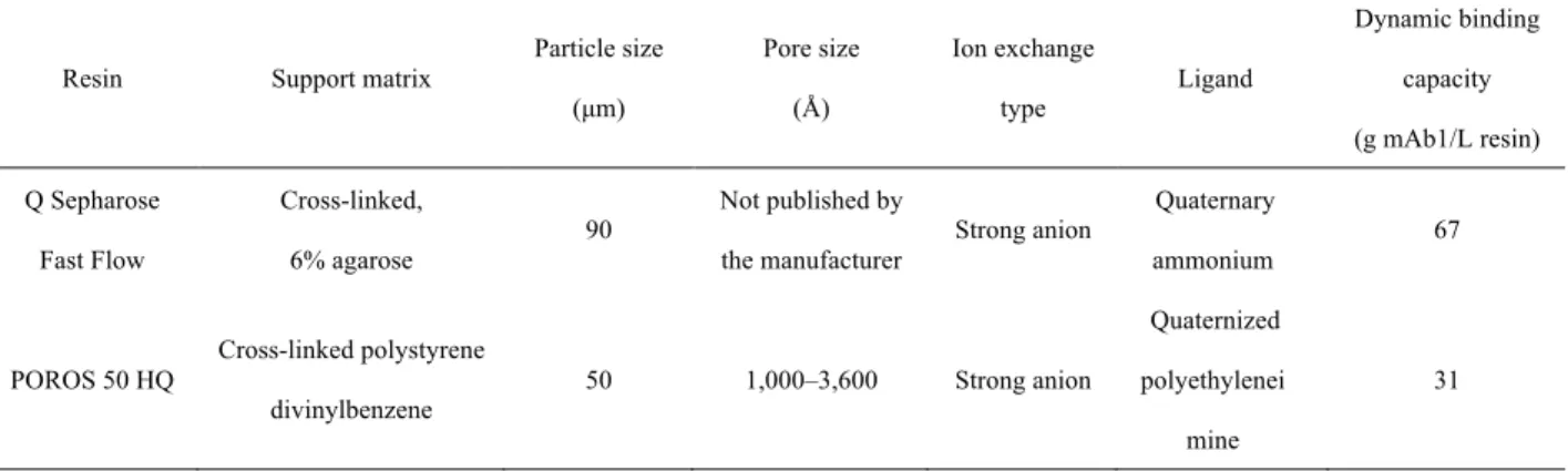
Table 1 AEX
Q Sepharose Fast Flow 67 g /L POROS 50 HQ 31 g
/L /
3 1/4 (Q Sepharose Fast Flow ) 1/10
(POROS 50 HQ ) /
Table 1. (5% breakthrough)
Resin Support matrix
Particle size (µm)
Pore size (Å)
Ion exchange
type Ligand
Dynamic binding capacity (g mAb1/L resin) Q Sepharose
Fast Flow
Cross-linked,
6% agarose 90
Not published by
the manufacturer Strong anion
Quaternary
ammonium 67
POROS 50 HQ Cross-linked polystyrene divinylbenzene
50 1,000–3,600 Strong anion
Quaternized polyethylenei
mine
31
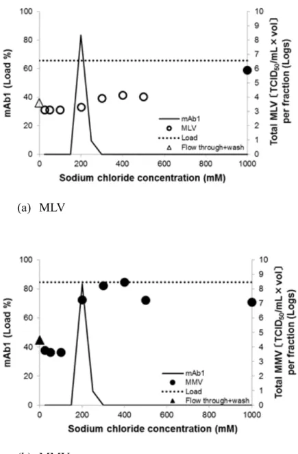
3.3.1.4 AEX A
AEX
Fig. 3 pH 7.5 / A
200 mM
MLV 500 mM
1,000 mM
A MLV MMV
(25 mM)
MMV A 200 mM MMV
A (200mM
) AEX MLV 3.90 log10 MMV
53
1.20 log10 (Table 2) / AEX
MLV × MMV
54
(a) MLV
(b) MMV
Fig. 3 AEX A
(Q Sepharose Fast Flow)
Open symbol
Table 2. Q Sepharose Fast Flow / 200 mM
Virus Viral clearance (log reduction value) a
MLV 3.90 ± 0.25
MMV 1.20 ± 0.37
a 95%
55
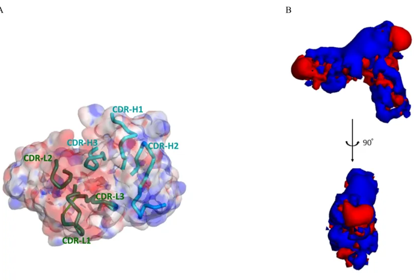
3.3.2
AEX A
A B C Fv pH 7.5
B C A
(Fig. 4A) A isovalue surface
(Fig. 4B)
IEX IEX
IEX
18-20
15 IEX 16
Lesins Ruckenstein 17
"steering effect" (Fig. 6) AEX
AEX 22 Fig. 4B isovalue surface
A Fv N
(Fig. 4B, Fig. 5)
56 A
(a) A (b) B (c) C
B
Fig. 4 pH 7.5 Fv
A
B A −1 +1 kBT/e
57
A B
Fig. 5 A Fv complementarity determining region (CDR) A
A A Fv CDR
B A −1 +1 kBT/e
58
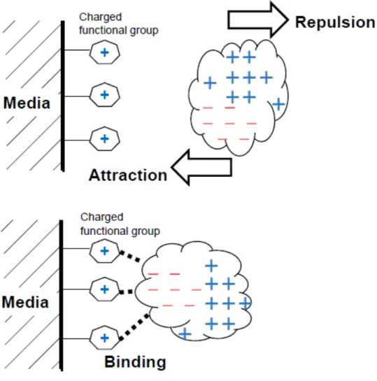
Fig. 6 "steering effect"
"steering effect"
AEX AEX
59
3.3.3 AEX NatriFlo HD-Q A
Kopaciewicz IEX
ion-exclusion layer "double layer"
22 double layer
A
/ pI
" " double layer
A NatriFlo
HD-Q ( 0.40 µm
)
A 5% A pH 7.5 NatriFlo HD-
Q (Fig. 7)
NatriFlo HD-Q
RT = 3 NatriFlo HD-Q RT = 4
NatriFlo HD-Q A
Fig. 8 A (4
NatriFlo HD-Q) (3 AEX
60
) NatriFlo HD-Q
A RT = 4 96.8% RT = 3 96.7%
A NatriFlo HD-Q
Fig. 7 NatriFlo HD-Q A
(a) Residence time 4 sec
Flow through + Wash HSW
Flow through + Wash HSW
61
(b) Residence time 3 min
Fig. 8 NatriFlo HD-Q A
3.3.4 NatriFlo HD-Q
A pH 7.5 NatriFlo HD-Q
A ( )
AEX
NatriFlo HD-Q
(Fig. 9) A NatriFlo HD-Q
MLV 500 mM
1,000 mM NatriFlo HD-Q
AEX MLV
A MMV
(25 mM) NatriFlo HD-
Q MLV 3.31 log10 MMV 2.26 log10
(Table 3) NatriFlo HD-Q MMV
/ LRV
Flow through + Wash HSW
62
(a) MLV
(b) MMV
Fig. 9 AEX A
(NatriFlo HD-Q)
Open symbol
Table 3. NatriFlo HD-Q
Virus Viral clearance (log reduction value) a
MLV 3.31 ± 0.25
MMV 2.26 ± 0.37
a 95%
63
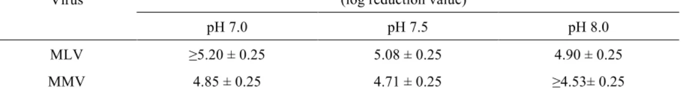
3.3.5 NatriFlo HD-Q pH
NatriFlo HD-Q A
NatriFlo HD-Q
AEX HCP DNA
pH
23,24 NatriFlo HD-Q pH
A
pH 7.0 7.5 8.0 Tris
pH MLV MMV Fig. 10
Table 4 MLV pH 500
mM 1,000 mM
MLV pH pH
NatriFlo HD-Q MLV NatriFlo HD-Q MLV
pH MMV
pH pH 7.0 7.5
MMV 25 mM 1,000 mM
pH 8.0 400 mM
MMV MMV
pH pH 7.0 pH 8.5
pH 7.5 A
(MLV 3.31 MMV 2.26 Table 3) A (MLV 5.08 MMV
4.71 Table 4)
6
HCP DNA 25 NatriFlo
HD-Q pH
64
65
(a) MLV, pH 7.0 (b) MMV, pH 7.0
(c) MLV, pH 7.5 (d) MMV, pH 7.5
(e) MLV, pH 8.0 (f) MMV, pH 8.0
Fig. 10 NatriFlo HD-Q pH
Open symbol
66
Table 4. NatriFlo HD-Q pH
Virus
Viral clearance (log reduction value) a
pH 7.0 pH 7.5 pH 8.0
MLV ≥5.20 ± 0.25 5.08 ± 0.25 4.90 ± 0.25
MMV 4.85 ± 0.25 4.71 ± 0.25 ≥4.53± 0.25
a 95%
‘>’
LVP
67
3.3.6
NatroFlo HD-Q A
" " double layer
3.4
A AEX
AEX
A
NatriFlo HD-Q
A A
3.5
1. Liu HF, Ma J, Winter C, Bayer R. Recovery and purification process development for monoclonal antibody production. MAbs. 2010;2:480-499.
2. Shukla AA, Hubbard B, Tressel T, Guhan S, Low D. Downstream processing of monoclonal antibodies-Application of platform approaches. J Chromatogr B.
68
2007;848:28-39.
3. CMC biotech working group. A-Mab: a case study in bioprocess development. Version 2.1 30th October 2009. Available at:
https://cdn.ymaws.com/www.casss.org/resource/resmgr/imported/A- Mab_case_study_Version_2-1.pdf
4. Miesegaes G, Lute S, Brorson K. Analysis of viral clearance unit operations for monoclonal antibodies. Biotechnol Bioeng. 2010;106:238-246.
5. Strauss DM, Gorrell J, Plancarte M, Blank GS, Chen Q, Yang B. Anion exchange chromatography provides a robust, predictable process to ensure viral safety of biotechnology products. Biotechnol Bioeng. 2009;102:168-175.
6. Weaver J, Husson SM, Murphy L, Wickramasinghe SR. Anion exchange membrane adsorbers for flow-through polishing steps: Part II. Virus, host cell protein, DNA clearance, and antibody recovery. Biotechnol Bioeng. 2013;110:500-510.
7. Goyon A, D'Atri V, Colas O, Fekete S, Beck A, Guillarme D. Characterization of 30 therapeutic antibodies and related products by size exclusion chromatography : Feasibility assessment for future mass spectrometry hyphenation. J Chromatogr B.
2017;1066:35-43.
8. Miesegaes GR, Lute SC, Read EK, Brorson KA. Viral clearance by flow-through mode ion exchange columns and membrane adsorbers. Biotechnol Prog. 2014;30:124-131.
9. Somasundaram B, Pleitt K, Shave E, Baker K, Lua LHL. Progression of continuous downstream processing of monoclonal antibodies : Current trends and challenges.
Biotechnol Bioeng. 2018;115:2893-2907.
10. Ichihara T, Ito T, Kurisu Y, Galipeau K, Gillespie C. Integrated flow-through purification for therapeutic monoclonal antibodies processing. MAbs. 2018;10:325-334.
11. Ichihara T, Ito T, Gillespie C. Polishing approach with fully connected flow-through purification for therapeutic monoclonal antibody. Eng Life Sci. 2019;19:31-36.
69
12. Miesegaes G, Lute S, Brorson K. Analysis of viral clearance unit operations for monoclonal antibodies. Biotechnol Bioeng. 2010;106:238-246.
13. Ishihara T, Kadoya T. Accelerated purification process development of monoclonal antibodies for shortening time to clinic: Design and case study of chromatography processes. J Chromatogr A. 2007;1176:149-156.
14. Ishihara T, Kadoya T, Yoshida H, Tamada T, Yamamoto S. Rational methods for
predicting human monoclonal antibodies retention in protein A affinity chromatography and cation exchange chromatography Structure-based chromatography design for monoclonal antibodies. J Chromatogr A. 2005;1093:126-138.
15. Janson JC, Ryden L. Ion-exchange chromatography. In: Protein purification: Principles, high-resolution methods, and applications (2nd edition). New York: Wiley-VCH, Inc., 1998;145-205.
16. Regnier FE. The role of protein structure in chromatographic behavior.
Science.1987;238: 319-323.
17. Lesins V, Ruckenstein E. Chromatographic probing of protein–sorbent interactions. J.
Colloid Interface Sci.1989;132:566-577.
18. Chicz RM, Regnier FE. Single amino acid contributions to protein retention in cation- exchange chromatography: Resolution of genetically engineered subtilisin variants. Anal Chem. 1989;61:2059-2066.
19. Gill DS, Roush DJ, Willson RC. Presence of a preferred anion-exchange binding site on cytochrome b5: structural and thermodynamic considerations. J Chromatogr A.
1994;684.55-63.
20. Chung WK, Hou Y, Freed A, Holstein M, Makhatadze GI, Cramer SM. Investigation of protein binding affinity and preferred orientations in ion exchange systems using a homologous protein library. Biotechnol Bioeng. 2009;102:869-881
21. Chirino AJ, Mire-Sluis A. Characterizing biological products and assessing
70
comparability following manufacturing changes: Nat Biotechnol. 2004;22:1383-1391.
22. Kopaciewicz W, Rounds MA, Fausnaugh J, Regnier FE. Retention model for high- performance ion-exchange chromatography. J Chromatogr. 1983;266:3-21.
23. Hou Y, Brower M, Pollard D, Kanani D, Jacquemart R, Kachuik B, Stout J. Advective hydrogel membrane chromatography for monoclonal antibody purification in
bioprocessing. Biotechnol Prog. 2015;31:974-982.
24. Merck KGaA, NatriFlo® HD-Q Membrane Adsorber - Application Note. Lit. No.
MK_AN1719EN Ver. 1.0. 2018/07
25. Roush D. Viral clearance using traditional, well-understood unit operations: Session 1.2.
Anion exchange chromatography; and Session 1.3. Protein A chromatography. PDA J Pharm Sci and Tech. 2015;69:154-162
71
72
4.1
50 570
CHO
CHO HMWS HCP
DNA
CHO
CHO
Fc Protein A
Protein A 2
3
(AEX) (CEX)
CEX AEX
73
CEX AEX
CEX
AEX
CEX /
HMWS HCP
CEX
2,000 g /L
MLV PRV Reo 3
/
/
pH
/ 20
/ CEX
74
HMWS HCP CEX
CEX
AEX
AEX
A AEX
A AEX
Fv
AEX
A
AEX
75
76
4.2
Masuda Y, Tsuda M, Hashikawa-Muto C, Takahashi Y, Nonaka K, Wakamatsu K,
“Cation exchange chromatography performed in overloaded mode is effective in removing viruses during the manufacturing of monoclonal antibodies”, Biotechnology Progress, 35, e2858 (2019).
Masuda Y, Ogino Y, Yamaichi K, Takahashi Y, Nonaka K, Wakamatsu K, “The prevention of an anomalous chromatographic behavior and the resulting successful removal of viruses from monoclonal antibody with an asymmetric charge distribution by using a membrane adsorber in highly efficient, anion-exchange chromatography in flow- through mode”, Biotechnology Progress (DOI 10.1002/btpr.2955)
Furukawa K, Okuno K, Onai S, Sugimura K, Yoko-o Y, Ishibashi-Masuda Y, Oshima T, Tsuruoka N, Magota K, Tanaka S, Ohsuye K, “Production of an α-amidating enzyme (α- AE) in recombinant CHO cells”, Animal Cell Technology: Basic & Applied Aspects, Vol.
5, 493-499 (1993)
4143300 20 6 20
77
CTD- 2 ( 2) ( )
2.3 ( )
2018 5
( )
“Meeting Planova™ and Selecting BioEX for Our Monoclonal Antibody Manufacturing Process” (Asahi Kasei Bioprocess)
The 21st PLANOVA™ WORKSHOP, 2018/10/11-12, San Francisco PLANOVA™ SEMINAR 2019 CHINA, 2019/4/11-12, Hangzhou PLANOVA™ SEMINAR in TOKYO, 2019/7/2, Tokyo
(ViSpot ) 2019 7
26
78
4.3