Correlations between Extranodal Metastasis and Prognosis in Patients with Squamous Cell Carcinoma of the Esophagus
Koji O
TSUKA, Masahiko M
URAKAMI, Yusuke T
AJIMA, Kimiyasu Y
AMAZAKI,Satoru G
OTOHand Tetsuo S
AWATANIAbstract : Background Extranodal metastasis (EM) has been reported in car- cinomas of many organs. However, the clinicopathological significance of EM in squamous cell carcinoma of the esophagus remains unclear, and this study sought to clarify this issue. Methods This study included 220 patients who underwent an esophagectomy with lymphadenectomy for primary esophageal carcinoma from 1996 to 2008. EM was defined as the presence of cancer cells in the soft tissue that were discontinuous with the primary lesion, or in the perinodal soft tissue distinct from the lymph nodes. Results EM was detected in 25 (9.6%) of the 220 patients, and in 56 (0.7%) of the 8,186 nod- ules retrieved as ʻlymph nodesʼ. The incidence of EM was significantly higher in patients who had tumors of a larger size (diameter ≥ 4 cm) , lymphatic vessel invasion, lymph node metastasis, a high pathological stage, infiltrative growth pattern, or a high pT-stage. The 5-year overall survival rates in N0-1 patients with EM were significantly lower than in the patients without EM
(P = 0.005) . Conclusion EM is closely associated with the development and aggressiveness of esophageal carcinoma, and the presence of EM can be useful for predicting prognosis after surgery in N0-1esophageal carcinoma patients.
Key words : esophageal cancer, extranodal metastasis, surgery, chemotherapy, squamous cell carcinoma
Introduction
Lymph node metastasis is a form of tumor spreading that is recognized as an important prognostic factor in several types of cancers, including squamous cell carcinoma of the esophagus. However, the spread of cancer cells to extracapsular connective tissues sur- rounding the lymph nodes, called extranodal metastasis (EM)
1-3), is also often found during detailed histological investigations of resected lymph nodes in squamous cell carcinoma of the esophagus. EM has also been reported in carcinomas of the stomach
4-7), rectum
8, 9), thyroid
10, 11), breast
12, 13), vulva
14), and lung
15, 16), and such metastasis has been linked to the rate of disease progression. However, few studies have examined the impact of EM on prognosis in patients with squamous cell carcinoma of the esophagus
2, 17), prompting this
Department of Surgery, Division of General and Gastroenterological Surgery, Showa University School of Medicine,
1-5-8, Hatanodai, Shinagawa-ku, Tokyo 142-8666, Japan.
Original
study aimed at clarifying the clinicopathological significance of EM in this patient group.
Materials and Methods
Two hundred and twenty patients with thoracic esophageal cancer were admitted to Showa University Hospital from November 1996 to May 2008 ; all of these underwent video-assisted thoracoscopic surgery for esophageal cancer (VATS-E) . Ninety-three patients underwent VATS-E with en bloc lymphadenectomy of cervical, mediastinal, and abdominal lymph nodes (three-field lymphadenectomy) . The remaining 131 patients underwent VATS- E with en bloc lymphadenectomy of the mediastinal and abdominal lymph nodes (two-field lymphadenectomy) . The criteria for three-field lymphadenectomy included patients without serious systemic disease, localization of the tumor to the upper thoracic esophagus, and other localizations with metastasis of recurrent nerve lymph nodes detected by intraoperative histopathological examination. The median follow-up period after resection ranged from 1.5 to 153 months (median : 32.4 months) .
Immediately after surgery, a surgeon separated the excised specimens, which comprised the esophagus, gastric fundus, and surrounding soft tissues including lymph nodes dissected en bloc. The isolated lymph nodes, which contained some of the surrounding fat tissue, were grouped according to the Guidelines for the Clinical and Pathological Studies of Carcinoma of the Esophagus (10
thedition)
18). The number of dissected lymph nodes ranged from 7 to 169 (median 39.2) per patient, and a total of 8,186 lymph nodes were examined in the present study. All resected specimens were fixed in 10% formalin, embedded in paraffin, and stained with hematoxylin and eosin. All solid structures in adipose connective tissue resected with the esophagus were retrieved, including the lymph nodes and any areas of extranodal metastasis (EM) . In this study, EM was defined as the presence of cancer cells in soft tissue that was discontinuous with the primary lesion, or in perinodal soft tissue dis- tinct from the lymph nodes. The clinicopathological features of the tumors are summarized in Table 1.
Statistical analysis
The correlation between EM and clinicopathological features was determined using a Fisherʼs exact test, χ
2test or Mann-Whitney U test. The cumulative overall and disease- free survival rates were calculated using the Kaplan-Meier method and compared using the log rank test. A multivariate analysis was performed using the Cox proportional hazard model together with factors described previously. A P-value of < 0.05 was considered to be significant.
Results
EM was detected in 25 (9.6%) of the 220 patients and in 56 (0.7%) of the 8,186 nod-
ules retrieved as ʻlymph nodesʼ (Fig. 1) . We investigated the clinicopathological factors of
186 patients after surgery except for patients who died of causes unrelated to esophageal cancer. Of these 25 patients with EM, 24 (96.0%) were histopathologically diagnosed as being positive for lymph node metastasis.
We found no significant differences in sex, tumor location, venous invasion, intramural metastasis, or histopathological type between the EM-positive group and EM-negative groups.
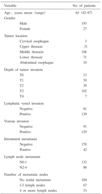
Table 1. Patient demographics and tumor characteristics
Variable No. of patients
Age ; years, mean(range) 65
(42-87)Gender
Male 193
Female
27Tumor location
Cervical esophagus
3Upper thoracic
31Middle thoracic 106
Lower thoracic
71Abdominal esophagus
10Depth of tumor invasion
T0
13T1
59T2
39T3 102
T4
7Lymphatic vessel invasion
Negative
81Positive 139
Venous invasion
Negative
91Positive 129
Intramural metastasia
Negative 178
Positive
42Lymph node metastasis
N0-1 132
N2-4
88Number of metastatic nodes
No nodal metastasis 100
1-3 lymph nodes
674 or more lymph nodes
53Clinical and pathological characteristics were grouped
according to the guidelines for the clinical and pathological
studies of carcinoma of the esophagus
(10thedition)
However, a univariate analysis of the clinicopathological features showed that the incidence of EM was significantly higher in patients with the following : lesions more than 40 mm in diameter, lesions with lymphatic vessel invasion, lymph node metastasis, lesions with a larger number of lymph node metastases, lesions with higher pathological stage, and lesions with an infiltrative growth pattern. Furthermore, the incidence of EM was also significantly higher in patients with pT3-pT4 tumors than in those with pT0-pT2 tumors (Table 2) . The Kaplan-Meier survival curves of the estimated overall survival and disease-free survival are shown in Figures 2 and 3. The 5-year overall survival rates were significantly lower in patients with EM than in patients without EM at 15.7% and 52.9%, respectively.
Similarly, the 5-year disease-free survival rate in patients with EM was 20% compared to 50.3% in patients without EM.
Multivariate analyses revealed that tumor location, depth of tumor invasion, intramural metastasis, and pathological stage were statistically significant, independent factors influencing the overall survival and disease-free survival in the present series (Tables 3, 4) , while lymph node metastasis was not identified as an independent prognostic factor in the present study.
However, it is known that lymph node metastasis worsens the prognosis in esophageal cancer
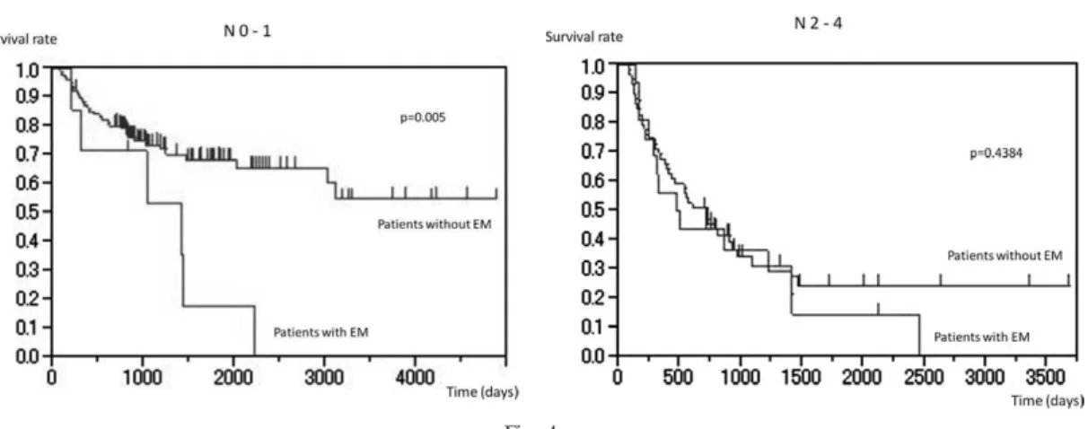
19-21). Thus, to eliminate this as a factor from our analysis, we divided the patients into two groups according to levels of lymph node metastasis (N0-1 vs. N2-4) , and then re- analyzed the estimated overall survival and disease-free survival (Fig. 4a, b) . The 5-year overall survival rates in N0-1 patients with EM were still significantly lower than in N0-1 patients without EM (Fig. 4a) ; however, the 5-year overall survival rates in N2-4 patients with EM were not significantly different from those in N2-4 patients without EM (P = 0.4384) (Fig. 4b) . These results suggested that EM influences the prognosis in N0-1 lymph node metastasis. The 5-year disease-free survival rates with or without EM showed no significant difference between N0-1 (P = 0.686) and N2-4 patients (P = 0.165) (Fig 5a, 5b) . Table 5 lists the sites of recurrence after surgery in patients with squamous cell carcinoma
Fig. 1. Hematoxylin and eosin staining shows extranodal metastasis in squamous cell carcinoma of the esophagus. Tumor cells are scattered in the perinodal soft tissue
(a)and lymphatic vessels
(b)are distinct from the metastatic lymph node. Original magnification a 100, b 200
Table 2. Relationship between clinicopathological factors and EM in 186 patients with squamous cell carcinoma of the esophagus
Variable No. of patients
EM Negative n
=161
EM Positive n
=25
P
=value
Age ; years, mean 65.0 64.5
Gender
Male 140 20
Female 21 5 not significant
Tumor size
(cm)<
4cm 87 7
≥ 4cm 74 18 P
=0.0154
Tumor location
Upper thoracic 28 2
Middle thoracic 73 14
Lower thoracic 60 9 not significant
Depth of tumor invasion
pT0-pT2 77 6
pT3-pT4 84 19 P
=0.0272
Lymphatic vessel invasion
Negative 64 3
Positive 97 22 P
=0.0066
Venous invasion
Negative 73 8
Positive 88 17 not significant
Intramural metastasis
Negative 146 20
Positive 15 5 not significant
Lymph node metastasis
N0-1 102 7
N2-4 59 18 P
=0.0017
Number of metastatic nodes
No nodal metastasis 76 1
1-3 lymph nodes 49 10
4 or more lymph nodes 39 14 P
=0.0001
Stage
0 19 0
I 27 0
II 49 5
III 43 12
IVa 19 8
IVb 4 0 P
=0.003
Pathology
Well 39 6
Mod 92 15
Poor 23 1
Other 7 3 not significant
Infiltrative growth
a 46 3
b 111 19
c 4 3 P
=0.0222
Clinical and pathological characteristics were grouped according to the guidelines for
the clinical and pathological studies of carcinoma of the esophagus
(10thedition)
of the esophagus. The incidence of recurrence in the liver and recurrence in the lymph nodes was significantly higher in patients with EM than in patients without EM.
Discussion
EM can be identified by detailed histological examination of surgical specimens obtained by radical esophagectomy. This distinctive type of tumor spread has also been reported in several types of carcinomas, and in this study, the incidence of EM among patients with esophageal carcinoma was 11.8%, which is similar to that reported previously
17). Non-resect- ed cancer cells in the adipose connective tissue may be associated with tumor recurrence after surgery. Therefore, a relatively high incidence of EM suggests that en bloc lymph
Fig. 2. Survival curves in patients with EM and those without EM
The 5-year overall survival rates were significantly lower in patients with EM than in patients without EM
(P =0.0005) .
Fig. 3. Disease-free intervals in patients with EM and those without EM
The 5-year disease-free survival rates were significantly lower in patients with EM than in patients without EM
(P=
0.0011) .
Table 3. Prognostic factors for the 5-year survival after surgery
Variable P
=value HR 95% CI
EM 0.2893 0.736 0.418-1.297
Age 0.5713 0.993 0.970-1.017
Gender 0.5371 1.202 0.667-2.175
Tumor location 0.0038 0.621 0.449-0.857
Tumor size 0.8366 0.952 0.600-1.513
Depth of tumor invasion 0.0139 2.057 1.158-3.654
Lymphatic vessel invasion 0.0685 1.289 0.981-1.693
Venous invasion 0.6598 1.062 0.813-1.386
Intramural metastasis 0.0185 2.070 1.130-3.792
Lymph node metastasis 0.722 0.892 0.475-1.676
Stage 0.0013 1.750 1.245-2.460
Infiltrative growth
a
0.376 0.599 0.193-1.863
b
0.6915 0.808 0.281-2.319
Pathology 0.4868 1.111 0.826-1.495
Number of metastatic nodes 0.3483 0.819 0.540-1.243 HR : hazards ratio ; CI : confidence intervals
Table 4. Prognostic factors for a disease-free interval after surgery
Variable P
=value HR 95% CI
EM 0.2893 0.710 0.390-1.292
Age 0.2047 0.985 0.963-1.008
Gender 0.3777 1.301 0.725-2.337
Tumor location 0.0017 0.608 0.445-0.830
Tumor size 0.9427 0.984 0.630-1.536
Depth of tumor invasion 0.0374 1.833 1.036-3.245
Lymphatic vessel invasion 0.1141 1.246 0.948-1.637
Venous invasion 0.9326 0.989 0.765-1.278
Intramural metastasis 0.0021 2.520 1.399-4.536
Lymph node metastasis 0.723 1.129 0.577-2.209
Stage 0.0057 1.605 1.148-2.246
Infiltrative growth
a
0.3958 0.618 0.204-1.877
b
0.8862 0.928 0.332-2.593
Pathology 0.0880 1.304 0.961-1.768
Number of metastatic nodes 0.6440 0.904 0.588-1.389
HR : hazards ratio ; CI : confidence intervals
Table 5. Site of recurrence after surgery
EM Negative EM Positive P-value
Liver 6 5 p
=0.0013
Lung 16 4 p
=0.3627
Lymph nodes 50 13 p
=0.0395
Others 5 0 p
=0.3717
Fig. 4.
a : Survival curves in N0-1 patients with EM and those without EM
The 5-year overall survival rates were significantly lower in N0-1 patients with EM than in patients without EM
(P =0.005) .
b : Survival curves in N2-4 patients with EM and those without EM
The 5-year overall survival rates in N2-4 patients with or without EM showed no significant difference
(P =0.4384) .
Fig. 5.
a : Disease-free intervals in N0-1 patients with EM and those without EM
The 5-year disease-free survival rates in N0-1 patients with or without EM showed no significant difference
(P=
0.686) .
b : Disease-free intervals in N2-4 patients with EM and those without EM
The 5-year disease-free survival rates in N2-4 patients with or without EM showed no significant difference
(P=
0.1646) .
node dissection could be essential for improving the prognosis of patients with esophageal carcinoma.
Tanabe et al
17)showed that EM in esophageal carcinoma was significantly associated with the depth of tumor invasion, intramural metastasis, lymph node metastasis, and the number of metastatic nodes. In this study, EM was significantly associated with the depth of tumor invasion, tumor size, lymphatic vessel invasion, number of metastatic nodes, pathological stage, and growth pattern. Together these results implicate EM in the development and aggressiveness of esophageal carcinoma.
Burn
22)hypothesized that the phenomenon of lymphaticovenous communication, when a lymph vessel is obstructed, occurs when cancer cells metastasize to a lymph node or lymph vessel and obstruct the flow of lymphatic fluid. He referred to the possibility of liver metastasis occurring via a lymphatic route. Tanaka et al
23)also suggested a relationship between liver metastasis and lymphatic involvement in gastric carcinoma. They reported that the lymphatic system was closely related to the establishment of liver metastasis ; in particular, they found that EM was a significant risk factor for liver metastasis. In our study, hematogenous (liver or lung) metastasis occurred in 9 out of the 26 cases with EM.
On the other hand, lymph node metastasis was detected in 13 cases. Therefore, EM was significantly associated with both hematogenous metastasis and lymph node metastasis (Table 5) . These data support the hypothesis that EM may be related to lymphaticovenous com- munication.
In many types of cancer, EM is known to be a pathological factor contributing to poor prognosis. Particularly in the cases of gastric cancer, close associations of EM with liver and peritoneal metastases have been reported
8, 23). For example, Baba et al
3)analyzed 131 patients who underwent complete resection of esophageal cancer, and found that the 5-year survival rate for 33 patients without involved nodes or perinodal tissue extension was 59.7%, compared to 14.0% for 43 patients with perinodal fat involvement (EM) , suggesting that the surgical cure of patients with EM is very difficult ; the present study reported similar rates in patients with and without EM. Together, these data support the notion that a finding of EM indicates a poor prognosis.
The disease-free survival rate was also significantly lower in patients with EM than in
patients without EM, a finding that could be directly related to the differences in the
overall survival rate between the groups. Although lymph node metastasis was not prognos-
tically significant in this study, we know clinically that it is closely related to survival. Thus,
we classified the extent of lymph node metastasis (N0-1 and N2-4) , and concluded that
EM is significantly associated with the survival of N0-1, but not N2-4 patients. Therefore,
this study showed that EM could be useful for predicting the prognosis of N0-1 patients
with esophageal carcinoma after surgery, and further suggests that adjuvant therapy such as
aggressive chemotherapy or chemoradiotherapy might be required to decrease postoperative
recurrence in these patients, even after R0 resection of esophageal cancer.
To clarify the impact of EM on patient prognosis, Tanabe et al
17)conducted a multivariate analysis in patients with esophageal carcinoma and found that EM was not a significant independent prognostic factor, as observed in our study. Instead, tumor location, depth of tumor invasion, presence of intramural metastasis, and pathological stage were identified as significant independent prognostic factors. The discrepancy between the uni- and multi- variate analyses could be explained by the possibility that variables such as the depth of invasion, intramural metastasis, tumor stage, and tumor location might all negatively affect prognosis, working as intermediate variables in the multivariate analyses, and thus negating or ameliorating the effect of EM as a prognostic factor.
In conclusion, this study provided important information that will help us to better understand EM. Our results show that EM is closely associated with the development and aggressiveness of esophageal carcinoma in N0-1 patients, and that EM can be useful for predicting the prognosis of esophageal carcinoma patients after surgery.
References
1) Watanabe H, Kato H, Tachimori Y, Yamaguchi H and Itababashi M : Characteristics of the spread pattern in esophageal carcinoma. Jpn J Thorac Surg 42:682-689
(1989)(in Japanese)2) Nishimaki T, Suzuki T, Suzuki S, Kuwabara S and Hatakeyama K : Outcomes of extended radical esophagec- tomy for thoracic esophageal cancer. J Am Coll Surg 186:306-312
(1998)3) Baba M, Aikou T, Yoshinaka H, Natsugoe S, Fukumoto T, Shimazu H and Akazawa K : Long-term results of subtotal esophagectomy with three-field lymphadenectomy for carcinoma of the thoracic esophagus. Ann Surg
219:310-316 (1994)4) Kumagai K, Tanaka T, Yamagata K, Yokoyama N and Shimizu K : Liver metastasis in gastric cancer with particular reference to lymphatic advancement. Gastric Cancer 4:150-155
(2001)5) Etoh T, Sasako M, Ishikawa K, Katai H, Sano T and Shimoda T : Extranodal metastasis is an indicator of poor prognosis in patients with gastric carcinoma. Br J Surg 93:369-373
(2006)6) Nakamura K, Okamoto Y, Matsui H, Makuuchi H and Ogoshi K : Impact of difference in the definition of extranodal spread on the outcome of node-positive patients with gastric cancer. Langenbecks Arch Surg 395:
211-216
(2010)7) Nakamura K, Ogoshi K and Makuuchi H : Subclassification of extranodal involvement in gastric cancer patients. Hepatogastroenterology 57:968-974
(2010)8) Ueno H, Mochizuki H and Tamakuma S : Prognostic significance of extranodal microscopic foci discontinuous with primary lesion in rectal cancer. Dis Colon Rectum 41:55-61
(1998)9) Shimada Y and Takii Y : Clinical impact of mesorectal extranodal cancer tissue in rectal cancer : detailed patho- logical assessment using whole-mount sections. Dis Colon Rectum 53:771-778
(2010)10) Yamashita H, Noguchi S, Murakami N, Kawamoto H and Watanabe S : Extracapsular invasion of lymph node metastasis is an indicator of distant metastasis and poor prognosis in patients with thyroid papillary carcinoma.
Cancer 80:2268-2272 (1997)
11) Ito Y, Fukushima M, Tomoda C, Inoue H, Kihara M, Higashiyama T, Uruno T, Takamura Y, Miya A, Kobayashi K, Matsuzuka F and Miyauchi A : Prognosis of patients with papillary thyroid carcinoma having clinically apparent metastasis to the lateral compartment. Endocr J 56:759-766
(2009)12) Mignano JE, Zahurak ML, Chakravarthy A, Piantadosi S, Dooley WC and Gage I : Significance of axillary lymph node extranodal soft tissue extension and indications for postmastectomy irradiation. Cancer 86:1258- 1262
(1999)13) Altinyollar H, Berberoglu U, Gulben K and Irkin F : The correlation of extranodal invasion with other prog-
nostic parameters in lymph node positive breast cancer. J Surg Oncol 95:567-571
(2007)14) van der Velden J, van Lindert AC, Lammes FB, ten Kate FJ, Sie-Go DM, Oosting H and Heintz AP : Extra- capsular growth of lymph node metastases in squamous cell carcinoma of the vulva.The impact on recurrence and survival. Cancer 75:2885-2890
(1995)15) Theunissen PH, Bollen EC, Koudstaal J and Thunnissen FB : Intranodal and extranodal tumour growth in early metastasised non-small cell lung cancer : problems in histological diagnosis. J Clin Pathol 47:920-923
(1994)16) Bell AM, DeYoung BR and Weydert J : Extranodal extension in metastatic non-small cell lung cancer. Chest
132:2058-2059 (2007)17) Tanabe T, Kanda T, Kosugi S, Ikeda Y, Makino S, Komukai S, Ohashi M and Suzuki T : Extranodal spreading of esophageal squamous cell carcinoma : clinicopathological characteristics and prognostic impact. World J Surg
31:2192-2198 (2007)18) Japan Esophageal Society : Japanese classification of esophageal cancer, tenth edition : parts II and III. Esopha-
gus 6:71-94 (2009)19) Reeh M, Nentwich MF, von Loga K, Schade J, Uzunoglu FG, Koenig AM, Bockhorn M, Rosch T, Izbicki JR and Bogoevski D : An attempt at validation of the Seventh edition of the classification by the International Union Against Cancer for Esophageal Carcinoma. Ann Thorac Surg 93:890-896
(2012)20) Prenzel KL, Holscher AH, Drebber U, Agavonova M, Gutschow CA and Bollschweiler E : Prognostic impact of nodal micrometastasis in early esophageal cancer. Eur J Surg Oncol 38:314-318
(2012)21) Liu J, Hu Y, Xie X and Fu J : Subcarinal node metastasis in thoracic esophageal squamous cell carcinoma.
Ann Thorac Surg 93:423-427 (2011)
22) Burn JI : Obstructive lymphopathy. Ann R Coll Surg Engl 42:93-113
(1968)23) Tanaka T, Kumagai K, Shimizu K, Masuo K and Yamagata K : Peritoneal metastasis in gastric cancer with particular reference to lymphatic advancement : extranodal invasion is a significant risk factor for peritoneal metastasis. J Surg Oncol 75:165-171
(2000)[Received February 20, 2012 : Accepted August 1, 2012]