パッチコロイド粒子の自己組織化構造の解明および 制御
野口, 朋寛
http://hdl.handle.net/2324/2236021
出版情報:Kyushu University, 2018, 博士(理学), 課程博士 バージョン:
権利関係:
Elucidation and Control of Self-assembled Structures of Patchy Colloidal Particles
Tomohiro Noguchi
2019
Abstract
When a colloidal particle has heterogeneity in their physical / chemical properties on its surface, the surface-surface interaction between particles or a particle and an object becomes anisotropic. Such particles thus behave as “colloidal molecules” that bind to each other only at specific sites on their surface, exhibiting various types of condensed phases according to their anisotropy. These condensed phases are also nano-microstructures composed of colloidal particles as building blocks, having been utilized in nanotechnology as a novel pathway to design self-assembled structures.
There have been extensive theoretical and numerical studies to elucidate the relation- ships between the anisotropy of colloidal molecules and their self-assembly or phase behavior. However, parameters characterizing the particle, such as the number and arrangements of the bonding sites and bonding energy, are enormous, and thus the relationships are far from being thoroughly elucidated. In particular, it is difficult to appropriately control the anisotropic structure and interaction of particles in experi- ments. The experimental studies are therefore strongly required from both academic and application fields. We therefore worked on experimentally controlling the inter- action between one-patch particles by the anisotropy of the surface properties and elucidating the self-assembled structures with the anisotropic structure of the par- ticles. One-patch particles are simplest colloidal molecules, having one distinctive region (patch) with different physical or chemical properties on its surface.
First, we used amphiphilic one-patch particles (Janus particle) having a hydrophilic and a hydrophobic hemisphere and investigated the structures formed in the particle- water-oil ternary system. The emulsification, i.e. stabilization of dispersed state of water in oil orvice versa, with Janus particles as surfactants has been investigated in several works. However, we observed and elucidated various self-assembled structures depending on amphiphilicity for the first time by our one-particle-level observations.
When the volume of water was smaller than that of the particles, a capillary bridge of water bonded between the hydrophilic hemispheres of the Janus particles, and the particles formed rod-like micelle structures. When the volume of water was large, spherical emulsion droplets formed, and the mechanism of the structure formation was similar to that of Pickering emulsions by particles having a uniform surface prop-
the aggregation structures were solid-like in the sense that thermal fluctuation was not effective, and “ideal surface active behavior” in which all particles are adsorbed on the water-oil interface was observed.
Next, we aimed to control the anisotropy of DLVO interactions, which is the most fundamental interaction working between colloidal particles, as a technique to intro- duce anisotropy into various colloidal particle systems. DLVO interactions consists of van der Waals attraction working between the same materials and electric double layer repulsion derived from surface charge, and the attraction is very strong between metal surfaces. Therefore, when dielectric particles with metal patches are used, it is expected that selective adsorption will occur between the patches of particles and between the patch and the surface of a metal object. In our experiments, the attrac- tion and repulsion were independently controlled by the thickness of the metal patch and by the surface electric potential of the patches, respectively. We succeeded in realizing a high adsorption selectivity in the particle dispersion system; the patchy particles were not adsorbed each other, and the metal patches were only adsorbed on the surface of a metal object. This selectivity induced a monolayer of patchy particles on the surface of large metal particles, which can be regarded as a solid dielectric layer preventing large particles from aggregation between them.
Our study elucidated the self-assembled structures of amphiphilic colloidal molecules by one-particle-level observation, and developed colloidal molecules whose anisotropic interaction inducing unique self-assembly can be tuned by the most typical and useful interaction in colloidal systems, i.e., DLVO interactions. The high surface activity of amphiphilic patchy particles has a great potential for applications and controlling DLVO interactions is useful for realizing experimental systems of colloidal molecules.
Contents
ChapterI General introduction 8
1 Colloidal particles and their dispersions 8
2 Colloidal particles 9
2.1 Isotropic particles . . . 9 2.2 Anisotropic particles . . . 10 3 Structure formation of colloidal particles: Effect of anisotropy by patches 13 3.1 Phase behavior of isotropic particles . . . 13 3.2 Phase behavior of patchy particles . . . 14
4 Surface-surface interactions 14
4.1 Mesoscopic structure, its physical property and functionality . . . 22
5 Motivations and purposes 25
5.1 Motivations . . . 25 5.2 Purposes . . . 26 5.3 Thesis outline . . . 26
ChapterII Elucidation of self-assembled structures in amphiphilic
Janus particles-water-oil ternary system 28
1 Introduction 28
1.1 Pickering emulsions . . . 28 1.2 Surface activity of amphiphilic Janus particles and Pickering emulsions
with them . . . 34 1.3 Differences and similarities between AJP and amphiphilic molecules . 36 1.4 Motivation and Purpose . . . 38
2 Experimental section 38
3 Results and Discussion 41 3.1 The aggregates and the change in structure . . . 41 3.2 Detail of micelle-like cluster and the formation mechanism . . . 43 3.3 Detail of spherical droplet and the formation mechanism . . . 46
4 Summary and conclusion 53
ChapterIII Control of adsorption behavior of metal-patchy dielectric particles utilizing anisotropy in DLVO interaction 55
1 Introduction 55
1.1 DLVO interaction . . . 55 1.2 DLVO interaction of metallodielectric particle . . . 62 1.3 Motivation and purpose . . . 63
2 Experimental section 64
2.1 Preparation of MDPs . . . 64 2.2 Adsorption of MDPs to an Au-patterned substrate . . . 65 2.3 Adsorption of MDPs to half Au-coated large particles . . . 68
3 Results and discussion 69
3.1 Adsorption of MDPs to Au-patterned substrate . . . 69 3.2 Adsorption of MDPs to thick Au films on large particles . . . 73
4 Summary 75
ChapterIV Concluding remarks 76
Acknowledgment 78
References 79
General introduction
1 Colloidal particles and their dispersions
Colloidal particles are fine particles of ∼ 10 nm to µm in size. There are various colloidal dispersion systems where colloidal particles are stably dispersed in gases, liquids, or solids. In such systems, the dispersed particles are called dispersoids, and the continuous phase is called dispersion medium. There are many colloidal dispersion systems around us. For example, when colloidal particles are solid and the dispersion medium is gas, liquid, and solid, the examples of the dispersions are smoke (aerosol), ink (suspension), and colored glass (solid sol), respectively (Figure 1). In addition, it is known that colloidal dispersions exhibit nonlinear responses to external stimuli since they possess large internal degrees of freedom due to the enormous number of colloidal particles. In other words, in a colloidal dispersion, not only the physical property and functionality of colloidal particles, e.g. color, light scattering ability, and catalytic ability, but also their dispersion states or self-assembled structures play important roles in the behavior and properties of the system. For example, when particles form a network structure and gel, the system exhibits elasticity, and when the particles are conductive, the system becomes conductive. In addition to the academic interest in the characteristic physical properties of colloidal dispersions around us, the study of colloidal particles and their dispersions has also been carried out for developing functional nano/microstructured materials.
Figure 1. Examples of colloidal dispersions. From the left, smoke, ink, and colored glass.
2 COLLOIDAL PARTICLES
2 Colloidal particles
2.1 Isotropic particles
Isotropic particles are those without anisotropy in e.g. shape and composition.
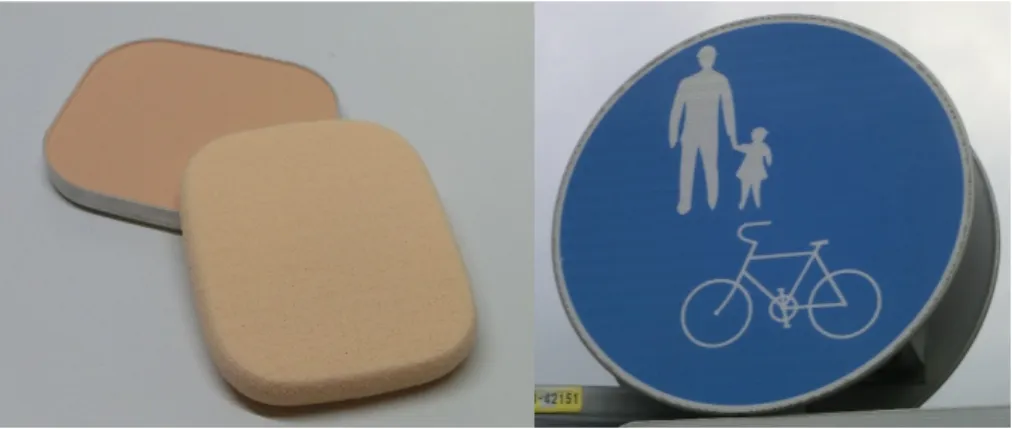
There are various kinds of isotropic particles including core-shell particles, gel balls, even liquid droplets and more, but the most typical and widely used one is a hard sphere of homogeneous material compositions. Isotropic and spherical particles are also found in our daily life. For example, spherical colloidal particles are added to a foundation of cosmetic products in order to impart light scattering/irregular re- flection ability, and to a road sign, where the particles are used to make its surface retroreflective (Figure 2). Isotropic monodisperse hard spheres have also been em- ployed in theoretical and experimental studies. Colloidal dispersions are classified into a complex system because of the numerous internal parameters such as physi- cal and chemical properties, volume fraction, and spatial arrangements of particles.
Therefore, the use of the simplest colloidal particle, e.g. hard sphere, is useful for elucidating the fundamental and essential features of colloidal dispersions.
Figure 2. Application of spherical isotropic colloidal particles. Left; foundation, right; road sign.
2.2 Anisotropic particles
In nature, anisotropic colloidal particles and their dispersions are abundant (Figure 3). In recent years, studies on anisotropic particles have attracted attention of re- searchers in soft matter [1–4]. In particular, experimental studies have become more and more practical in this decade according to the development of particle fabrication techniques.
(a) (b) (c)
2 µm
Figure 3. Examples of shape anisotropic particles. (a) A SEM image of a mineral dust particle found in a soil sample collected in northern Sahara[5]. (b) A schematic image of red blood cells. Their typical size is 7-8 µm. (c) A SEM image of halloysite clay particles [6].
The anisotropy of colloidal particles can be classified into two types; anisotropy in shape and substance (Figure 4) [1]. Anisotropy in substance is that in the spatial distribution of substance in one particle, and thereby such a particle has anisotropy in physical and/or chemical properties. These anisotropic particles show not only the anisotropy in the interaction but also active motion by consuming free energy by themselves (Figure 5) [7]. As described above, anisotropic colloidal particles ex- hibit essential difference from isotropic ones, and thus gain great attention from both experimental and theoretical points of view.
Figure 6 shows examples of shape-anisotropic particles [8]. There are variety of shapes from relatively simple ellipses and disk particles with different aspect ratios to complex polyhedral and branched particles.
As shown in Figure 4, it is possible to impart various types of anisotropy in phys- ical / chemical properties by the anisotropy in substance, such as catalytic ability, amphiphilicity, conductivity, surface charge, color, etc.
2 COLLOIDAL PARTICLES
Figure 4. Various anisotropies in particles[1].
Figure 5. Schematic drawing of a catalytic tube particle self-propelling while decomposing H2O2 [7].
Figure 6. Various shape-anisotropic particles[8].
Patchy particles
Particles having distinctive regions (patches) with different physical / chemical properties on the particle surface are called patchy particles [9, 10]. Since the inter- action between colloidal particles strongly depends on the properties of the particle surface, the anisotropy in the surface-surface interaction between patchy particles can be tuned by the patch. Patchy particles have been utilized in many theoreti- cal studies of condensed phases, and various patchy particles have been experimen- tally realized. Various combinations of patchy anisotropic properties can be consid- ered, such as hydrophilic-hydrophobic [11, 12], positive charge-negative charge [13], transparent-opaque [14], metal-dielectric [15], etc., inducing various anisotropic in- teractions depending on the properties of the patch and other surfaces. In addition, particles composed of multiple materials are called hybrid particles, being studied as functional nano particles for application [16].
3 STRUCTURE FORMATION OF COLLOIDAL PARTICLES: EFFECT OF ANISOTROPY BY PATCHES
3 Structure formation of colloidal particles: Effect of anisotropy by patches
The structures of aggregates formed by colloidal particles naturally depend on the interactions between them. Solid particles with shape anisotropy exhibit anisotropic hard-body interaction. Anisotropy of surface properties induces anisotropic surface- surface interaction in patchy particles. Due to these anisotropic interactions, anisotropic particles are expected to form characteristic structures which cannot be formed by isotropic particles. Therefore, by designing the anisotropic interaction between the particles, it is possible to obtain desired aggregated structures [16]. For example, attractively interacting patches, e.g. sticky patches [16], and the patches that bond to each other only for particular combinations of multiple patches [17], have been realized.
3.1 Phase behavior of isotropic particles
Colloidal particles exhibit thermal motion and thus can exhibit equilibrium phases in their condensed systems. A condensed system of colloidal particles has actually been used as a model experimental system to explore the phase behavior of general condensed systems. In condensed systems of isotropic particles, it is known that several phases appear depending on the interaction between particles, temperature, and volume fraction of particles (Figure 7). For example, monodispersed spherical particles only show face-centered-cubic and hexagonal close-packed structures as their crystalline phases in three dimensions. In addition, the interaction between particles is often sufficiently short-ranged (typically≲10 nm) compared with the particle size.
When the interaction is short-ranged, no gas-liquid phase separation appears and particles crystalize from the fluid phase[18]. In addition, it is known that a glassy state appears in the condensed system when the particles are not monodisperse. Figure 7 shows the dependence of phase behavior on the volume fraction of particles [19].
Figure 7. Phase behaviors of condensed systems of isotropic particles against particle den- sity. ϕis the volume fraction of particles. Solid green arrows indicate equilibrium states, and dashed arrows indicate metastable non-equilibrium states. Images are confocal micrographs of colloidal particle suspensions with 5% size dispersion[19].
3.2 Phase behavior of patchy particles
When a particle possesses sticky patches, number of binding sites (valence number), arrangements of them, and directions of bonds are limited in comparison with isotropic particles. Thus, such patchy particles are also called “colloidal molecules”[20, 21]. In condensed systems of particles with sticky patches, various phase behaviors different from those of isotropic particles have been observed[22–28]. For example, as shown in Figure 8 (a-d), when a particle having tetrahedrally-arranged four patches, diamond crystal phase, body centered cubic phase, and gas-liquid phase separation appear, exhibiting complicated phase diagrams depending on the strength of the interaction and size of patches [29]. The phase diagram also depends on the valence number (Figure 8 (i)). In a two-dimensional system, as shown in Figure 9, triblock particles form a kagome lattice in the experiment [30].
4 Surface-surface interactions
Various interactions work between colloidal particles in a solvent depending on the physical / chemical properties of the particles and the solvent. The magnitude of the interaction is generally compared with thermal energy. Multiple types of interactions
4 SURFACE-SURFACE INTERACTIONS
Figure 8. Phase diagrams of three- and four-patch particles, depending on the density, temperature, binding energy, width of binding sites, and the valence number[29].
Figure 9. Fluorescence microscope image of a lattice formed by triblock (two-patches) particles, with its Fourier transformation image and schematic diagram. The scale bar is 4 µm[30].
Derjaguin-Landau-Verwey-Overbeek (DLVO) interactions
DLVO interaction is one of the most typical interactions considered and actually works in colloidal particles in an aqueous solution, and thus often used for discussing the dispersion stability and aggregation behavior of colloidal particles [18]. DLVO interaction is the sum of the repulsion by the electric double layer (EDL) produced by surface charge and the van der Waals (vdW) attraction between materials. A characteristic feature of DLVO interaction is that the potential profile against the interparticle distance drastically changes by the properties of particles and solvent.
Figure 10 shows the scanning electron microscope and fluorescence microscope images of gold-polystyrene patchy particles and their clusters irreversibly aggregated by the vdW attraction between gold patches [31]. DLVO interaction is described in more detail in Sec.1.1 of Chapter III..
Figure 10. Gold-polystyrene patchy particles and their clusters. Green and red regions in (d-f) are gold patches and polystyrene surfaces, respectively[31].
4 SURFACE-SURFACE INTERACTIONS
Electrostatic interaction
An electrostatic interaction can be introduced between particles by modifying their surface by dissociable group. It is possible to impart not only positive or negative charge to a particle but also both positive and negative charges, i.e. a particle can be dipolar. Figure 11 shows the cluster formation of one-patch particles with a positively charged and negatively charged hemisphere in phosphate-buffered saline (PBS) buffer (pH: 6) with 1 mM ionic strength, together with the result of Monte Carlo simulations [13]. The cluster structures in the experiments (Column A) and in the simulations (Column B) agree, where the surfaces with the opposite sign, yellow hemisphere - red hemisphere, are adsorbed.
Figure 11. Clusters formed by charge dipolar particles in PBS buffer (pH: 6) with 1 mM ionic strength. Column A, column B, and column C are the epifluorescence observation images, the result of Monte Carlo simulations, and the charge distributions in the clusters of the B column. The numbers of particles constituting the clusters are shown in column C[13].
Hydrophilic / hydrophobic interaction
An attractive force works between hydrophobic surfaces in a polar solvent and between hydrophilic surfaces in an apolar solvent. These are called hydrophilic or hy- drophobic interaction. Patches having the same hydrophilic / hydrophobic property attract each other, being sticky. Figure 12 shows the experimental results of the clus- ter formation of amphiphilic one-patch particles whose hydrophilic and hydrophobic surfaces are hemispherical and corresponding results of Monte Carlo simulations[32].
In the simulations, the yellow and blue hemispherical surfaces are the hydrophilic and hydrophobic surfaces, respectively. The particles showed a monomer state, small clus- ters, and long, branched wormlike strings at 0, 1, and 5 mM of KNO3 consentrations, respectively.
Capillary interaction
When two particles are bridged by a small liquid droplet wetting their surfaces, an attractive (capillary) interaction occurs due to the interfacial tension of the droplet.
This attraction is generally very strong compared with the vdW attraction at the scale of colloidal particles. When the liquid of a droplet is polar / apolar, the droplet prefers to wet hydrophilic / hydrophobic surfaces, and therefore the attraction is large between the surface of similar hydrophilicity. Figure 13 is an experiment in which three-dimensional clusters of amphiphilic one-patch particles are formed by the wetting-induced interaction[33]. Droplets of the polar phase are formed by the phase separation of binary liquid mixture (water - 2,6-lutidine). The droplets can wet only the hydrophilic hemispheres of the one-patch particles (the white hemisphere). When the volume of the polar phase decreases, clusters in which the hydrophilic surfaces of the particles face each other are formed (Figure 13 (a)). Figure 13 (b) shows optical microscope images of the cluster formation process and its schematic diagram. Figure 13 (c) shows optical microscope images of clusters of various numbers of particles.
Interaction induced by solvent criticality
An attractive interaction works between the hydrophilic or hydrophobic particles in the vicinity of the critical demixing point of a solvent between the polar and apolar phases. When the binary liquid mixture solvent is in the one phase region near the
4 SURFACE-SURFACE INTERACTIONS
Figure 12. Aggregate structures formed by amphiphilic patchy particles in KNO3aqueous solution[32]. The left and right panels are epifluorescence observation images and simulation results, respectively.
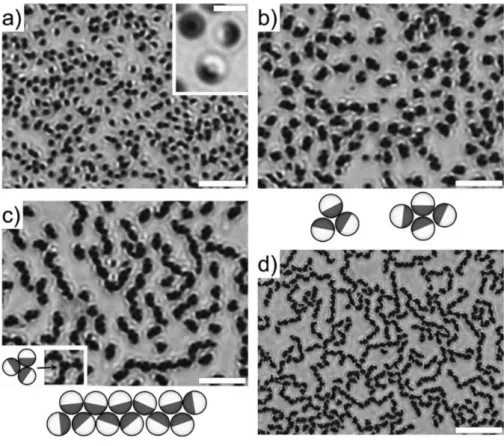
critical demixing point, the composition of the solvent at the particle surface is biased so that the ratio of the liquid component having high affinity to the particle surface becomes high. In addition, the concentration fluctuation between the particles is spatially limited. These effects cause an attractive interaction between the surfaces of the same polarity. Figure 14 shows the aggregate structures of one-patch particles having a moderate and highly hydrophilic hemispherical surface near the critical point
Figure 13. Three dimensional cluster formation of amphiphilic one-patch particles utiliz- ing the phase transition of water - 2,6-lutidine binary mixture. The black and the white hemispheres of the particle are the hydrophobic and hydrophilic hemisphere, respectively.
(a) A schematic drawing of the process forming and resolving clusters of the particles with the phase transition of solvent. (b) Optical and schematic images of the 4-particle cluster in the process of internal phase dissolving. Scale bar is 5 µm. (c) Three-dimensional clusters formed by many particles[33].
4 SURFACE-SURFACE INTERACTIONS
of a binary liquid mixture (water - 2,6-lutidine)[34]. As the temperature approaches the critical point (Figure 14 (a) to (d)), the particles, first in a dispersed state (Figure 14 (a)), form trimers or tetramers (Figure 14 (b)), and then form tetramer chain structures (Figure 14 (c)). In the structures, the highly hydrophilic hemispherical surfaces (black surfaces) are adsorbed each other, and the structural development with temperature is driven the increase of attraction with approaching the critical point. In Figure 14 (d), which is very close to the critical point, the white hemispheres are also adsorbed each other and form a network structure.
Figure 14. Temperature dependence of the aggregate structures formed by one-patchy par- ticles composed of moderate and highly hydrophilic hemispheres near the critical demixing point of water - 2,6-lutidine binary mixture. The temperature differences from the critical point are -0.5, -0.2, -0.1, and -0.01 K respectively from (a) to (d). The scale bars are 2µm in the inset in (a), 20µm in (d), and 10 µm for the others[34].
Depletion interaction
In a system in which colloidal particles and non-adsorptive macromolecules are dis- persed, an entropy-driven attractive interaction, depletion interaction, works between the particles. This interaction is generally weak, but it can be a dominant interac- tion in colloidal dispersions. Figure 15 is a schematic diagram of the key (red) and lock (green) particles, and an optical microscope image in an aqueous solution of poly ethylene oxide with a molecular weight of 600,000[35]. Figure 15 (a) shows the region in which the polymers are excluded and the overlap of the excluded space of the key and lock particles, respectively. When the curvature of the key particle and that of the lock particle are exactly the same, the overlap of the excluded volumes, and thus the depletion interaction, is maximized. Figure 15 (b) shows the combining process of a key and lock particle; key particles entering the key hole of the lock particle are bound (green arrow), but key particles are not bound to the other part (red arrow).
Figure 15 (c) shows the process in which a coupled key-lock particles combines with another lock particle.
Specific binding by DNA ligand
DNA chains are representative molecules having specificity in their binding target defined by the sequence of bases. By modifying two patches, A and B, with DNA chains having complemental single strand, selective binding between A and B patches are realized. Figure 16 shows the aggregated structures formed by particles having complemental A and B patches [36]. The left column presents optical microscope images of the aggregated structures, the middle column fluorescence microscopic im- ages, and the right column schematic diagrams of the aggregation structures. A and B patch are selectively adsorbed, and various structures are formed depending on the arrangement of the patches.
4.1 Mesoscopic structure, its physical property and functionality
The characteristic length scale of the internal structure in a colloidal dispersion is naturally mesoscopic because of the particle size. In different from the structures with microscopic length scale, i.e. structures composed of atoms or molecules, and those
4 SURFACE-SURFACE INTERACTIONS
Figure 15. Schematic drawing and optical microscope images of the combination of key and lock particles in aqueous solution of poly ethylene oxide with a molecular weight of 600,000.
The red and green particles in (a) are key and lock particles, respectively. The scale bar is 2 µm[35].
with macroscopic length scale around us, these structures with mesoscopic length scale exhibit characteristic features; the drastic increase in surface area compared with macroscopic structures, a relatively easy-change of internal structures in response to external input compared with microscopic structures, etc. These features induce unique physical properties and functionalities. For example, changes in the internal network structure give frequency dependence of viscoelasticity. The drastic increase in surface area dramatically increases the catalytic ability. In addition, mesoscopic structures often have a characteristic length in the order of visible light wavelength and thus exhibit characteristic optical properties such as absorption and reflection of light with specific wavelength. It is therefore expected that self-assembled structures of patchy particles can contribute to the creation of highly functional mesostructured materials [37].
Figure 16. Aggregation structure of patchy particles with complemental DNA ligands. The scale bar is 2 µm[36].
5 MOTIVATIONS AND PURPOSES
5 Motivations and purposes
5.1 Motivations
Patchy particles have extensive parameter space as colloidal molecules, such as the number and arrangements of bonding sites and the strength of bonds. Thus, the whole aspects of the role of the patchy surface anisotropy is not elucidated. Theoretical and numerical studies of condensed systems of patchy particles have greatly advanced in these decades because of the improvement of simulation methods and performance of computers [38–44]. In contrast, experimental studies are still advancing; it is not easy to appropriately control the anisotropic structure and interaction of particles in a real system. Deeper understanding of patchy particles in real, experimental systems is therefore strongly required from both academic and application point of view. We are therefore interested in the two following research themes.
Spherical amphiphilic one-patch particles have one hydrophilic and another hy- drophobic region on the surface. Since this particle has qualitatively the same struc- ture as a simple amphiphilic molecule, it is expected to have high surface activity and emulsification ability, stabilizing dispersion state of immiscible liquids such as water and oil. In addition, since various self-assembled structures and their ordered phases are known to appear in amphiphilic molecular dispersion systems in water or a water-oil mixture, similar structures and phases are expected to be formed in am- phiphilic patchy particle systems. Nevertheless, such studies with amphiphilic patchy particles have not been reported, and self-assembled structures and their formation mechanisms are still to be elucidated. Clarification of these fundamental behaviors of patchy particles as surface active agents would contribute not only to their use as a superior and/or unique surfactant compared to surfactant molecules, but also to their application to designing self-assembled functional mesostructures.
In order to realize anisotropic bonding between patchy particles, various interactions have been utilized as described above. However, there are few researches introducing anisotropy to DLVO interaction of patchy particles and controlling their self-assembly with it, though DLVO interaction is the most fundamental one working between col- loidal particles. When controlling anisotropy of DLVO interaction is realized, the
action to various experimental systems of colloidal particles compared with the other methods introducing the other types of interactions described above. Our method would thus be useful for various fundamental studies with colloidal molecules and applications for producing functional nanostructures by patchy particles.
5.2 Purposes
In order to elucidate self-assembled structures and their formation mechanisms in amphiphilic patchy particle dispersion systems, we carried out experiments on the structure formation in amphiphilic Janus particles-water-oil ternary systems. Am- phiphilic Janus particle is the simplest and symmetrical amphiphilic patchy particle composed of a hydrophilic hemisphere and a hydrophobic hemisphere, and thus it is suitable for capturing the essential features of their self-assembly. The details of the aggregation structures formed in this ternary system were observed at one-particle level, and the formation mechanisms were discussed with the typical interactions working in such particle-water-oil mixtures.
In order to realize the control of anisotropic DLVO interaction using patchy parti- cles, the dependence of the adsorption behavior of metallodielectric particles to metal surface and dielectric surface on salt concentration was investigated. A metallodi- electric particle is a type of patchy particle whose particle surface is composed of a metal and a dielectric part. We introduced the anisotropy in DLVO interaction to the system by tuning the repulsion by the surface charge of a patch and the strong vdW attraction by the thickness of a patch. The dependence of the adsorption behavior on the experimental conditions was studied with a flat dielectric substrate possessing thick metal films, and the self-assembly was observed with a freely dispersed large particle possessing a thick metal film.
5.3 Thesis outline
In Chapter II, we first describe the surface activity and self-assembly of colloidal particles and surfactants. Next, our fabrication method of amphiphilic one-patch (Janus) particles and experimental method are described. Then, the results of the
5 MOTIVATIONS AND PURPOSES
self-assembled structures in the amphiphilic Janus particles-water-oil ternary system explored by the single particle-level observation are explained. The analysis of the structures reveals their formation mechanisms.
In Chapter III, following the description of the DLVO interaction between colloidal particles, the idea of inducing anisotropy to DLVO interaction by metallodielectric patchy particles is described. Next, our particle fabrication and experimental method are described. Then, we show the results of the selective adsorption of the particles to metal surface. Finally, we describe the formation of the coated structure of metal particles by patchy particles.
In Chapter IV, we summarize the whole study and describe the future perspectives of structure formation using patchy particles.
Elucidation of self-assembled structures in amphiphilic Janus particles-water-oil ternary system
1 Introduction
1.1 Pickering emulsions
When immiscible fluids such as water and oil, and water and air are mixed finely, the respective fluid domains coalesce immediately and the fluids separate macroscop- ically. When a colloidal particle exhibits partial wetting to the interface between immiscible fluids, it can be stably adsorbed on the interface. The dense colloidal particles adsorbed on the interface prevent the fusion of the interface (cf. Figure 17) and stabilize the dispersion state of immiscible fluids; that is, the fluids are emulsified with the particles [45–48]. Such an emulsion stabilized by solid colloidal particles is called Pickering(-Ramsden) emulsion [49]. Pickering emulsions appear in various materials in nature and industry such as foods and cosmetics, playing characteristic roles in their physical properties and functionalities [50–52]. In general, Pickering emulsions have superior interface stability to conventional emulsion stabilized by sur- factant molecules[53, 54].
Here we consider the adsorption of isotropic spherical particles to the interface between immiscible polar liquid (“water”) and apolar liquid (“oil”) (Figure 18). As shown in Figure 18, the area of the water-oil interface decreases by the adsorption of the particle to the interface. When the particle exhibits partial wetting at the water- oil interface, the interfacial energy of the system including the energy at the particle surface decreases by the adsorption of the particle from the water or oil phase. Since the interfacial energy is the product of the interfacial tension and the area, the change of the energy of the system is expressed as
∆E =E1−E0 =−πr2γ(1±cosθ)2, (1)
1 INTRODUCTION
Figure 17. A spherical shell of densely-packed colloidal particles (colloidosome) at an interface[55].
whereE0 andE1 are the interfacial energy of the system before and after the adsorp- tion of the particle, andr,γ, θ are the radius of the particle, interfacial tension of the water-oil interface, and the contact angle between the particle surface and water-oil interface, respectively. In equation (1), ± corresponds to the adsorption from the two liquid phases, and usually the one with 1±cosθ <1 is taken as the adsorption energy of a particle. The decrease of the water-oil interface by a colloid, e.g. a meso- scopic scale particle whose diameter is typically tens of nanometers to micrometers is much larger than that of molecules. Therefore, the adsorption energy of a colloidal particle is much larger than that of molecules. For example, the adsorption energy is −2750kBT, when a spherical particle with a radius of 10 nm are adsorbed on a water-toluene interface (γ = 36 mN/m) at a contact angle of 90◦. kB and T are the Boltzmann constant and absolute temperature, respectively. Since the adsorption en- ergy is much larger than the thermal energy, the adsorption of particles is thermally
emulsions.
oil phase
water phase ݎ
ߠ
Figure 18. Adsorption of isotropic spherical particle to water-oil interface.
When the oil phase becomes droplets the system is called oil in water (oil/water) type emulsion and vice versa. An important point in the formation of Pickering emulsions is that the emulsion type depends not only on the hydrophilicity of the particles and polarities of the solvents but also on the volume ratio of the ternary system and the emulsification process. Majority liquid phase or the liquid phase by to which particles are more wettable tend to be the external, continuous liquid phase of the emulsions generally. Whether the particles are easily wetted by water or oil can be understood from the contact angle between the particle surface and water-oil interface. For example, if the angle between the particle-water interface and water-oil interface is smaller than that between the particle-oil interface and water-oil interface, the particles are more wettable by water. This contact angle θ is determined by the balance of the interfacial tension γsw at the particle-water interface, the interfacial tension γso at the particle-oil interface, and the interfacial tension γ at the water-oil interface (Figure 19) described as
γsw+γcosθ =γso. (2)
This is the Young’s equation. Figure 20 shows the Pickering emulsion types and the contact angles θow (the angle between the solid-water interface and the water-oil interface) in various combinations of solid, water, and oil. The contact angles greatly depend on the combination. The position of a particle at the water-oil interface depends on the contact angle, and the interface covered densely by the particles has
1 INTRODUCTION
spontaneous curvature when their contact angle is not 90◦ (Figure 21).
particle surface
ߛ ௦௪ ߛ ௦
ߛ ߠ
Figure 19. Balance of interfacial tensions at the contact point between water, oil, and particle surfaces.
As described above, since the adsorption energy of particles is much larger than the energy of thermal fluctuation, the free energy of Pickering emulsion systems can be approximated by interfacial energy. When the water-oil interface is covered with solid spherical particles, there is always a gap between the particles (Figure 22), and water- oil interface exists in the gap. The effective tension of the interface densely covered with particles is always positive. The total free energy of a Pickering emulsion is thus minimized when the total area of the particle-adsorbed interface is minimized, that is, when water and oil phase macroscopically separated. This is the thermodynamically most stable, equilibrium state. In general, Pickering emulsions are not at equilibrium but metastable state, and are kinetically stabilized because of the particle layer at the water-oil interface that becomes a steric barrier and prevents coalescence of liquid domains.
Because Pickering emulsions are kinetically stabilized, the formation process of the system also affects the resulting state. Limited coalescence is a typical process of structural development in Pickering emulsions (Figure 23) [57, 58]. The volumes of water, oil, and particles are preserved, and the initial state is that a minority liquid phase is thoroughly divided into a large number of droplets which are sufficiently small and the particles are isolated. Then, the particles are adsorb on the liquid-liquid interface and the droplets repeatedly coalesce and grow in size. Since the droplet
Figure 20. Contact angleθow and emulsion type in various combinations of solid particle, oil, and water[56].
Figure 21. The relation between contact angle θow and spontaneous curvature for the interface adsorbing particles densely[56].
1 INTRODUCTION
Figure 22. The interstitial gap (arrow) between the densely-packed particles at a droplet surface [55].
surface is not fully covered with the particles at the early stage, they can coalesce by their fusion. The coverage of the particles at a droplet surface increases by the coalescence because the total surface area of the droplets decreases by coalescence whereas the number of the adsorbed particles is preserved. When the surfaces of droplets are densely covered by particles with the progress of coarsening, coarsening halts since the particles are strongly adsorbed on the interface. That is, the system reaches a kinetically stable state.
Figure 23. Schematic drawing of limited coalescence process.
1.2 Surface activity of amphiphilic Janus particles and Pickering emulsions with them
Colloidal particles having a hydrophilic and hydrophobic surface exhibit am- phiphilicity as known for surface active molecules. When the hydrophilic and hydrophobic surface are both hemispherical, the particle is called amphiphilic Janus particle (AJP) (Figure 24). This particle is the first patchy particle designed and fabricated [59]. This section explains the surface activity and Pickering emulsions of AJPs, and they are compared with the case with isotropic particles.
water oil hydrophilic
hydrophobic
Figure 24. Schematic drawing of amphiphilic Janus particle.
Here we describe the adsorption energy of an AJP (Figure 25). When the hy- drophilic/hydrophobic hemispherical surfaces of the amphiphilic Janus particles are
1 INTRODUCTION
completely wetted by water/oil respectively, the change of the interfacial energy by the adsorption of an AJP to water-oil interface is written as
∆EJanus≤ −3πr2γ, (3)
whererandγ are the radius of the amphiphilic Janus particle and the water-oil inter- facial tension, respectively, and ∆EJanus is difined as in equation (1). This equation is derived from the relation between the interfacial energies for complete wetting, γso/sw ≥ γsw/so+γ, and ∆E = −3πr2γ when θ = 0◦ or 180◦ in the Young’s equa- tion (2)[60, 61]. Comparing equation (3) with equation (1), the adsorption energy of an AJP can be more than three times as large in magnitude as that of an isotropic spherical particle.
Figure 25. Schematic image of AJPs at water-oil interface and in water phase.
When AJPs are adsorbed on the water-oil interface, if the hydrophilicity and hy- drophobicity of each hemisphere are sufficiently high, the three phase contact line is on the hydrophilic-hydrophobic boundary (Figure 26) [62]. At the boundary a range of the contact angle of the water-oil interface is possible, and the arrangement of an adsorbed AJP does not change for some variation in the polarity of the liquid phases (Figure 26).
Next, we consider the stability of Pickering emulsion stabilized by AJPs. When comparing Pickering emulsions by spherical AJPs with those by isotropic spherical particles, the difference in interfacial energy at the particle surface affects the emul- sified state, because their shapes are the same. As described, the equilibrium state of the Pickering emulsions with isotropic spherical particles is the macroscopic sepa- ration (Sec.1.1 of Chapter II). On the other hand, Aveyard have theoretically shown that, when the amphiphilicity of an AJP is sufficiently large, the free energy of the
Figure 26. Schematic drawings of AJP (right), hydrophobic particle (center), and hy- drophilic particle (left) adsorbed on the interface[62].
Pickering emulsion becomes minimum when all AJPs are adsorbed on the water-oil interface [63]. It indicates Pickering emulsion by AJPs can be thermodynamically most stable (equilibrium state). Because of the high stability of such emulsions, AJP are used in emulsion polymerization or emulsion-based microreactors [64–67].
1.3 Differences and similarities between AJP and amphiphilic molecules
Amphiphilic molecules possess a hydrophilic and hydrophobic group in one molecule (Figure 27). As shown in Figure 27, when amphiphilic molecules are dispersed in a liq- uid phase, self-assembled structures such as spherical micelles and bilayer membranes appear by hydrophobic/hydrophilic interaction between the molecules. Furthermore, ordered structures composed of these supramolecular unit structures can form. These structures and ordered states depend on the molecular structure such as the size of the hydrophilic head group and the hydrophobic tail. In the amphiphilic molecules- water-oil ternary system, the structures depend also on the composition of the three components.
As described above, AJPs and amphiphilic molecules possess structural similar- ity. Therefore, the self-assembled structures of AJPs in their dispersions may also strongly depend on the composition of the system and the amphiphilic structure as in amphiphilic molecule systems. It is expected that various aggregate structures and ordered structures can form [34, 68–70]. However, there are also significant dif- ferences between AJPs and amphiphilic molecules. Due to the mesoscopic size of the particle, AJPs are adsorbed on the water-oil interface much more strongly than
1 INTRODUCTION
amphiphilic molecule
hydrophilic part hydrophobic part
spherical micelle
rod-like micelle
bilayer reverse micelle
sponge lamella vesicle
Figure 27. Examples of self-assembled structures by amphiphilic molecules.
amphiphilic molecules, and the adsorption of an AJP is irreversible. In contrast, amphiphilic molecules are at dynamic equilibrium between their adsorption to and desorption from the interface by thermal fluctuations. When AJPs are densely packed in water-oil interface, the hard-body interaction between the particles makes the par- ticle layer solid. In contrast, the molecular layer at the water-oil interface usually exhibits fluidity and becomes a soft membrane. As described above, since small gaps exist between the spherical particles in the adsorbed layer, and water and oil are in contact with each other there, it is possible to exchange substances through the gaps.
There are many differences between the colloidal and molecular system, and thus it can also be expected that the AJPs form unique self-assembled structures and/or ordered structures which are different from those of the amphiphilic molecules.
1.4 Motivation and Purpose
As described above, it is expected that AJPs would be superior surfactants to isotropic particles. In addition, AJPs are expected to form self-assembled struc- tures reflecting the amphiphilic structure of the particles, which cannot be formed by isotropic particles. However, most of the experiments with AJP-water-oil ternary systems have focused on the emulsification ability and the stability of the emulsions in macroscopic systems [71–75]. Therefore, the details of the self-assembled struc- tures, their dependence on the experimental conditions, and the structure formation mechanism are still to be clarified.
In order to clarify the whole aspects of the self-assembly in the ternary system, we studied the structures for a wide range of composition of the components by single particle-level observation, and worked on clarifying the structure formation mechanism.
2 Experimental section
2.1 Prepareing AJPs
We prepared AJPs composed of hydrophilic and hydrophobic hemispheres by coat- ing a hemisphere of spherical silica particles with Au and modifying the surface of the Au layer with 1-octadecanethiol (90.0 %, Wako) to make the surface hydropho- bic. First, a monolayer of monodisperse silica particles (Hyprecica, UEXC), diameter d = 3.01µm with size dispersion ∼ 1.5 % coefficient of variation, was prepared at an air-water interface and transferred onto a hydrophilized glass slide[76]. A 50-nm- thick gold layer was thermally deposited onto the monolayer, following a 3-nm-thick chromium layer. This gold layer formed a hemispherical gold cap on the particle[73].
To render the gold surface hydrophobic, the particles were soaked in a 2 mM ethanol solution of 1-octadecanethiol for 24 h. We rinsed the particles (volume ≃ 10µL) three times by dispersing them in 50 mL of ethanol and collecting them with a cen- trifuge.These particles possessed high amphiphilicity. They show clear amphiphilic Janus nature not only at pure water (18.2 MΩ, Millipore)–n-dodecane (99.0 %, Wako) interface (Figure 28) used in subsequent experiments, but also at an interface where
2 EXPERIMENTAL SECTION
the difference in polarity was smaller than that of water-n-dodecane. The interface was formed in the mixture composed with ethanol (99.5 %, Wako), dodecane, and a small quantity of pure water. The volume ratio of ethanol and dodecane was 25:75 and the water content was less than 1 % of the total volume. Because of the small water content, this mixture phase separated into ethanol- and dodecane-rich phases at room temperature, roughly at the volume fractions of the ethanol and dodecane content. In Figure 29, AJP can be seen attached to the interface between the two liquid phases, with their hydrophobic Au hemisphere immersed in the dodecane-rich phase and their silica hydrophilic hemisphere in the ethanol-rich phase. Thus, the fabricated AJP had a clear Janus nature with high amphiphilicity.
n -dodecane water
Au silica
(a) (b)
Figure 28. Amphiphilic Janus particles attaching to a liquid-liquid interface. (a) Au optical microscope image of AJP at a water-dodecane interface. The black and white regions of a particle are, respectively, the hydrophobic part (Au-coated surface) and hydrophilic part (silica surface). The scale bar is 5 5µm. (b) A scanning electron microscope image of a spherical silica particle with a hemisphere coated with Au. The white and gray regions of the particle are, respectively, the Au-coated surface and the silica surface. The radius of the particle was 2.6 µm for visual clarity.
2.2 Structure formation and observation
We prepared a ternary system comprising AJP, pure water, and n-dodecane. We used a volume ratio of water to the particles,α =VW/ Vp , over the range 0.00 – 8.28, where bothVW andVp were precisely measured and were up to a few microliters. The volume of dodecane was 100 – 200µL. The water was thus the minority liquid phase
Figure 29. An optical microscope image of amphiphilic Janus particles attached to the interface between dodecane-rich and ethanol-rich phases. The black and white regions of a particle are, respectively, the hydrophobic part (Au-coated surface) and the hydrophilic part (silica surface). The scale bar is 5µm.
in all samples so that the water domain was isolated and enclosed by the particles.
For each sample preparation, the amount of AJP was weighed and the value was converted to volume with the mass densities of the particle and gold. The AJPs were dispersed in dodecane in a polypropylene tube with an ultrasound bath (38 kHz, 100 W)(US-102, SND Inc.) for ∼ 5 min. Next, when VW ≥ 0.5µL, the required amount of water was added to the AJP-dispersed dodecane with a micropipette, followed by the ultrasonic mixing for ∼ 60 s. When VW < 0.5µL, a larger amount of water was first dispersed in dodecane homogeneously by ultrasonication, and then the calculated amount of the water-dispersed dodecane containing the required VW was added to AJP-dispersed dodecane, followed by ultrasonic mixing for∼60 s. This process enables precise control ofVW and through break-up of such a tiny amount of water (cf. Figure 30). After the self-assembled structure formation in a sample under gentle stirring by hand, some amount of the sample, ∼20µL, was taken and diluted in dodecane ∼ 100µL to isolate each cluster/emulsion droplet. This diluted sample was observed with optical microscopy: BX50 (Olympus) with objective lenses Plan 40x/0.65 and Ach 20x/0.40, and TE2000-U (Nikon) with objective lenses PlanApo VC 100x/1.40, PlanApo VC 60x/1.40, PlanFluor 40x/0.75 Ph2 DLL and PlanFluor
3 RESULTS AND DISCUSSION
20x/0.50 Ph1 DLL, dependent on the required magnification. No noticeable effect of the dilution process on their structures was observed under the microscopy. We prepared the samples with differentα to investigate the dependence of self-assembled structures on it.
Figure 30. Optical microscopy images of water droplets wetting a glass coverslip in do- decane. Apparently, the diameter of the wetting droplets is ≲ 1µm: When the diameter is 1µm, the corresponding diameter of the floating droplet is 0.52µm. It follows that the diameter of water droplets after the agitation should have been less than 1µm typically. The scale bar is 1µm.
3 Results and Discussion
3.1 The aggregates and the change in structure
This section first qualitatively describes the change in structure of the ternary sys- tem with increasing water content,α. With increasingα, (inverse) small, micelle-like clusters appeared, followed by anisotropic growth into rod-like shapes, and finally spherical emulsion droplets were formed (Figure 31 (b-k)). At α = 0.00, random aggregates simply were formed by the van der Waals attraction between the particles (Figure 31 (a)). The attraction between bare silica particles is strong enough to irre- versibly aggregate in dodecane, explaining why there was no selectivity for bonding surfaces observed, although a stronger attraction was expected between Au surfaces than between Au and silica or between silica surfaces due to the large Hamaker con- stant of Au. Adding a small amount of water (α = 0.09) caused small clusters in which
structure (Figure 31 (b)). The micelle-like clusters coexisted with random aggregates.
Larger micelle-like clusters were observed with an increase in α. Simultaneously, the number ratio of the micelle-like clusters to random aggregates increased with α, and random aggregates were not observed for α > 0.27. When the sizes of small clusters exceeded about 15 particles, the clusters exhibited unidirectional anisotropy in shape.
The clusters with clear rod-like shapes appeared at α≥0.16 (Figure 31 (c-e)). With further increases in α, almost all clusters became rod-like and their widths slightly increased, as shown in Figure 31 (c-e). For α ≥ 0.39, spherical emulsion droplets (colloidosomes) with a water inner phase appeared. In Figure 31 (j), AJP can be seen attached to the water-oil interface, reflecting their amphiphilic Janus structure and forming a monolayer. With an increase in α, the size of the spherical emulsion droplets and the number ration of them to all aggregates increased. For α ≥ 0.66, finally, the system formed an emulsion, in which almost all structures were spherical droplets (Figure 31 (g-j)). A diagram of the α-ranges for each of the characteristic structures is shown in Figure 31 (k). At the intermediate concentration where rod-like micelles and spherical droplets coexisted, a structure was observed with a thickness that differed somewhat from other parts (Figure 31 (f)). This may have been an intermediate structure between rod-like and spherical droplets. The reason for the in- crease in thickness of the rod-like clusters observed in Figure 31 (c-e) is not only that more particles are attached to the cluster to eliminate hydrophilic surfaces exposed to the external environment, which is apparent by comparing Figure 31 (c) and (d), but also that the rods slightly swelled because of the inner water phase. However, the inner structure of micelle-like clusters has not been observed in detail at this stage.
The way in which thickness changed in Figure 31 (f) and the existence of swelling remain uncertain.
There is a similarity between the observed structural changes induced by increasing the water content for the AJP and those known for surfactant molecules. At low water content, the formed clusters were similar to the spherical and rod-like micelles that occur in a binary system of surfactant and solvent. Atα≳1, where clusters can swell, emulsions composed of spherical droplets appeared, which are well-known in (micro)-emulsions of surfactant systems. However, the particles showed no thermal motion in the clusters and emulsion droplets, indicating much larger interparticle and
3 RESULTS AND DISCUSSION
particle-interface interaction compared with thermal agitation. Thus, the structures with AJP did not relax thermally; that is, they are trapped deeply within metastable states, which differs from the dynamic equilibrium structures found in surfactant sys- tems including micro-emulsions. Such a metastability has often been observed in mesoscopic particle systems, such as in Pickering Emulsions formed with homoge- neous particles[45]. We therefore consider that the continuous change in the ratio of respective structures with the increase of α was predominantly caused by stochastic and/or kinetic formation of metastable structures and cannot be related directly to the transition between equilibrium states. It would be noteworthy that the emulsion droplets can be regarded as swollen spherical micelles of a micro-emulsion when the emulsion with AJP is (almost) at equilibrium, though it would not be the case here as discussed later.
3.2 Detail of micelle-like cluster and the formation mechanism
In this section, the formation mechanism of micelle-like clusters observed at low water content is discussed. From the microscopic observation of randomly sampled small clusters composed of two or three particles for α ≤ 0.27 (Figure 32 (a)), the ratio of bonds that were not formed between hydrophilic surfaces is large atα= 0.09, whereas most bonds were formed between hydrophilic surfaces at α = 0.27. Figure 32 (b) shows the orientation of hydrophobic surfaces in typical clusters over the α- range. With an increase inα, the hydrophobic surfaces of the particles tended to face outwards from the cluster, i.e. forming inverse micelle-like structures enclosing the hydrophilic surfaces, as observed in Figure 31. In inverse micelles formed with sur- factant molecules in apolar solvents, the condensation force arises from the attraction between the hydrophilic parts of the molecules (or the repulsion between hydrophilic parts and apolar solvents, or hydrophobic parts). For AJP in a polar solvent, the formation of micelle-like clusters through attraction between hydrophobic surfaces has been also reported [11, 32]. However, because the bonds between hydrophilic surfaces increased with α in this study (Figure 32 (a)), the formation of clusters can- not be explained simply by the attraction between hydrophilic surfaces in an apolar solvent. The results can be decently explained with the selective attraction between hydrophilic surfaces induced by water rather than hydrophilic-hydrophilic attraction.
(d) (e)
(f) (g)
(h)
(i) ( j )
α = 0.00 0.09 0.16
0.27 0.39
0.52 0.66
2.24
8.28 8.28
micelle-like aggregate
spherical droplet
α
(k)
emulsion
0.39 0.66
~ rod-like
0.09 0.27 0.52
Figure 31. α-dependence of the morphology in self-assembled structures. (a-i) Optical microscope images of typical structures formed at respectiveα. (a) Random aggregate. (b) Small micelle-like cluster. (c-e) Rod-shaped micelle-like clusters. (f) Structure observed at a value of α where rod-shaped micelle-like clusters and spherical droplets coexist. (g-i) Spherical droplets in emulsions. (i) Hemispherical droplet attached to the bottom of the observation cell. (j) Magnified image of the framed region in (i). (k) Diagram of the α- range of the observed structures. The scale bars are 5 µm in (a) and (b), 10 µm in (c)-(h) and (j), and 50 µm in (i).
3 RESULTS AND DISCUSSION
We speculate that this attractive force acts through a capillary bridge of a water droplet between hydrophilic surfaces as shown in Figure 32 (c). In the mixture of homogenous particles and two immiscible liquids, it is known that the minority liquid phase that is wetted by the particles “glues” them together by the strong attraction between them [77–80]. The capillary force is typically much stronger than other interparticle interactions such as van der Waals and electrostatic force in colloidal systems [77, 78]. The force is calculated as F = 2πr1γ + πr21∆P, where r1 is the neck radius of a capillary bridge and ∆P is the Laplace pressure.(Figure 33) When a capillary bridge is very small, its shape is approximated as in Figure 33 (a) for r1 ≫ r2. The force asymptotically approaches its maximum, Fmax = 2πaγcosθ, where a is the radius of the particles, with the decrease in the bridge size, where the Laplace pressure is dominant. The force decreases with the increase in the capillary bridge size. For a large cylindrical liquid bridge as shown in Figure 33 (b), e.g.
F = πaγcosθ; that is, the size dependence of the force is not significant compared with the differences between the capillary force and other typical inter-particle forces as shown in Ref.[77]. In our system, the capillary force between two silica surfaces in contact is described as Fc = 2πaγcosθ ≈400 nN, where the surface tension between water andn-dodecane at room temperatureγ = 53 mN/m [81]and the contact angle of water in dodecane on a silica surface θ ≈ 40◦ [56]. The typical value for the van der Waals force is described as FvdW = Aa/12s2 ≈ 1 nN, where the typical interparticle distance s = 1 nm is given by assuming the surface roughness is 1 nm and Hamaker constant A ≈ 10−20 J for silica particles in water. The force between gold hemispheres is described as FvdWgold =Aa/12s2 ≈50 nN, where A≈40×10−20J.
Thus, the capillary force must be generally much larger than the van der Waals forces in our system because the former does not significantly depend on the droplet volume.
During the structure formation process, water was initially broken into small- diameter droplets≲1µm. Water partially wets the silica surface in n-dodecane, thus the droplets would cover a relatively small area of the hydrophilic silica surface at a low water content (e.g. α = 0.09, see the left image in Figure 32 (d)). The number of possible capillary bridges between hydrophilic surfaces is therefore relatively small un- der such conditions, resulting in a low ratio of hydrophilic-hydrophilic bonding. With increasing water content, a larger area on the hydrophilic surface can be covered with water droplets (the right image in Figure 32 (d)), and hydrophilic-hydrophilic bonding
have reported that a rod-like structure is most stable when a hemisphere of Janus particles aggregates through a short-range attraction. This explains the formation of the rod-shaped micelle-like clusters when hydrophilic-hydrophilic bonding becomes predominant with increasing α.
3.3 Detail of spherical droplet and the formation mechanism
This section discuss the structure of emulsion droplets formed at high water content.
The size of the droplets apparently increases with water content (Figure 34 (a)). For α > 0.66, where almost all of the structures in the system were spherical droplets, their standard deviation and average radius ¯r (Figure 34 (b)), and the distribution of their radii (Figure 34 (c)) were obtained by image analysis. The definition of r is given in the schematic image shown in Figure 34 (b). The microscope images of the droplets shown in Figure 34 (a) include the particles attached to the interface and thus the observed radius r∗ is given as r∗ = r+a. ¯r was almost proportional to α, as shown in Figure 35 (b). The size distribution was narrow at all values of α, 0.66 - 8.28 as shown in Figure 34 (b) and (c). The average of coefficient of variation over the α-range was 24 %.
The linear relationship between ¯r and α, i.e. ¯r ∝ VW and ¯r ∝ Vp−1, appears to have arisen from the limited coalescence upon Pickering Emulsion formation [57] as follows. It is assumed that the minority liquid phase was initially fully broken into tiny droplets, and all particles became attached irreversibly to the liquid-liquid interface during the coalescence process. The droplets then coalesced to each other until the layer of attached particles became sufficiently dense to kinetically prevent their fusion.
Thus, the total surface area of droplets coincided with the liquid-liquid interfacial area that particles could possibly cover by forming the dense layer of attached particles, resulting in the proportionality mentioned above. The surface area density of droplets S(r) followed the log-normal distribution (see the inset in Figure 34 (c)), which also appears in the case of limited coalescence [57, 58]: S(r) ∝ √2πσr1 e−(ln(r−M))2/2σ2, where M and σ are the parameters whose combinations are related to the mean and variance of the distribution.
We compare our experimental results with the following model, which is based on
3 RESULTS AND DISCUSSION
Figure 32. Bonding between AJP in micelle-like clusters. (a) Ratio of bonds between hydrophilic hemispheres (silica surfaces) to the total bonds in small clusters. The number of analyzed particles were 30, 28 and 19, respectively, forα= 0.09, 0.16, and 0.27. (b) Orienta- tion of the hydrophilic surfaces. The broken lines represent the outlines of the particles. The arrow indicates the direction of the center of a hydrophobic hemisphere in two-dimensions.
(c) Schematic of Janus particles bonded by a capillary bridge between their hydrophilic surfaces. (d) Schematic of particles wetted by water droplets on their hydrophilic surfaces.
Figure 33. Schematics of a capillary bridge between particles at contact. (a) A small capillary bridge, wherer1≫r2. (b) A cylindrical bridge, wherer1=acosθ.
limited coalescence and takes into account the volume of the attached particles (the inset in Figure 34 (b)). We assume that (i) droplets are monodisperse (¯r = r in this model), (ii) the influence of curvature of the droplets can be ignored, (iii) the boundary between hydrophilic and hydrophobic surfaces of AJP lies at the water- dodecane interface, and (iv) the interfacial coverage of AJP, C, which is defined as the ratio of the area covered by particles to the lateral surface area of a droplet, 4πr2, is constant. The resultant relation betweenr and α is therefore
r = 4aCα+ 2aC (4)
where a = 1.5µm. For this calculation, we used the surface area and volume of the spherical body shown by the dashed circular line in the inset of Figure 34 (b), thus half of each attached particle is included in the body. The total surface area of the spherical body Sd is
Sd = 4πr2nd (5)
where nd is the number of droplets and ¯r = r here. In limited coalescence, all the particles attach to the surface to give coverage C. Thus, CSd gives the total cross- sectional area of the particles at the surface expressed as
CSd =πa2np = 3Vp
4a (6)
where np = Vp/vp is the number of AJPs; Vp is the total volume of AJPs and vp =
![Figure 5. Schematic drawing of a catalytic tube particle self-propelling while decomposing H 2 O 2 [7].](https://thumb-ap.123doks.com/thumbv2/123deta/9846077.1896856/12.892.194.700.732.994/figure-schematic-drawing-catalytic-tube-particle-propelling-decomposing.webp)
![Figure 6. Various shape-anisotropic particles [8].](https://thumb-ap.123doks.com/thumbv2/123deta/9846077.1896856/13.892.117.778.167.596/figure-various-shape-anisotropic-particles.webp)

![Figure 8. Phase diagrams of three- and four-patch particles, depending on the density, temperature, binding energy, width of binding sites, and the valence number [29].](https://thumb-ap.123doks.com/thumbv2/123deta/9846077.1896856/16.892.118.773.208.622/figure-diagrams-particles-depending-density-temperature-binding-binding.webp)
![Figure 10 shows the scanning electron microscope and fluorescence microscope images of gold-polystyrene patchy particles and their clusters irreversibly aggregated by the vdW attraction between gold patches [31]](https://thumb-ap.123doks.com/thumbv2/123deta/9846077.1896856/17.892.195.700.610.941/microscope-fluorescence-microscope-polystyrene-particles-irreversibly-aggregated-attraction.webp)

![Figure 12. Aggregate structures formed by amphiphilic patchy particles in KNO 3 aqueous solution [32]](https://thumb-ap.123doks.com/thumbv2/123deta/9846077.1896856/20.892.188.702.192.826/figure-aggregate-structures-formed-amphiphilic-particles-aqueous-solution.webp)