Fukushima Medical University
福島県立医科大学 学術機関リポジトリ
This document is downloaded at: 2021-11-08T01:11:57Z
Title
急性肺炎患者におけるバイオマーカーとしての血清syndecan-4の可能性( 本文 )
Author(s)
二階堂, 雄文Citation
Issue Date
2015-09-28URL
http://ir.fmu.ac.jp/dspace/handle/123456789/478Rights
© The Author 2015. This is a pre-copyedited, author-produced version of an article accepted for publication in [J Infect Dis.]
following peer review. The version of record [J Infect Dis.
2015 Nov 1;212(9):1500-8] is available online at:
https://doi.org/10.1093/infdis/jiv234.
DOI
Text Version
ETDSerum Syndecan-4 as a Possible Biomarker in Patients with Acute Pneumonia
(急性肺炎患者におけるバイオマーカーとしての血清 syndecan-4
の可能性)Takefumi Nikaido
1, Yoshinori Tanino
1, Xintao Wang
1, Suguru Sato
1, Kenichi Misa
1, Naoko Fukuhara
1, Yuki Sato
1, Atsuro Fukuhara
1, Manabu Uematsu
1, Yasuhito Suzuki
1, Tetsuhito Kojima
3, Mishie Tanino
4, Yuichi Endo
2, Kohsuke Tsuchiya
5, Ikuo Kawamura
5, Charles W. Frevert
6,7,8, and Mitsuru Munakata
1Departments of
1Pulmonary Medicine,
2Immunology, Fukushima Medical University School of Medicine, Fukushima,
3Department of Medical Technology, Nagoya University School of Health Sciences, Nagoya ,
4Department of Cancer Pathology, Hokkaido University Graduate School of Medicine, Sapporo, and
5
Department of Microbiology, Kyoto University Graduate School of Medicine, Kyoto, Japan;
6Division of Pulmonary/Critical Care Medicine, Department of Medicine,
7Comparative Pathology Program, Department of Comparative Medicine, and
8Center of Lung Biology, University of Washington School of Medicine, Seattle
Corresponding Author: Takefumi Nikaido, MD
Department of Pulmonary Medicine, Fukushima Medical University School of Medicine, 1 Hikarigaoka, Fukushima 960-1295, JAPAN
Tel: (+81)24-547-1360; FAX: (+81)24-548-9366; E-mail: [email protected] Running head: Syndecan-4 in Acute Bacterial Pneumonia
Key words: Syndecan-4, Acute bacterial pneumonia, S. pneumoniae, survival, biomarker
Conflict of interest: No conflicts exist for all authors.
Funding: Partly supported by a grant to the Diffuse Lung Diseases Research Group from the Ministry of Health, Labour and Welfare, Japan.
Nomination of prior abstract: ERS annual congress on Sep 19, 2010, Barcelona,
Spain.
Nikaido T, et al.
ABSTRACT
Background: Syndecan-4 is a transmembrane heparan sulfate proteoglycan expressed in a variety of cells, and glycosaminoglycan side chains of syndecan-4 bind to several proteins, suggesting several biological functions.
However, the role of syndecan-4 in acute bacterial pneumonia has not yet been elucidated.
Methods: Serumsyndecan-4 levels weremeasured in patients with acute pneumonia, and the relationships between serum syndecan-4 levels and clinical parameters were analyzed. Next, we treated wild-type and syndecan-4–deficient mice with Streptococcus pneumoniae intranasally and analyzed the phenotype of syndecan-4–deficient mice.
Results: In the patients with acute pneumonia, serumsyndecan-4 levels were significantly higher than in the healthy volunteers and correlated negatively with the pneumonia severity score. In addition, in patients who improved with
short-term antibiotic therapy, serum syndecan-4 levels were higher on admission and gradually increased during antibiotic therapy. Furthermore, in syndecan-4–deficient mice, the survival rate was significantly worse, and total neutrophil counts in bronchoalveolar lavage fluid, bacterial counts in blood, and plasma levels of inflammatory cytokines were significantly higher than in wild-type mice.
Conclusions: These results suggest that syndecan-4 has an anti-inflammatory
function in acute pneumonia and could serve as a useful biomarker in these
patients.
Nikaido T, et al.
ABBREVIATION LIST
ADAM17 = a disintegrin and metalloproteinase 17 BAL = bronchoalveolar lavage
CFU = colony forming unit CRP = C-reactive protein
HSPG = heparan sulfate proteoglycan GAG = glycosaminoglycan
LPS = lipopolysaccharide
MMP = matrix metalloproteinase
Sdc4 KO = syndecan-4 deficient
WBC = white blood cell count
WT = wild-type
Nikaido T, et al.
INTRODUCTION
Bacterial pneumonia continues to have a high mortality rate worldwide, with approximately 320 million persons dying each year from viral and bacterial pneumonia, making it the third most frequent cause of death in the world in 2011 [1]. Streptococcus pneumoniae is the most common cause of acute pneumonia, accounting for more than 25% of all cases of bacterial pneumonia. Both bacterial and host factors are involved in the pathogenesis of acute pneumonia. The bacterial factors include capsular polysaccharides, cell wall polysaccharides, and cell surface or intracellular proteins/enzymes. These pathogenic factors are recognized by the innate immune systems, leading to production of proinflammatory mediators and causing exaggerated inflammation and lung injury [2, 3].
Proteoglycans are glycoproteins consisting of a core protein with glycosaminoglycan (GAG) side chains. Several types of proteoglycans exist in the lung as components of extracellular matrices and were previously recognized only as a “simple glue” that provided structural support to tissues.
However, growing evidence suggests that proteoglycans have a variety of
biological activities and are involved in inflammation, wound healing,
development, and homeostasis [4–10]. Heparan sulfate is the most abundant
GAG in healthy lungs, and heparan sulfate proteoglycans (HSPGs) play a key
role in tissue inflammation. Syndecan is one of the HSPGs and consists of 4
isoforms. Syndecan-1, -2, and -3 specifically are expressed on surfaces of
epithelial cells or plasma cells, fibroblasts or endothelium, and nerve cells,
Nikaido T, et al.
respectively. On the other hand, syndecan-4 is expressed on a variety of cells, including alveolar macrophages, epithelial cells, endothelial cells, and fibroblasts [7, 11–14]. Syndecans are also reported to be cleaved from cell surfaces by matrix metalloproteinase 7, metalloproteinase 9, or a disintegrin and metalloproteinase 17 (ADAM17) [15–18] and exist as soluble forms. Heparan sulfate GAG side chains of syndecans bind to various cytokines, chemokines, and growth factors and mediate their biological activities [4, 7, 19, 20].
Previous work by Tanino et al [21] shows that the treatment of syndecan
4–deficient mice (Sdc4 knockout [KO]) with intratracheal lipopolysaccharide
(LPS) significantly increases the recovery of neutrophils and CXC chemokines
(Keratinocyte Chemoattractant [KC] and macrophage inflammatory protein
[MIP]-2) in bronchoalveolar lavage (BAL) fluid compared with results in wild-type
(WT) mice. Although these results suggest that syndecan-4 has an important
role in acute lung inflammation [21], the role of syndecan-4 in bacterial
pneumonia is not known. It is also not clear whether changes in syndecan-4
levels in biological samples occur in humans with bacterial pneumonia. The goal
of this study was to determine whether syndecan-4 actually has a protective role
in acute bacterial pneumonia.
Nikaido T, et al.
MATERIALS AND METHODS
Subjects
We reviewed the records and clinical course of 30 patients with acute pneumonia who had been admitted to our hospital. The patients were evaluated for clinical characteristics and laboratory data on the first day of their admission and treated with suitable intravascular antibiotics. Healthy controls were recruited voluntarily in our study group. The study was conducted after receiving approval of the Fukushima Medical University Ethics Committee.
Measurement of Syndecan-4
Syndecan-4 levels in human and mice were measured using commercially available kits (IBL and Wuhan Huamei Biotech), according to the manufacturers’
protocols.
Analysis of Serum Syndecan-4 and Clinical Parameters
At first, serum syndecan-4 levels measured in the patients with acute pneumonia at admission were compared with those in the healthy volunteers.
Thereafter, the relationship between serum syndecan-4 levels and clinical
parameters (white blood cell count, C-reactive protein level, pneumonia severity
score [A-DROP score], and clinical course) in the patients with acute pneumonia
were analyzed as described elsewhere [22]. The A-DROP scoring system uses
a 6-point scoring scale (0–5) including the following parameters: (1) age (≥70
years in male and ≥75 years in female subjects), (2) dehydration (serum urea
Nikaido T, et al.
nitrogen ≥21 mg/dL), (3) respiratory failure (arterial oxygen saturation ≤90% or partial pressure of oxygen, arterial, ≤60 torr), (4) orientation disturbance (confusion), and (5) low blood pressure (systolic blood pressure ≤90 mm Hg).
The scoring system was proposed by the Japanese Respiratory Society and was reported to correlate significantly with the Pneumonia Severity Index of the Infectious Disease Society of America [23–25]. The score ranges from 0 to 5, and higher scores indicate worse clinical status.
Analysis of Serum Syndecan-4 Levels and Clinical Course
We evaluated the changes in serum syndecan-4 levels (days 1, 8, and 15) during the course of antibiotic therapy in the patients with acute pneumonia and analyzed the relationship to severity scores and treatment effects. A-DROP and duration of intravenous antibiotic therapy were used for evaluating severity scores and treatment effects, respectively. Death during the hospital stay was considered the worst prognosis.
Syndecan-4 Deficient Mice
WT and Sdc4 KO mice (provided by T. Kojima, MD, PhD, University of Nagoya)
were maintained under specific pathogenfree conditions [26]. To confirm Sdc4
messenger RNA (mRNA) expression in lung tissues of WT and Sdc4 KO mice,
reversetranscription polymerase chain reaction (PCR) was performed using the
following primers, as described elsewhere [27]: forward,
5′-CGAGAGACTGAGGTCATCGAC-3′; reverse, 5′- GCGGTA
GAACTCATTGGTGG-3′.
Nikaido T, et al.
Preparation of Bacteria
Streptococcus pneumoniae D39 (the National Collection of Type Cultures [NCTC 7466]) stocked in 10% skimmed milk at −80°C was inoculated onto blood agar, followed by incubation for 20 hours in a carbon dioxide incubator at 37°C, and the colonies were collected and suspended in brain-heart infusion broth (Nikken Biomedical Laboratory), as described elsewhere [28]. With reading at 600 nm, the number of bacteria in the solution was calculated as 1 × 10
6colony-forming units (CFUs)/μL at an optical density of 38, and we prepared bacterial solution of the adequate density for further studies.
Mouse Model of Acute Bacterial Pneumonia
Mice were instilled with live S. pneumoniae intranasally, as described elsewhere [28]. Mice were anesthetized with pentobarbital, and 20 μL of bacteria solution (5 × 10
6CFUs per mouse) in brain-heart infusion broth was pipetted onto the nose of each mouse. In the preliminary experiment, survival of WT mice was assessed using various doses of S. pneumoniae. Survival rates at 7 days after S. pneumoniae instillation of 3.3 × 10
6, 6.6×10
6, and 1.0 × 10
7CFUs were 100%, 42.9%, and 20.0%, respectively. From these results, we decided to instill 5.0 × 10
6CFUs of S. pneumoniae for this study.
After determining the dosage, we first injected S. pneumonia intranasally into
WT mice to evaluate the expression of syndecan-4. Next, we compared survival
rates between the WT and Sdc4 KO mice. Furthermore, to clarify the role of
syndecan-4 in this model, we performed BAL in the left lungs and obtained right
Nikaido T, et al.
lungs and plasma 24 hours after instillation. The Animal Research Committees of Fukushima Medical University approved all animal experiments.
Isolation of RNA and Measurement of mRNA
RNA was isolated with the Absolutely RNA Miniprep Kit (Stratagene). Genomic DNA was digested with DNase I (Ambion), and RNA was reverse transcribed with the High Capacity cDNA Archive Kit (Applied Biosystems), as described elsewhere [21]. Quantitative PCR was performed using Power SYBR Green PCR Master Mix and an ABI PRISM 7000 (Applied Biosystems). The threshold cycle was calculated using threshold cycles for the target genes and 18S.
Relative mRNA expression was expressed as fold increase over values obtained from RNA from normal lungs, untreated cells, or human reference total RNA (Stratagene).
Measurement of the Levels of Total Protein and Inflammatory Cytokines The levels of total protein in BAL fluid were measured using the BCA Protein Assay Kit (Thermo Scientific). Multiplex Luminex assay (mouse cytokine/chemokine magnetic bead panel; EMD Millipore) was used to measure the levels of inflammatory cytokines (tumor necrosis factor α, interleukin 1β and 6, KC, and MIP-2) in BAL fluid and plasma, according to the manufacturer’s protocol.
Analysis of Bacterial Counts
Analysis of bacterial counts in lung tissues and blood was performed as
Nikaido T, et al.
described elsewhere [28]. Briefly, after collection of the right lungs, they were weighted and homogenized in 900 μL of brain-heart infusion broth. Viable bacteria counts were determined by inoculating the serial dilution of the lung homogenates on blood agar. Viable bacteria counts of blood were analyzed in the same way for lung homogenates.
Pathological Evaluation of Lung Sections
For pathological evaluation, the lungs were excised by opening the chest. The lungs were fixed by inflation at 25 cm H2O with a phosphate buffer (10 mmol/L;
pH 7.4) containing 10% formalin for 24 hours and then embedded in paraffin. A 5-μm-thick tissue section was prepared from the midportion of paraffinembedded whole lung tissue and stained with hematoxylineosin, as described elsewhere [29].
Statistical Analysis
Data are expressed as means with standard errors of the mean (SEM), unless
otherwise stated. We used t tests to compare syndecan-4 levels between the 2
groups. Pearson’s correlation coefficient was used to analyze correlations
between serumsyndecan-4 levels and clinical parameters. Analysis of variance
was used to analyze the time course of serum syndecan-4 levels. Survival
curves were made using Kaplan–Meier methods, and survival rates were
analyzed with the log-rank test. Unless otherwise indicated, theMann–Whitney U
test or analysis of variance with Fisher’s least significant difference was used to
compare the groups. Differences were considered statistically significant at P
Nikaido T, et al.
< .05.
RESULTS
Relationship between Serum Syndecan-4 Levels and Clinical Parameters Table 1 shows clinical characteristics of the patients with acute bacterial pneumonia (n = 30) in this study. Their mean age was 67.1 years old. Laboratory data showed increased inflammatory response, although A-DROP was relatively low. The causative organism was identified in 19 of the patients (S. pneumoniae [5], Haemophilus influenzae [6], Moraxella catarrhalis [2], Klebsiella pneumoniae [1], Enterococcus faecalis [1], Mycoplasma pneumoniae [1], and anaerobic bacteria [3]). However, the causative agent was not determined in the remaining 11 patients. Serum syndecan-4 levels at admission were significantly higher in all the patients (mean [SEM], 20.3 [8.9] ng/mL) compared with the healthy volunteers (15.1 [2.6] ng/mL) (Figure 1A).
In the patients with acute pneumonia, there was a significant negative correlation between serum syndecan-4 levels and the A-DROP score (Figure 1B) but not age (r = −0.184; P = .33), white blood cell count (r = −0.017; P = .58), C-reactive protein level (r = −0.032; P = .62), or duration of hospitalization (r =
−0.175; P = .36).
When serum syndecan-4 levels were evaluated according to severity of acute
pneumonia, the patients with mild pneumonia (A-DROP score, 0–1) showed
significantly higher serum syndecan-4 levels (mean [SEM], 24.7 [9.2] ng/mL)
compared with the healthy volunteers (15.1 [2.6] ng/mL). Interestingly, however,
Nikaido T, et al.
there was no difference in serum syndecan-4 levels between the patients with moderate or severe pneumonia (A-DROP 2–5; 14.5 [4.0] ng/mL) and the healthy volunteers (Figure 1C).
Serum Syndecan-4 Levels and Clinical Outcome
We next investigated the relationship between serum syndecan-4 levels and clinical course after admission. Serum syndecan-4 levels in the patients who improved with short-term antibiotic therapy (<14 days) were significantly higher (mean [SEM], 20.7 [9.9] ng/mL) than in the healthy volunteers (15.1 [2.6] ng/mL);
however, in the patients who required long-term antibiotic therapy (≥14 days), serum syndecan-4 levels (19.3 [6.9] ng/mL) were not different from those in the healthy subjects (Figure 1D).
In addition, we evaluated the relationship between the time course of serum syndecan-4 levels after admission and duration of antibiotic therapy. Because this study was retrospectively performed, no specific protocol regarding completion of antibiotic therapy had not been prepared. The practicing physicians decided whether antibiotic therapy should be continued or stopped by evaluating clinical parameters, such as vital signs, oxygenation, blood inflammatory markers, and chest images. In the patients who improved with short-term antibiotic therapy (<14 days), serum syndecan-4 levels gradually increased after admission (mean [SEM], 14.2 [5.6], 16.4 [7.6], and 23.0 [11.2]
ng/mL on days 1, 8, and 15, respectively) (Figure 2). In contrast, serum
syndecan-4 levels remained at similar levels after admission in the patients who
required long-term antibiotic therapy (≥14 days) (15.3 [3.3], 14.6 [6.6], and 14.8
Nikaido T, et al.
[1.4] ng/mL on days 1, 8, and 15, respectively). There was no difference in an initial A-DROP severity score and positive rate of causative pathogens between short- and long-term antibiotic therapy groups.
Phenotype of Syndecan-4–Deficient Mice in Acute Bacterial Pneumonia The results from the patients with acute pneumonia suggest that syndecan-4 has an anti-inflammatory property. To clarify the role of syndecan-4 in acute pneumonia, we used a murine model of acute bacterial pneumonia. At first, we instilled live S. pneumoniae intranasally into WT mice to evaluate the expression of syndecan-4 in the lungs and plasma. At 24 hours after instillation, mRNA expression and plasma levels of syndecan-4 in the WT mice were significantly higher than in the control mice (Figure 3).
After we confirmed the lack of Sdc4 expression in Sdc4 KO lungs (data not shown), we instilled S. pneumoniae intranasally into the WT or Sdc4 KO mice and compared the survival rates. As shown in Figure 4, the Sdc4 KO mice had a significantly higher mortality rate than the WT mice.
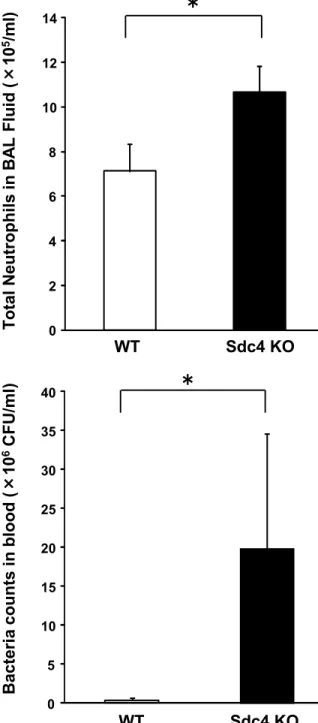
To clarify the mechanism responsible for the higher mortality rate in the Sdc4 KO mice, we evaluated the extent of inflammation and bacterial burden in the lungs and blood 24 hours after infection. Although bacterial counts in lung tissues did not differ between the 2 groups, total cell and neutrophil counts in BAL fluid were significantly higher (Figure 5A), and pulmonary inflammation was more pathologically severe (Figure 5B) in the Sdc4 KO mice than in the WT mice.
Blood bacterial counts were also higher in the Sdc4 KO mice than in the WT
Nikaido T, et al.
mice (Figure 5C). Furthermore, analysis of inflammatory cytokines showed that the plasma levels of interleukin 6, MIP-2, and KC were significantly higher in the Sdc4 KO mice than in the WT mice (Figure 6). On the other hand, the levels of inflammatory cytokines in BAL fluid did not differ between the 2 groups (data not shown).
DISCUSSION
We have demonstrated 5 major findings in this study: (1) patients with acute pneumonia had significantly higher serum syndecan-4 levels than healthy volunteers; (2) there was a negative correlation between serum syndecan-4 levels and severity score and significantly higher serum syndecan-4 levels in patients with acute pneumonia whose severity score was low or who improved with short-term antibiotic therapy; (3) there was a gradual increase of serum syndecan-4 levels in patients who improved with short-term antibiotic therapy;
(4) the mortality rate was significantly higher in Sdc4 KO mice than in WT mice after intranasal instillation of S. pneumoniae; and (5) Sdc4 KO mice had more severe pulmonary and systemic inflammation with higher blood bacterial counts than WT mice. Taken together, these findings suggest that syndecan-4 has a protective role in acute bacterial pneumonia.
Syndecan-4 is an HSPG expressed on surfaces of a variety of cells and
consists of a core protein and heparan sulfate GAG side chains, which bind
several cytokines and growth factors, leading to a variety of biological activities
[7, 11, 13]. Ishiguro et al [30] reported that syndecan-4 expression in vascular
Nikaido T, et al.
endothelial cells and monocytes was up-regulated in WT mice after intraperitoneal LPS injection, and mortality rates were significantly worse in Sdc4 KO mice than in the WT mice. Tanino et al [21] reported elsewhere that Sdc4 mRNA expression was significantly increased in lungs of mice treated with LPS and showed that the lack of syndecan-4 resulted in significantly more pulmonary inflammation and injury. Although these results suggested that syndecan-4 was involved in the pathogenesis of acute pulmonary inflammation, there was no previous information about the role of syndecan-4 in humans with acute pneumonia.
Our findings of elevated serum syndecan-4 levels in patients with acute pneumonia are consistent with previous reports showing increased Sdc4 expression in lungs of mice after intratracheal LPS instillation and in murine endothelial cells and monocytes after intraperitoneal LPS injection [21, 30].
Although the precise mechanism of increased syndecan-4 expression in acute pneumonia is not certain, stimulation of Toll-like receptor-2, -4, and -5 was reported to increase syndecan-4 expression via NF-κB in the gastric epithelium [14, 31]. These results suggest that syndecan-4 expression is increased through a stimulation of Toll-like receptors 2 and 4 by bacterial pathogenassociated molecular patterns, such as LPS or peptidoglycan in acute pneumonia.
Syndecan-4 is expressed on the surfaces of several types of cells in
membrane-bound forms and can be cleaved from cell surfaces by
metalloproteinase 7 or 9 or ADAM17, producing soluble forms [32]. The serum
syndecan-4 measured in this study was a soluble form. This study did not show
the precise source(s) of serum syndecan-4, but there are several possibilities,
Nikaido T, et al.
including lung tissues and other organs/cells apart from the lungs. Prior studies have shown that LPS stimulation increases syndecan-4 expression in the lungs and several other organs (eg, intestine, kidney, and liver) [21]. Moreover, endothelial cells, neutrophils, and lymphocytes are reported to express syndecan-4 [33]. To clarify the precise source(s) of syndecan-4 in acute pneumonia, further studies need to be conducted.
Interestingly, we found that serum syndecan-4 levels in patients with mild pneumonia, but not severe pneumonia, were higher than in the healthy volunteers. Furthermore, serum syndecan-4 levels gradually increased in patients who improved with short-termantibiotic therapy. These results suggest the possibility that serumsyndecan-4 can be used as a biomarker which predicts the clinical outcome of patients with acute pneumonia. Tanino et al [21] have reported elsewhere that pretreatment of recombinant syndecan-4 inhibits LPS-induced CXCL8 up-regulation in bronchial epithelial cells. We also showed that the mortality rate was significantly worse in the Sdc4 KO mice than in the WT mice after intranasal bacterial instillation. Furthermore, pulmonary and systemic inflammation was more severe, and blood bacterial counts were significantly higher in Sdc4 KO mice than in WT mice. These results suggest that syndecan-4might have a previously unrecognized anti-inflammatory effect in acute pneumonia.
Li et al demonstrated that neutrophil migration into the alveolar space was
impaired (ie, neutrophils remained in the interstitium and did not advance into
the alveolar space) after intratracheal bleomycin instillation in matrilysin-deficient
mice compared with WT mice [15]. This was associated with impaired formation
Nikaido T, et al.
of a CXC chemokine gradient because of the lack of syndecan-1 shedding by matrilysin. This study showed the importance of syndecan-1 and CXC chemokine binding for neutrophil migration into the alveolar space in injured lungs [11].Moreover, Xu et al [34] reported that shedded syndecan-1 inhibited pulmonary allergic inflammation by inhibiting T-cell migration by binding to CC chemokines. These results show that binding of syndecan-1 to chemokines contributes to inflammatory cell migration into the lung. In addition, Tanino et al [35] demonstrated that intratracheal instillation of mutant CXCL8, whose binding to GAG was weakened, showed increased migration of neutrophils into the lung compared with WT CXCL8. These data show that the binding of chemokine with GAG has critical roles in neutrophil migration into the lung.
Although the precise mechanism by which syndecan-4 inhibits acute lung inflammation is not clear, the GAG side chains of syndecan-4 expressed on alveolar macrophages, epithelial cells, and in the interstitium may bind to inflammatory mediators and regulate their activities. In previous studies [36–39], Sdc4 KO mice had higher sensitivity than WT mice to renal damage and sclerosis, hepatic damage, lung injury and fibrosis, and heart rupture after myocardial infarction, as well as showing delayed wound repair of skin [40].
These results suggest that syndecan-4 has important roles in inflammation and repair processes of injured organs. In this study, we showed that the mortality rate was significantly worse in Sdc4 KO mice after S. pneumoniae instillation. In addition, pulmonary and systemic inflammation was more severe in Sdc4 KO mice.
Interestingly, blood bacterial counts were significantly higher in Sdc4 KO mice.
Nikaido T, et al.
Although more severe pulmonary inflammation may lead to higher blood bacterial counts, the lack of syndecan-4 in the lungs might accelerate the movement of bacteria into the systemic circulation. Tanino et al [35] have demonstrated that the binding of CXCL8 to GAG regulated its movement from the alveolar space to systemic circulation, when CXCL8 was instilled into WT mice intratracheally. Because bacterial pathogens such as S. pneumoniae bind to heparan sulfate, a part of syndecan-4 [41], the lack of syndecan-4 in the lungs may allow the pathogens to move easily from the alveolar space to systemic circulation. Although further studies are necessary to clarify the exact mechanism, our results in the current study show that syndecan-4 plays a critical role in modulating the inflammatory response in bacterial pneumonia.
It has been demonstrated that intravenous administration of danaparoid, which
is composed mainly of HSPG, attenuates LPS-induced proinflammatory cytokine
production and organ dysfunction, including acute lung injury, and reduces
mortality rates in rats [42, 43]. In addition, a synthesized GAG analogue was
reported to inhibit delayed-type hypersensitivity and allergen-induced arthritis
[44]. It was also reported that intraperitoneal administration of endogenous
glucosamine, used for biosynthesis of GAG, inhibited LPS-induced acute lung
injury [45]. Overexpression of Sdc4 in rats has been reported to reduce the
decline of heart function after myocardial infarction [46]. These data suggest
that GAGs and proteoglycans, such as syndecan-4, might have therapeutic uses
in various inflammatory diseases. Taken together, these observations lead us to
conclude that syndecan-4 has a previously unrecognized protective effect in
acute bacterial pneumonia and that serum syndecan-4 might be useful as a
Nikaido T, et al.
biomarker to predict the clinical course in patients with acute pneumonia.
ACKNOWLEDGEMENTS
We thank Thomas Martin, MD, PhD, (Novartis Pharmaceuticals) for scientific input and review of the manuscript and Yoshiko Sato (Fukushima Medical University, Japan) for her excellent technical assistance.
CONTRIBUTIONS
T. N., Y. T., and M. M. contributed to the study design, data acquisition, data analysis, and data interpretation and drafting of the manuscript. X. W.
contributed to the study design, data acquisition, and data analysis. S. S., K. M., N. F., Y. S., A. F., M. U., and Y. S. contributed to sample collection and data interpretation. Y. E. contributed to study design, data acquisition, data analysis, and interpretation for animal experiments and drafting of the manuscript. T. K., M.
T., K. T., and I. K. contributed to study design, data analysis, and interpretation
for animal experiments. C. W. F. contributed to data analysis, data interpretation,
and critical revision of the manuscript. All authors approved the final version of
the manuscript. M. M. is guarantor of the manuscript.
Nikaido T, et al.
REFERENCES
1. World Health Organization. The top 10 causes of death in the world, 2000 and 2011. http://www.who.int/mediacentre/factsheets/fs310/en/
index.html. Accessed 9 January 2014.
2. Pitter JF, Mackersie RC, Martin TR, Matthay MA. Biological markers of acute lung injury: prognostic and pathogenetic significance. Am J Respir Crit Care Med 1997; 155:1187–205.
3. Segel GB, Halterman MW, Lichtman MA. The paradox of the neutrophil’s role in tissue injury. J Leukoc Biol 2011; 89:359–72.
4. Esko JD, Lindahl U. Molecular diversity of heparan sulfate. J Clin Invest 2001; 108:169–73.
5. Bishop JR, Schuksz M, Esko JD. Heparan sulphate proteoglycans finetune mammalian physiology. Nature 2007; 446:1030–7.
6. Gill S, Wight TN, Frevert CW. Proteoglycans: key regulators of pulmonary inflammation and the innate immune response to the lung infection.
Anat Rec (Hoboken) 2010; 293:968–81.
7. Parish CR. The role of heparan sulphate in inflammation. Nat Rev Immunol 2006; 6:633–43.
8. Celie JW, Beelen RH, van den Born J. Heparan sulfate proteoglycans in extravasation: assisting leukocyte guidance. Front Biosci (Landmark Ed) 2009; 14:4932–49.
9. Bartlett AH, Hayashida K, Park PW. Molecular and cellular mechanisms of syndecans in tissue injury and inflammation. Mol Cells 2007; 24:153–6.
10. Alexopoulou AN, Multhaupt HA, Couchman JR. Syndecans in wound
Nikaido T, et al.
healing, inflammation and vascular biology. Int J Biochem Cell Biol 2007; 39:505–28.
11. Götte M. Syndecans in inflammation. FASEB J 2003; 17:575–91.
12. Saphire AC, Bobardt MD, Zhang Z, David G, Gallay PA. Syndecans serve as attachment receptors for human immunodeficiency virus type 1 on macrophages. J Virol 2001; 75:9187–200.
13. Bernfield M, Kokenyesi R, Kato M, et al. Biology of the syndecans: a family of transmembrane heparan sulfate proteoglycans. Annu Rev Cell Biol 1992; 8:365–93.
14. Okuyama E, Suzuki A, Murata M, et al. Molecular mechanisms of syndecan- 4 upregulation by TNF-α in the endothelium-like EAhy926 cells.
J Biochem 2013; 154:41–50.
15. Li Q, Park PW, Wilson CL, Parks WC. Matrilysin shedding of syndecan- 1 regulates chemokine mobilization and transepithelial efflux of
neutrophils in acute lung injury. Cell 2002; 111:635–46.
16. Brule S, Charnaux N, Sutton A, et al. The shedding of syndecan-4 and syndecan-1 from HeLa cells and human primary macrophages is accelerated by SDF-1/CXCL12 and mediated by the matrix metalloproteinase-9. Glycobiology 2006; 16:488–501.
17. Pruessmeyer J, Martin C, Hess FM, et al. A disintegrin and metalloproteinase 17 (ADAM17) mediates inflammation-induced shedding of syndecan-1 and -4 by lung epithelial cells. J Biol Chem 2010; 285:
555–64.
18. Ramnath R, Foster RR, Qiu Y, et al. Matrix metalloproteinase 9-mediated
Nikaido T, et al.
shedding of syndecan-4 in response to tumor necrosis factor α: a contributor to endothelial cell glycocalyx dysfunction. FASEB J 2014; 28:4686–99.
19. Frevert CW, Kinsella MG, Vathanaprida C, et al. Binding of interleukin-8 to heparan sulfate and chondroitin sulfate in lung tissue. Am J Respir Cell Mol Biol 2003; 28:464–72.
20. Muramatsu T, Muramatsu H, Kojima T. Identification of proteoglycanbinding proteins. Methods Enzymol 2006; 416:263–78.
21. Tanino Y, Chang MY,Wang X, et al. Syndecan-4 regulates early neutrophil migration and pulmonary inflammation in response to lipopolysaccharide.
Am J Respir Cell Mol Biol 2012; 47:196–202.
22. Inokoshi Y, Tanino Y, Wang X, et al. Clinical significance of serum hyaluronan in chronic fibrotic interstitial pneumonia. Respirology 2013;
18:1236–43.
23. Kohno S, Seki M, Watanabe A. Evaluation of an assessment system for the JRS 2005: A-DROP for the management of CAP in adults. Intern Med 2011;
50:1183–91.
24. Kasamatsu Y, Yamaguchi T, Kawaguchi T, et al. Usefulness of a semiquantitative procalcitonin test and the A-DROP Japanese prognostic scale for predicting mortality among adults hospitalized with community- acquired pneumonia. Respiration 2012; 17:330–6.
25. Kono S, Seki M, Takehara K, et al. Prediction of requirement formechanical ventilation in community-acquired pneumonia with acute respiratory failure; a multicenter prospective study. Respiration 2013; 85:27–35.
26. Ishiguro K, Kadomatsu K, Kojima T, et al. Syndecan-4 deficiency impairs
Nikaido T, et al.
focal adhesion formation only under restricted condition. J Biol Chem 2000;
275:5249–52.
27. Wang X, Ohtsuka Y, Kimura K, et al. Mannose-binding lectin gene polymorphisms and the development of coal workers’ pneumoconiosis in Japan. Am J Ind Med 2008; 51:548–53.
28. Endo Y, Takahashi M, Iwaki D, et al. Mice deficient in ficolin, a lectin complement pathway recognition molecule, are susceptible to Streptococcus pneumoniae infection. J Immunol 2012; 189:5860–6.
29. Tanino Y, Makita H, Miyamoto K, et al. Role of macrophage migration inhibitory factor in bleomycin-induced lung injury and fibrosis in mice. Am J Physiol Lung Cell Mol Physiol 2002; 283:L156–62.
30. Ishiguro K, Kadomatsu K, Kojima T, et al. Syndecan-4 deficiency leads to high mortality of lipopolysaccharide-injected mice. J Biol Chem2001;
276:47483–8.
31. Smith MF Jr, Novotny J, Carl VS, Comeau LD. Helicobacter pylori and Toll-like receptor agonists induce syndecan-4 expression in an NF-κ-dependent manner. Glycobiology 2006; 16:221–9.
32. Tan X, Khalil N, Tesarik C, et al. Th1cytokine-induced syndecan-4 shedding by airway smooth muscle cells independent on mitogen-activated protein kinases. Am J Physiol Lung Cell Mol Physiol 2012; 302:L700–10.
33. Kaneider NC, Egger P, Dunzendorfer S,Wiedermann CJ. Syndecan-4 as antithrombin receptor of human neutrophils. Biochem Biophys Res Commun 2001; 287:42–6.
34. Xu J, Park PW, Kheradmand F, Corry DB. Endogenous attenuation of
Nikaido T, et al.
allergic lung inflammation by syndecan-1. J Immunol 2005; 174: 5758–65.
35. Tanino Y, Coombe DR, Gill SE, et al. Kinetics of chemokine-glycosaminoglycan interaction control neutrophil migration into the airspaces of the lung. J Immunol 2010; 184:2677–85.
36. Ishiguro K, Kadomatsu K, Kojima T, et al. Syndecan-4 deficiency increased susceptibility to κ-carrageenan–induced renal damage. Lab Invest 2001;
81:509–16.
37. Cevikbas F, Schaefer L, Uhlig P, et al. Unilateral nephrectomy leads to up-regulation of syndecan-2- and TGF-beta-mediated glomerulosclerosis in syndecan-4 deficient male mice. Matrix Biol 2008; 27:42–52.
38. Kon S, Ikesue M, Kimura C, et al. Syndecan-4 protects hepatic injury by masking functional domains of osteopontin. J Exp Med 2008; 205:25–33.
39. Matsui Y, Ikesue M, Danzaki K, et al. Syndecan-4 prevents cardiac rupture and dysfunction after myocardial infarction. Circ Res 2011; 108:1328–39.
40. Echtermeyer F, Streit M, Wilcox-Adelman S, et al. Delayed wound repair and impaired angiogenesis in mice lacking syndecan-4. J Clin Invest 2001;
107:R9–17.
41. Tonnaer EL, Hafmans TG, Van Kuppevelt TH, Sanders EA, Verweij PE, Curfs JH. Involvement of glycosaminoglycans in the attachment of pneumococci to nasopharyngeal epithelial cells. Microbes Infect 2006;
8:316–22.
42. Hagiwara S, Iwasaka H, Hidaka S, Hishiyama S, Noguchi T. Danaparoid
sodium inhibits systemic inflammation and prevents endotoxininduced acute
lung injury in rat. Crit Care 2008; 12:R43.
Nikaido T, et al.
43. Iba T, Miyasho T. Danaparoid sodium attenuates the increase in inflammatory cytokines and preserves organ function in endotoxemic rats.
Crit Care 2008; 12:R86.
44. Severin IC, Soares A, Hantson J, et al. Glycosaminoglycan analog as a novel anti-inflammatory strategy. Front Immunol 2012; 3:293.
45. Chuang KH, Peng YC, Chien HY, Lu ML, Du HI, Wu YL. Attenuation of LPS-induced lung inflammation by glucosamine in rat. Am J Respir Cell Mol Biol 2013; 49:1110–9.
46. Xie J, Wang J, Li R, et al. Syndecan-4 over-expression preserves cardiac function in a rat model of myocardial infarction. J Mol Cell Cardiol 2012;
53:250–8.
FIGURE LEGENDS
Figure 1: A, Serum syndecan-4 levels in healthy volunteers (HVs) and patients with acute pneumonia. Serum syndecan-4 levels were significantly higher in patients with acute pneumonia (n=30) than in HVs (n=11) *P=.006. B, Correlation of serum syndecan-4 levels with A-DROP score in acute pneumonia.
Serum syndecan-4 levels correlated negatively with A-DROP score (r=−0.391;
P=.03). C, Relationship between serum syndecan-4 levels and severity of acute
pneumonia at admission. Serum syndecan-4 levels in patients with mild
pneumonia (A-DROP score, 0 or 1; n=17), but not those with moderate or severe
Nikaido T, et al.
pneumonia (A-DROP score, 2–5; n=13), were significantly higher than in HVs (n=11). *P=.001 (vs HVs);
†P<.001 (vs moderate/severe pneumonia). D, Relationship between serum syndecan-4 levels and duration of antibiotic therapy. Serum syndecan-4 levels in patients with acute pneumonia who improved with short-term antibiotic therapy (<14 days; n=20), but not in those who required long-term antibiotic therapy (≥14 days; n=10), were significantly higher than in HVs (n=11). *P =.02 (vs HVs). Values represent means with standard errors of the mean.
Figure 2: Time course of serum syndecan-4 levels in patients with acute pneumonia who improved with short-term antibiotic therapy. Levels were gradually increased in patients with acute pneumonia who improved with short-term antibiotic therapy (<14 days). *P = .005.
Figure 3: Syndecan-4 expression in the lungs after intranasal Streptococcus pneumoniae instillation. Messenger RNA (mRNA) expression of syndecan-4 in lung tissues (A) and the levels of syndecan-4 in bronchoalveolar lavage (BAL) fluid (B) were significantly higher in Sdc4 knockout mice than in wild-type mice 24 hours after instillation. *P<.05.
Figure 4: Survival of wild-type (WT) and Sdc4 knockout (KO) mice
afterintranasal instillation of Streptococcus pneumoniae. The mortality rate in
Sdc4 KO mice was significantly worse than that in WT mice after intranasal
instillation of S. pneumoniae (5.0 × 10⁶ colony-forming units) (P = .04).
Nikaido T, et al.
Figure 5: Pulmonary inflammation and bacterial burden in blood and lung tissues. A, Total neutrophils in bronchoalveolar lavage (BAL) fluid was significantly higher in Sdc4 knockout (KO) mice than in wild-type (WT) mice. B, Pathological findings showed more severe inflammation in Sdc4 KO mice (×200, Hematoxylin and Eosin Staining). Each photomicrograph is the representative of 4 mice. C, D, Bacterial counts in blood (C), but not lung tissues (D), were significantly higher in Sdc4 KO mice 24 hours after instillation. *P<.05.
Abbreviations: CFUs, colony-forming units; NS, not significant.
Figure 6: Plasma levels of inflammatory cytokines. A, B, C, Interleukin 6 (IL-6)
(A), Keratinocyte Chemoattractant (KC) (B), and macrophage inflammatory
protein (MIP)-2 (C) levels were significantly higher in Sdc4 knockout (KO) mice
than in wild-type (WT) mice 24 hours after instillation. D, Tumor necrosis factor
(TNF) α levels did not differ between the groups. *P<.05. Abbreviation: NS, not
significant.
Healthy volunteer Acute pneumonia P value
Subjects ( n ) 11 30
Age ( yrs ) 50.1 ± 4.8 67.1 ± 3.1* P < 0.05
Gender ( M/F ) 6/5 20/10 NS
WBC ( /μL ) NA 12626 ± 630.4 NS
CRP ( mg/dl ) NA 20.1 ± 1.4 NS
A- DROP 0 ± 0 1.57 ± 0.25 P < 0.05
PF ratio NA 1444 ± 241 NS
The A-DROP scoring system is a 6-point scoring scale (0–5) and considers the following parameters: 1) Age (male ≥ 70years, female ≥ 75 years), 2) Dehydration (BUN ≥ 21 mg/dL), 3) Respiratory failure (SaO2 ≤ 90% or PaO2 ≤ 60 Torr), 4) Orientation disturbance
(confusion) and 5) Low blood pressure (systolic blood pressure ≤ 90 mm Hg). * vs HV,
Table 1 . Clinical Characteristics of Healthy Volunteers and
Patients with Acute Pneumonia
*
Se rum Sy nd ecan -4 (ng / mL )
HV Short-term antibiotic
therapy
Long-term antibiotic
therapy
Figure 1
A B
C D
r = -0.391 p = 0.033
Serum Syndecan-4 (ng/mL)
A- D R O P (poi nt )
Ser um S yn decan -4 (ng / mL )
HV
✝
Mild
pneumonia Moderate/Severe pneumonia
*
Ser um S yn decan -4 ( ng / mL )
HV Acute pneumonia
*
Ser um Sy nd ecan -4 (n g/ mL )
Day 15 Day 8
Day 1
*
0 5 10 15 20 25 30 35 40 45
Figure 2
Figure 3
A B
* *
Sy nd ec an -4 Re la tiv e m RNA in l ung ( Fol d)
0 1 2 3 4
Control Infected mice 0
50 100 150 200 250 300 350 400 450
Sy nd ec an - 4 in BA LF ( ng /m l)
Control Infected mice
Sdc4 KO (n = 22) WT (n = 21)
P = 0.035
Sur vival R at e
Time (Day) 0
0.2 0.4 0.6 0.8 1.0
0 1 2 3 4 5 6 7
Figure 4
Figure 5
A B
C D
WT Sdc4 KO
0 2 4 6 8 10 12 14
Tot al N eut rophi ls in B AL Fl ui d ( × 10
5/ml ) *
WT Sdc4 KO
0 5 10 15 20 25 30 35 40
B act er ia count s i n bl ood ( × 10
6CF U/ m l) *
WT Sdc4 KO
5B act er ia count s i n l ung t issues ( × 10 CF U/ m g) NS
0 1 2 3 4 5 6
WT Sdc4 KO
0 10000 20000 30000 40000 50000
Figure 6
A B
C D
*
IL -6 in pl as m a ( pg/ m l) K C in pl as m a ( pg/ m l)
WT Sdc4 KO
010000 20000 30000 40000 50000 60000
*
WT Sdc4 KO
0 500 1000 1500 2000 2500 3000
*
MI P- 2 i n pl as m a (pg/ m l)
WT Sdc4 KO
050 100 150 200 250 300