分担研究報告書
- 26 -
フタル酸エステル類(DEHP)胎児期曝露による臍帯血 DNA メチル化の網羅的解析と 出生時体格との関連
Association between cord blood DNA methylations by fetal exposure of phthalates and ponderal index at birth in epigenome-wide study
研究代表者 岸 玲子 北海道大学環境健康科学研究教育センター 特別招へい教授 研究分担者 三浦 りゅう 北海道大学環境健康科学研究教育センター 特任助教 研究分担者 荒木 敦子 北海道大学環境健康科学研究教育センター 特任准教授 研究分担者 松浦 英幸 北海道大学大学院・農学研究院 教授 研究分担者 篠原 信雄 北海道大学大学院・医学研究院 教授
A. 研究目的
Phthalates are widely used plasticizers (Koch et al.
2013) for consumer products including toys, food packages, personal care products, and other household items, leading to widespread exposure to these chemicals through diet, inhalation, and dermal adsorption (Ait Bamai et al. 2015; Jensen
et al. 2015). Phthalates have potential for endocrine-disrupting chemicals (EDCs) and have been found to be associated with multiple adverse effects on human health. In particular, exposure in utero has been linked to adverse birth outcomes such as decreased birth size (Minatoya et al. 2017;
Song et al. 2018; Whyatt et al. 2009), preterm birth (Ferguson et al. 2017; Huang et al. 2014), pregnancy loss (Gao et al. 2017), and reduced anogenital distance of infants (Swan et al. 2015).
Prenatal exposure can also affect childhood health 研究要旨
Exposure to phthalate in utero is associated with adverse health outcome of the offspring.
Differential DNA methylation at specific CpG sites may link phthalate exposure to health impacts. We examined the association of prenatal Di-2-ethylhexyl phthalate (DEHP) exposure with genome-wide DNA methylation changes in cord blood in 203 mother-child pairs in the Hokkaido Study on Environment and Children’s Health, using the Illumina HumanMethylation 450 BeadChip. We found that the primary metabolite of DEHP: mono (2-ethylhexyl) phthalate (MEHP) levels in maternal blood were predominantly associated with hypermethylation in cord blood DNA. The genes annotated to hypermethylated CpGs associated with maternal MEHP levels were enriched for pathways related to metabolism, endocrine system, and signal transduction. Among them, hypermethylated CpGs involved in metabolism were inversely associated with offspring’s ponderal index (PI). Further, mediation analysis suggested that multiple hypermethylation changes may jointly mediate the association between prenatal DEHP exposure and offspring’s PI. Although additional studies are needed to determine the functional consequences of these changes, our findings imply differential DNA methylation may link
研究協力者
増田 秀幸 (北海道大学環境健康科学研究教
育センター,特任助教)
分担研究報告書
- 27 -
outcomes such as behavioral problems (Engel et al.
2009; Engel et al. 2010; Minatoya et al. 2018b;
Tellez-Rojo et al. 2013), obesity (Buckley et al.
2016; Kim and Park 2014), and allergic disease (Ait Bamai et al. 2018; Jaakkola and Knight 2008;
Whyatt et al. 2014). Early-life exposure to phthalates may contribute to fetal origins of disease; however, actual mechanisms accounting for long-term effects remain unclear.
As phthalates are rapidly metabolized and excreted, epigenetic modification, such as DNA methylation, may be a potential mechanism by which phthalate exposure in utero exerts the long-term effects.
Accumulating evidence suggests that epigenetic alternations may link developmental EDC exposure with susceptibility to diseases later in life (Barouki et al. 2018; Ho et al. 2017; McLachlan 2016; Tapia-Orozco et al. 2017). Animal studies have demonstrated the association between developmental phthalate exposure and DNA methylation changes in the offspring (Abdel-Maksoud et al. 2015; Manikkam et al.
2013; Martinez-Arguelles and Papadopoulos 2015;
Rajesh and Balasubramanian 2015; Sekaran and Jagadeesan 2015; Wu et al. 2010). Serval human cohort studies showed that prenatal phthalate exposure was associated with DNA methylation in selected candidate genes using placenta (LaRocca et al. 2014; Zhao et al. 2015; Zhao et al. 2016) or cord blood samples (Huang et al. 2018; Huen et al.
2016; Montrose et al. 2018; Tindula et al. 2018).
Recently, a few epigenome-wide association studies (EWASs) that can allow a hypothesis-free assessment of epigenetic alterations in relation to the environmental factors (Christensen and Marsit 2011) were published. One study reported phthalate exposure altered placental methylome and identified epidermal growth factor receptor
(EGFR) as a critical candidate gene mediating the effects of phthalates on early placental function (Grindler et al. 2018). Several differential methylation regions in cord blood associated with prenatal phthalate exposure were identified (Solomon et al. 2017). Genes with those regions were involved in inflammatory response, cancer, endocrine function, and male fertility. Another study also examined the association of genome-wide DNA methylation in cord blood with prenatal exposure to the most common phthalates, Di-2-ethylhexyl phthalate (DEHP) and suggested that DNA methylation in genes involved in the androgen response, spermatogenesis, and cancer-related pathway may be affected by prenatal phthalate exposure (Chen et al. 2018). Although existing evidence supports the role of prenatal phthalate exposure in modifying DNA methylation, little is known about potential effects of the exposure-associated methylation on fetal development and later in life.
Using epigenome-wide approach, we aimed to explore association between prenatal DEHP exposure and DNA methylation changes in cord blood collected from the participants of the Hokkaido study. Furthermore, we studied whether the DNA methylation at identified loci mediated the effect of DEHP exposure in utero on ponderal index at birth as an indicator of fetal growth.
B. 研究方法
Study population.Participants were enrolled in the Sapporo cohort of the Hokkaido Study on Environment and Children s Health (Kishi et al. 2011; Kishi et al.
2013; Kishi et al. 2017). Briefly, we recruited
pregnant women at 23–35 weeks of gestation
between 2002 and 2005 from the Toho Hospital
分担研究報告書
- 28 -
(Sapporo, Japan). After the second trimester during their pregnancy, the participants completed the self-administered questionnaire containing baseline information including family income, educational level, parity history, and pregnancy health information including smoking status, alcohol consumption, and caffeine intake.
Information on pregnancy complications, gestational age, infant sex, and birth size was obtained from medical records.
Measurement of the primary metabolite of DEHP
: Maternal blood samples were obtained at the time of their hospital examination and stored at −80℃
prior to analysis. Levels of mono (2-ethylhexyl) phthalate (MEHP) were measured in maternal blood by gas chromatography mass spectrometry (GC-MS) at Nagoya university as described previously (Araki et al. 2014; Araki et al. 2017; Jia et al. 2015). The detection of limit (LOD) was 0.28 ng/mL.
450K DNA methylation analysis.
Umbilical cord blood samples were taken immediately after birth, and then stored at −80℃.
After DNA extraction using a Maxwell® 16 DNA
Purification Kit (Promega, Madison, WI, USA), cord blood DNA methylation at 485,577 CpGs was quantified using the Infinium HumanMethylation 450 BeadChip (Illumina Inc., San Diego, CA, USA) by G&G Science Co., Ltd.
(Fukushima, Japan). Details for the 450K methylation analysis are described elsewhere (Miura et al. 2018). Samples were run across five plate batches and were assigned randomized location across plates. After quality control (Aryee et al. 2014), functional normalization (Fortin et al.
2014) was applied to the raw data, and normalized
beta (β) values, ranging from 0-1 for 0% to 100%
methylated, were obtained for the 292 cord blood samples. Probes with a detection p-value >0.05 in more than 25% of samples, single nucleotide polymorphism (SNP)-affected probes, cross-reactive probes identified by Chen et al.
(Chen et al. 2013), and probes on sex chromosomes were removed. As a result, 426,413 CpG probes were included in the working set. We applied the ComBat method on M-values (logit-transformedβ-values) to adjust methylation data for the sample plate to reduce a potential bias due to batch effects (Leek et al. 2012). The M-values were back-transformed to β-values that were used for subsequent data analyses.
Data analysis.
Among the 514 participants of the Sapporo Cohort
Study, 203 mother-infant pairs had both exposure
and DNA methylation data. Cord blood cell
proportion was estimated by the method
implemented in the R/Bioconductor package
minfi (Bakulski et al. 2016). Using limma package
in R, robust linear regression analysis (Fox and
Weisberg 2011) and empirical Bayesian method
(Smyth 2004) were applied to determine the
associations of β-value at each CpG site with
MEHP natural log (ln)-transformed
concentrations, adjusted for maternal age,
educational levels, pre-pregnancy body mass index
(BMI), smoking during pregnancy, blood
sampling periods, gestational age, infant sex, and
cord blood cell estimates for CD4
+T cells, CD8
+T cells, granulocytes, monocytes, B cell and
nucleated red blood cells. Adjustment covariates
were selected from factors previously reported to
be associated with exposure or cord blood DNA
methylation. For multiple comparisons, p-values
分担研究報告書
- 29 -
were adjusted by a false discovery rate (FDR) to obtain q-values. Because of too few FDR-significant findings, we evaluated the differentially methylated CpGs (DMCpGs) with uncorrected p-value <2.5E-04. We also assessed hypermethylated DMCpGs (hyper-DMCpGs) for functional enrichment with KEGG pathways (Kanehisa et al. 2002) via the gometh function in the missMethyl package in R/Bioconductor (Phipson et al. 2016). Statistical analyses were performed using minfi, sva, and limma packages in R ver. 3.3.2 and Bioconductor ver. 3.3.
The Spearman s correlation test, Mann-Whitney U-test, and Kruskal-Wallis test were applied to determine whether maternal and offspring characteristics were associated with MEHP levels.
We examined associations between methylation levels at hyper-DMCpGs and ponderal index (PI) at birth by a multivariate regression model adjusted by maternal age, educational levels, parity, pre-pregnancy BMI, smoking during pregnancy, gestational age, and infant sex, using JMP Pro 14 (SAS Institute Inc., Cary, NC, USA). PI was calculated as follows; PI (kg/m
3) = birth weight (kg) / (birth length (m))
3. Then, we tested the CpGs for mediation in the association between MEHP levels in maternal blood and PI using PROSESS (Hayes 2013), a macro implemented in SPSS (IBM, Armonk, NY, USA). In addition to the same possible cofounders as named above, blood sampling periods were included as covariate in the mediator and outcome regression models.
Ethics.
Written informed consents were obtained from all participants. The institutional Ethical Board for human gene and genome studies at the Hokkaido University Graduate School of Medicine and the
Hokkaido University Center for Environmental and Health Science approved the study protocol.
All experiments were performed in accordance with relevant guidelines and regulations.
C. 研究結果
Study characteristics
The characteristics of the participants with the corresponding median MEHP concentrations in cord blood are described in Table 1. The median of MEHP concentration was 10.3 ng/mL (Interquartile range (IQR): 5.8
– 15.3 ng/mL)with 100% of detection rate. The average ± standard deviation (s.d.) age of the mothers was 29.8 ± 4.9 years. Maternal blood sampling periods were significantly associated with MEHP levels.
Of the 203 newborns, 94 (46.3%) were male. The mean gestational age was 39.9 weeks, birth weight was 3137.5 g, and birth length was 48.5 cm. The MEHP level was negatively correlated to PI (ρ=
-0.133, p=0.059).
Epigenome-wide association study of DEHP exposure in utero
In adjusted robust linear regression models, there were 2 CpGs with epigenome-wide significant methylation changes (FDR q-value < 0.05): one located in 200 bases from transcription start site (TSS200) of
PARP12(cg26409978), and another mapped to
SDK1(cg00564857) as shown in Figure 1A. DEHP exposure was more frequently associated with hypermethylation than with hypomethylation as seen in volcano plots (Figure 1B). For instance, of 271 DMCpGs with uncorrected p-value <2.5E-04, 253 CpGs (93.4%) were hypermethylated (Figure 1B). We examined the location of the hyper-DMCpGs with p-value
<2.5E-04 in gene features and CpG islands. As
分担研究報告書
- 30 -
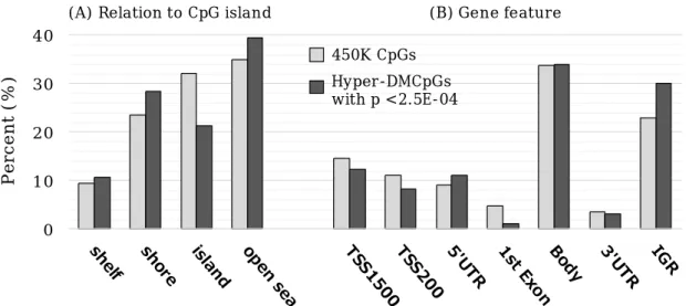
shown in Figure 2, there were statistically significant differences associated with DEHP exposure compared with the expected proportions (for gene features, Χ2 P-value = 0.004; for CpG islands, Χ
2P-value = 0.01). Decrease in island and increase in the intergenic region (IGR) were particularly observed.
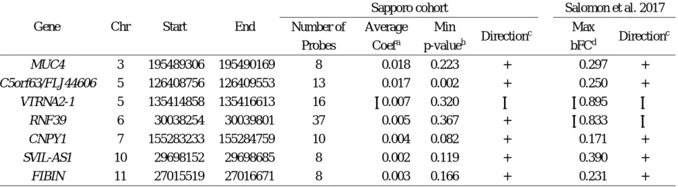
Next, we considered our results in relation to a published study of association between prenatal phthalate exposure and cord blood DNA methylation using Illumina HumanMethlation450 Beads chips (Solomon et al. 2017). In the study, the authors identified seven differentially methylated regions (DMRs) associated with MEHP levels in maternal urine at 26 weeks gestation by using two differential approaches. We examined the direction of methylation changes in the DMRs identified by Solomon et al. in our data set (Table 2), in which we averaged methylation levels of each CpG site because the CpGs included in each region showed the same direction of methylation changes. Although no CpG reached statistical significance in our cohort, six of the seven DMRs showed the same direction as those identified by Solomon et al. (Table 2), of which five DMRs mapped to
MUC4,
C5orf63,
CNPY1,
SVIL-AS1, and
FIBINwere hypermethylated, suggesting that prenatal DEHP exposure would predominantly induce hypermethylation.
Gene Ontology (GO) analysis
To investigate the underlying biology that may be affected by DEHP-associated hypermethylation changes, we tested for Kyoto Encyclopedia Genes and Genomes (KEGG) pathways (Kanehisa et al.
2002) enrichment among the 253 hyper-DMCpGs with p <2.5E-04. We observed twelve enriched pathways with FDR <0.05. GO analyses of the
data obtained using 450K chip are known to be biased for cancer-related genes (Harper et al.
2013); therefore, the enriched pathways excluded cancer and human disease pathways are listed in Table 3. The most significant pathway was metabolic pathway with FDR = 2.4E-08. We also observed three pathways involved in endocrine system: GnRH signaling pathway, Renin secretion, and Cortisol synthesis and secretion, and two pathways involved in signal transduction: mitogen-activated protein kinase (MAPK) signaling pathway and Notch signaling pathway.
The methylation for mediation in the association between prenatal DEHP exposure and offspring s PI at birth
First, we performed multiple regression analyses to examine the association between PI and methylation levels at sixteen hyper-DMCpGs on the genes involved in metabolic pathways (as shown in Table 3). Of those, methylation levels at twelve hyper-DMCpGs were inversely related to PI (Figure 3). In particular, methylation levels at cg27433759:
PIK3CG, cg10548708:
ACAA1, and cg07002201:
FUT9were associated with PI with p-value <0.1. Then, we considered averaged methylation levels at the three CpGs and observed mediate effect with Sobel test p-value <0.05 (Table 4), which explained 30.4 % of the effect of MEHP levels on PI.
D. 議論
We examined the effect of prenatal DEHP exposure on DNA methylation in cord blood and found that maternal MEHP levels were predominantly associated with hypermethylation.
The genes annotated to hyper-DMCpGs were
分担研究報告書
- 31 -
enriched for pathways related to metabolism, endocrine system, and signal transduction.
Further, mediation analysis suggested that a part of hypermethylation may mediate the association between prenatal DEHP exposure and offspring s ponderal index.
As we described previously (Araki et al. 2014), the levels of MEHP in this cohort (median = 10.3 ng/mL) were higher than those in pregnant women at 18 weeks (median = 1.18 ng/ml).
Additionally, in most cases, the levels of phthalate metabolites were considerably higher in urine samples (Frederriksen et al. 2010). We found two DMCpGs with FDR < 0.05: cg26409978 located in TSS200 of
PARP12(poly(ADP-Ribose) polymerase family member 12; previous name:
zinc finger CCCH-type domain containing 1 (
ZC3H1)) and cg00564857 mapped to
SDK1(Sidekick Cell Adhesion Molecule 1). Both CpGs showed hypermethylation. We also observed the preference of hypermethylation associated with MEHP levels with p-value <2.5E-04. In the previous study using the 450K platform, Salomon et al. (Solomon et al. 2017) reported the seven DMRs associated with MEHP levels in maternal urine at 26 weeks gestation (n=332, median: 3.63
µg/g-creatinine). Our study differs in sample size,matrices, collecting timing, and analysis methods;
nonetheless, when we evaluated the direction of methylation changes in those DMRs, hypermethylation in the five DMRs were replicated in our data set (Table 2).
Phthalate-induced hypermethylation was also consistent with a previous study that demonstrated a positive association between prenatal levels of high molecular weight phthalate and cord blood methylation region of
MEG3(Tindula et al. 2018). It is plausible that maternal
MEHP would predominantly induce offspring s hypermethylation. However, others on cord blood methylation alterations reported prenatal phthalate-induced hypomethylation. One study demonstrated an inverse association between prenatal concentrations of monoethyl phthalate, a metabolite of diethyl phthalate (DEP), with cord blood methylation of Alu repeats, and a similar but weaker association with
LINE-1methylation (Huen et al. 2016). Maternal urinary mono-n-butyl phthalate (MBP) and monobenzyl phthalate (MBzP) were negatively associated with Alu methylation (Huang et al. 2018). Another study showed that maternal phthalate concentrations were negatively associated with methylation on
LINE-1and metabolism-related genes;
IGF2and
PPARA(Montrose et al. 2018).
The differences in metabolite type, the measuring time, and level of phthalates may account for the disparities.
We also observed an enrichment of hyper-DMCpGs in the IGR, with decrease within CpG island (Figure 2). Disease associated- and environmentally induced-DMCpGs, such as obesity or exercise intervention, have been shown to be enriched within the IGR or open seas (Grundberg et al. 2013; Huang et al. 2015; Ronn et al. 2013; Zhu et al. 2018), suggesting that DNA methylation may also be dynamically regulated outside CpG islands. The enrichment of DMCpGs with in the IGR may affect functional process of regulatory elements, such as enhancers or insulators, located within the IGR. Recent study showed that the methylation levels at CpGs in the IGR were anticorrelated with nearest gene expression (Zhu et al. 2018).
GO analysis showed that DEHP-associated
hypermethylation was associated with metabolic
分担研究報告書
- 32 -
pathway, endocrine system, and MAPK signaling pathway. This is consistent with previous work.
For instance, epidemiological studies showed that phthalate exposure in utero has been associated with fetal metabolic outcomes, such as birth size (Minatoya et al. 2017; Watkins et al. 2016; Whyatt et al. 2009) and adipokine levels, markers of metabolic function, in cord blood (Ashley-Martin et al. 2014; Minatoya et al. 2017;
Minatoya et al. 2018a). Prenatal exposure has also been linked to steroid hormone levels in infants (Araki et al. 2014; Araki et al. 2017; Lin et al.
2011). Recently, an experimental study showed that MEHP has an impact on MAPK pathways as well as an effect on peroxisome proliferator–
activated receptor
(PPAR
) transcriptional activity, which together promote disturbances in lipid metabolism and in human villous cytotrophoblast differentiation (Shoaito et al.
2019).
Given the accumulation of DEHP-induced hypermethylations in metabolic pathway, we hypothesized that those methylation changes would disrupt fetal growth. We examined the association between methylation levels at sixteen hyper-DMCpGs in metabolic pathways and PI at birth, an indicator for fetal growth, and found that methylation levels at twelve CpGs were negatively associated with PI (Figure 3). Among them, three CpGs; cg27433759:
PIK3CG, cg10548708:
ACAA1
, and cg07002201:
FUT9, approached significance (p-value <0.1).
PIK3CG(Phosphatidylinositol-4,5-bisphosphate 3-kinase) encodes a class I catalytic subunit of PI3K (Phosphoinositide 3-kinase) that phosphorylates inositol lipid and is related to the pathway affecting insulin-like growth factor (IGF1)-Akt signaling (Matheny et al. 2017) and development
erythropoietin (EPO)-induced Jak-STAT pathway (Cokic et al. 2012).
ACAA1(Acetyl-CoA Acyltransferase 1) encodes an enzyme operative in the beta-oxidation system of the peroxisomes and is involved in fatty acid metabolism (Islam et al.
2019). FUT9 (Fucosyltransferase 9) belongs to the glycosyltransferase family and is related to glycosphingolipid biosynthesis (Ogasawara et al.
2011). Although each CpG did not show significant mediation in the association between prenatal DEHP exposure and offspring s PI, the averaged methylation levels at the three CpGs represented significant mediate effect (Sobel test p-value <0.05) and explained 30.4 % of the effect of MEHP levels on PI (Table 4). This suggests that multiple hyper-DMCpGs may jointly contribute to effects of DEHP exposure in utero on fetal development. We assumed that there would be more DM-CpGs related to PI as not all genes hit KEGG pathways. Among thirty-eight hyper-DMCpGs with FDR < 0.25 (Supplementary Table S3), seven CpGs; cg05836256 (
LMF1: Lipase Maturation Factor 1), cg21491711 (
DBN1: Drebrin 1), cg01142096 (
ERICH1: Glutamate Rich 1), cg12651645 (
PCSK6: Proprotein Convertase Subtilisin/Kexin Type 6 ), cg04849589 (
CSNK1G3: Casein Kinase 1 Gamma 3), cg01560642 (
TTC34: Tetratricopeptide Repeat Domain 34), cg02735381 (
ALPK1: Alpha Kinase 1) were related to PI with p-value <0.05, and two CpGs; cg26684601 (
JPH3: Junctophilin 3), cg22493212 (
MIR4277: MicroRNA4277), were with p-value <0.1 (Supplementary Figure S1).
Notably, these genes are linked to metabolism, cell growth and development. LMF1 is related to lipoprotein metabolism (Hosseini et al. 2012).
DBN1
encodes a cytoplasmic actin-binding
protein thought to play a role in the process of
分担研究報告書
- 33 -
neuronal growth (Shirao and Sekino 2017). DMR at
ERICH1was identified in multiple sclerosis patients by 450K platform (Maltby et al. 2017).
There is a link between PCSK6 and obesity (Du et al. 2016; Levenson et al. 2017). CSNK1G3 is involved the Hedgehog (Hh) signaling pathway that has numerous roles in the control of cell proliferation, tissue patterning, stem cell maintenance and development. TTC34 contains tetratricopeptide repeat domain which can regulate diverse biological processes, such as organelle targeting, protein import, and vesicle fusion (Zeytuni and Zarivach 2012). microRNAs are involved in post-transcriptional regulation in gene expression in multicellular organisms by affecting both the stability and translation of mRNAs. JPH3 provides a structural foundation for functional cross-talk between cell surface and intracellular ion channels. Although not significant mediate effect, each CpG explained 12.5 % - 23.2 % of the effect of MEHP levels on PI.
The following limitations of this study should be considered. We measured MEHP levels only once from second to third trimester. There have been concerns about using a single MEHP measurement as a representation of the long-term prenatal exposure due to the short half-life of MEHP. In addition, among several metabolites of DEHP, only MEHP levels were measured. MEHP is the primary metabolite of DEHP. Other secondary metabolites, such as mono(2-ethyl-5-hydroxyhexyl) phthalate and mono(2-ethyl-5-carboxyl) phthalate which have been detected in maternal serum (Hart et al.
2014), also be considered in future studies.
Second, we used blood samples for exposure assessment as urine samples were not available in this study. The majority of the recent studies
assessed phthalate levels in urine samples as urine samples can avoid the influence of external contamination. In this assay, all samples were handled carefully to avoid ex vivo hydrolysis of DEHP and external contamination. We measured background levels of MEHP and confirm that the influences of external contamination were null.
Third, DNA methylation was measured using unfractionated cord blood. DEHP is known to affect multiple tissues. Whether the associations observed in this study may reflect associations between prenatal DEHP exposure and the methylation at target tissues is unknown. Lastly, we included participants for whom cord blood samples were available, thus limiting the scope only to mothers who delivered vaginally. It is thus possible that relatively healthier children were included in our analysis, and we may have underestimated the effects of DEHP exposure.
Despite these potential limitation, this epigenome-wide study identified hypermethylation changes associated to prenatal DEHP exposure. The DEHP-associated hypermethylations may jointly contribute to effects of prenatal exposure on fetal development.
Further studies are needed to confirm our findings and to investigate their relevance to infant long-term outcomes.
E.
研究発表
1.論文発表1)
Miura R, Araki A, Minatoya M,
et al. An epigenome-wide analysis of cord blood DNA methylations reveals sex-specific effect of exposure to bisphenol A. Sci Rep. 9 12369, 2019.
2)
Kishi R., Araki A., Minatoya M, Itoh S,
Goudarzi H, Miyashita C; Birth cohorts in
分担研究報告書
- 34 -
Asia: The importance, advantages, and disadvantages of different-sized cohorts.
Science of the Total Environment. 615 1143-1154, 2018.
3)
Miura R, Araki A, Miyashita C,
et al. An epigenome-wide study of cord blood DNA methylations in relation to prenatal perfluoroalkyl substance exposure: the Hokkaido study. Environment International.
115: 21-28, 2018.
4)
Minatoya M, Araki A, Miyashita C,
et al. Association between prenatal bisphenol A and phthalate exposures and f
et almetabolic related biomarkers: The Hokkaido Study on Environment and Children's Health. Environ Res. 161 505-511, 2018.
5)
Minatoya M, Itoh S, Yamazaki K,
et al. Prenatal exposure to bisphenol A and phthalates and behavioral problems in children at preschool age: The Hokkaido Study on Environment and Children's Health Environtal Health and Preventive Medicine.
23:43 2018.
2.学会発表
1)
Minatoya M., Araki A., Miyashita C., itoh S., Kobayashi S., Yamazaki K., Ait Bamai Y., Miura R., Tamura N., Kishi R.; Update and the recent findings from the Hokkaido Study.
The 9th International Conference on Children's Health and the Environment (INCHES). Hoam Faculty House, Seoul National University, Seoul, Korea.
(2018.6.27-29)
2)
小林澄貴, 佐田文宏, 宮下ちひろ, 三浦りゅ
う, ゴウダルジ・ホウマヌ, 荒木敦子, 梶原淳 睦, 堀就英, 岸玲子; 胎児期の PCB 類曝露に よる児の H19・LINE-1 の DNA メチル化へ
の影響:北海道スタディ. 第 88 回日本衛生学 会学術総会. 東京工科大学蒲田キャンパス
(東京都大田区). (2018.3.22-24)
参考文献
1. Abdel-Maksoud FM, Leasor KR, Butzen K, Braden TD, Akingbemi BT. 2015. Prenatal exposures of male rats to the environmental chemicals bisphenol a and di(2-ethylhexyl) phthalate impact the sexual differentiation process. Endocrinology 156:4672-4683.
2. Ait Bamai Y, Araki A, Kawai T, Tsuboi T, Yoshioka E, Kanazawa A, et al. 2015.
Comparisons of urinary phthalate metabolites and daily phthalate intakes among japanese families. International journal of hygiene and environmental health 218:461-470.
3. Ait Bamai Y, Miyashita C, Araki A, Nakajima T, Sasaki S, Kishi R. 2018. Effects of prenatal di(2-ethylhexyl) phthalate exposure on childhood allergies and infectious diseases:
The hokkaido study on environment and children's health. The Science of the total environment 618:1408-1415.
4. Araki A, Mitsui T, Miyashita C, Nakajima T, Naito H, Ito S, et al. 2014. Association between maternal exposure to di(2-ethylhexyl) phthalate and reproductive hormone levels in fetal blood: The hokkaido study on environment and children's health.
PloS one 9:e109039.
5. Araki A, Mitsui T, Goudarzi H, Nakajima T,
Miyashita C, Itoh S, et al. 2017. Prenatal
di(2-ethylhexyl) phthalate exposure and
disruption of adrenal androgens and
glucocorticoids levels in cord blood: The
hokkaido study. The Science of the total
environment 581-582:297-304.
分担研究報告書
- 35 -
6. Aryee MJ, Jaffe AE, Corrada-Bravo H,
Ladd-Acosta C, Feinberg AP, Hansen KD, et al. 2014. Minfi: A flexible and comprehensive bioconductor package for the analysis of infinium DNA methylation microarrays.
Bioinformatics (Oxford, England) 30:1363-1369.
7. Ashley-Martin J, Dodds L, Arbuckle TE, Ettinger AS, Shapiro GD, Fisher M, et al.
2014. A birth cohort study to investigate the association between prenatal phthalate and bisphenol a exposures and fetal markers of metabolic dysfunction. Environmental health : a global access science source 13:84.
8. Bakulski KM, Feinberg JI, Andrews SV, Yang J, Brown S, S LM, et al. 2016. DNA methylation of cord blood cell types:
Applications for mixed cell birth studies.
Epigenetics 11:354-362.
9. Barouki R, Melen E, Herceg Z, Beckers J, Chen J, Karagas M, et al. 2018. Epigenetics as a mechanism linking developmental exposures to long-term toxicity. Environment international 114:77-86.
10. Buckley JP, Engel SM, Braun JM, Whyatt RM, Daniels JL, Mendez MA, et al. 2016. Prenatal phthalate exposures and body mass index among 4- to 7-year-old children: A pooled analysis. Epidemiology (Cambridge, Mass) 27:449-458.
11. Chen CH, Jiang SS, Chang IS, Wen HJ, Sun CW, Wang SL. 2018. Association between fetal exposure to phthalate endocrine disruptor and genome-wide DNA methylation at birth. Environmental research 162:261-270.
12. Chen YA, Lemire M, Choufani S, Butcher DT, Grafodatskaya D, Zanke BW, et al. 2013.
Discovery of cross-reactive probes and polymorphic cpgs in the illumina infinium humanmethylation450 microarray.
Epigenetics 8:203-209.
13. Christensen BC, Marsit CJ. 2011.
Epigenomics in environmental health.
Frontiers in genetics 2:84.
14. Cokic VP, Bhattacharya B, Beleslin-Cokic BB, Noguchi CT, Puri RK, Schechter AN. 2012.
Jak-stat and akt pathway-coupled genes in erythroid progenitor cells through ontogeny.
Journal of translational medicine 10:116.
15. Du Y, Li S, Cui CJ, Zhang Y, Yang SH, Li JJ.
2016. Leptin decreases the expression of low-density lipoprotein receptor via pcsk9 pathway: Linking dyslipidemia with obesity.
Journal of translational medicine 14:276.
16. Engel SM, Zhu C, Berkowitz GS, Calafat AM, Silva MJ, Miodovnik A, et al. 2009. Prenatal phthalate exposure and performance on the neonatal behavioral assessment scale in a multiethnic birth cohort. Neurotoxicology 30:522-528.
17. Engel SM, Miodovnik A, Canfield RL, Zhu C, Silva MJ, Calafat AM, et al. 2010. Prenatal phthalate exposure is associated with childhood behavior and executive functioning.
Environmental health perspectives 118:565-571.
18. Ferguson KK, Chen YH, VanderWeele TJ, McElrath TF, Meeker JD, Mukherjee B. 2017.
Mediation of the relationship between maternal phthalate exposure and preterm birth by oxidative stress with repeated measurements across pregnancy.
Environmental health perspectives 125:488-494.
19. Fortin JP, Labbe A, Lemire M, Zanke BW,
分担研究報告書
- 36 -
Hudson TJ, Fertig EJ, et al. 2014. Functional normalization of 450k methylation array data improves replication in large cancer studies.
Genome biology 15:503.
20. Fox J, Weisberg S. 2011. Robust regression in r. Thousand Oaks, CA.:Sage.
21. Gao H, Zhang YW, Huang K, Yan SQ, Mao LJ, Ge X, et al. 2017. Urinary concentrations of phthalate metabolites in early pregnancy associated with clinical pregnancy loss in chinese women. Scientific reports 7:6800.
22. Grindler NM, Vanderlinden L, Karthikraj R, Kannan K, Teal S, Polotsky AJ, et al. 2018.
Exposure to phthalate, an endocrine disrupting chemical, alters the first trimester placental methylome and transcriptome in women. Scientific reports 8:6086.
23. Grundberg E, Meduri E, Sandling JK, Hedman AK, Keildson S, Buil A, et al. 2013.
Global analysis of DNA methylation variation in adipose tissue from twins reveals links to disease-associated variants in distal regulatory elements. American journal of human genetics 93:876-890.
24. Harper KN, Peters BA, Gamble MV. 2013.
Batch effects and pathway analysis: Two potential perils in cancer studies involving DNA methylation array analysis. Cancer epidemiology, biomarkers & prevention : a publication of the American Association for Cancer Research, cosponsored by the American Society of Preventive Oncology 22:1052-1060.
25. Hayes AF. 2013. Introduction to mediation, moderation, and conditional process analysis.
In: A regression-based approach. New York, NY, USA:Guilford Press.
26. Ho SM, Cheong A, Adgent MA, Veevers J,
Suen AA, Tam NNC, et al. 2017.
Environmental factors, epigenetics, and developmental origin of reproductive disorders. Reproductive toxicology (Elmsford, NY) 68:85-104.
27. Hosseini M, Ehrhardt N, Weissglas-Volkov D, Lai CM, Mao HZ, Liao JL, et al. 2012.
Transgenic expression and genetic variation of lmf1 affect lpl activity in mice and humans.
Arteriosclerosis, thrombosis, and vascular biology 32:1204-1210.
28. Huang LL, Zhou B, Ai SH, Yang P, Chen YJ, Liu C, et al. 2018. Prenatal phthalate exposure, birth outcomes and DNA methylation of alu and line-1 repetitive elements: A pilot study in china.
Chemosphere 206:759-765.
29. Huang RC, Garratt ES, Pan H, Wu Y, Davis EA, Barton SJ, et al. 2015. Genome-wide methylation analysis identifies differentially methylated cpg loci associated with severe obesity in childhood. Epigenetics 10:995-1005.
30. Huang Y, Li J, Garcia JM, Lin H, Wang Y, Yan P, et al. 2014. Phthalate levels in cord blood are associated with preterm delivery and fetal growth parameters in chinese women. PloS one 9:e87430.
31. Huen K, Calafat AM, Bradman A, Yousefi P, Eskenazi B, Holland N. 2016. Maternal phthalate exposure during pregnancy is associated with DNA methylation of line-1 and alu repetitive elements in mexican-american children. Environmental research 148:55-62.
32. Islam N, Bates PD, Maria John KM, Krishnan HB, Z JZ, Luthria DL, et al. 2019.
Quantitative proteomic analysis of low
分担研究報告書
- 37 -
linolenic acid transgenic soybean reveals perturbations of fatty acid metabolic pathways.
Proteomics 19:e1800379.
33. Jaakkola JJ, Knight TL. 2008. The role of exposure to phthalates from polyvinyl chloride products in the development of asthma and allergies: A systematic review and meta-analysis. Environmental health perspectives 116:845-853.
34. Jensen MS, Anand-Ivell R, Norgaard-Pedersen B, Jonsson BA, Bonde JP, Hougaard DM, et al. 2015. Amniotic fluid phthalate levels and male fetal gonad function.
Epidemiology (Cambridge, Mass) 26:91-99.
35. Jia X, Harada Y, Tagawa M, Naito H, Hayashi Y, Yetti H, et al. 2015. Prenatal maternal blood triglyceride and fatty acid levels in
relation to exposure to
di(2-ethylhexyl)phthalate: A cross-sectional study. Environ Health Prev Med 20:168-178.
36. Kanehisa M, Goto S, Kawashima S, Nakaya A.
2002. The kegg databases at genomenet.
Nucleic acids research 30:42-46.
37. Kim SH, Park MJ. 2014. Phthalate exposure and childhood obesity. Annals of pediatric endocrinology & metabolism 19:69-75.
38. Kishi R, Sasaki S, Yoshioka E, Yuasa M, Sata F, Saijo Y, et al. 2011. Cohort profile: The hokkaido study on environment and children's health in japan. International journal of epidemiology 40:611-618.
39. Kishi R, Kobayashi S, Ikeno T, Araki A, Miyashita C, Itoh S, et al. 2013. Ten years of progress in the hokkaido birth cohort study on environment and children's health: Cohort profile--updated 2013. Environ Health Prev Med 18:429-450.
40. Kishi R, Araki A, Minatoya M, Hanaoka T,
Miyashita C, Itoh S, et al. 2017. The hokkaido birth cohort study on environment and children's health: Cohort profile-updated 2017. Environ Health Prev Med 22:46.
41. Koch HM, Lorber M, Christensen KL, Palmke C, Koslitz S, Bruning T. 2013.
Identifying sources of phthalate exposure with human biomonitoring: Results of a 48h fasting study with urine collection and personal activity patterns. International journal of hygiene and environmental health 216:672-681.
42. LaRocca J, Binder AM, McElrath TF, Michels KB. 2014. The impact of first trimester phthalate and phenol exposure on igf2/h19 genomic imprinting and birth outcomes.
Environmental research 133:396-406.
43. Leek JT, Johnson WE, Parker HS, Jaffe AE, Storey JD. 2012. The sva package for removing batch effects and other unwanted variation in high-throughput experiments.
Bioinformatics (Oxford, England) 28:882-883.
44. Levenson AE, Shah AS, Khoury PR, Kimball TR, Urbina EM, de Ferranti SD, et al. 2017.
Obesity and type 2 diabetes are associated with elevated pcsk9 levels in young women.
Pediatric diabetes 18:755-760.
45. Lin LC, Wang SL, Chang YC, Huang PC, Cheng JT, Su PH, et al. 2011. Associations between maternal phthalate exposure and cord sex hormones in human infants.
Chemosphere 83:1192-1199.
46. Maltby VE, Lea RA, Sanders KA, White N,
Benton MC, Scott RJ, et al. 2017. Differential
methylation at mhc in cd4(+) t cells is
associated with multiple sclerosis
independently of hla-drb1. Clinical
分担研究報告書
- 38 -
epigenetics 9:71.
47. Manikkam M, Tracey R, Guerrero-Bosagna C, Skinner MK. 2013. Plastics derived endocrine disruptors (bpa, dehp and dbp) induce epigenetic transgenerational inheritance of obesity, reproductive disease and sperm epimutations. PloS one 8:e55387.
48. Martinez-Arguelles DB, Papadopoulos V.
2015. Identification of hot spots of DNA methylation in the adult male adrenal in response to in utero exposure to the ubiquitous endocrine disruptor plasticizer di-(2-ethylhexyl) phthalate. Endocrinology 156:124-133.
49. Matheny RW, Jr., Carrigan CT, Abdalla MN, Geddis AV, Leandry LA, Aguilar CA, et al.
2017. Rna transcript expression of igf-i/pi3k pathway components in regenerating skeletal muscle is sensitive to initial injury intensity.
Growth hormone & IGF research : official journal of the Growth Hormone Research Society and the International IGF Research Society 32:14-21.
50. McLachlan JA. 2016. Environmental signaling: From environmental estrogens to endocrine-disrupting chemicals and beyond.
Andrology 4:684-694.
51. Minatoya M, Araki A, Miyashita C, Sasaki S, Goto Y, Nakajima T, et al. 2017. Prenatal di-2-ethylhexyl phthalate exposure and cord blood adipokine levels and birth size: The hokkaido study on environment and children's health. The Science of the total environment 579:606-611.
52. Minatoya M, Araki A, Miyashita C, Ait Bamai Y, Itoh S, Yamamoto J, et al. 2018a.
Association between prenatal bisphenol a and phthalate exposures and fetal metabolic
related biomarkers: The hokkaido study on environment and children's health.
Environmental research 161:505-511.
53. Minatoya M, Itoh S, Yamazaki K, Araki A, Miyashita C, Tamura N, et al. 2018b. Prenatal exposure to bisphenol a and phthalates and behavioral problems in children at preschool age: The hokkaido study on environment and children's health. Environ Health Prev Med 23:43.
54. Miura R, Araki A, Miyashita C, Kobayashi S, Kobayashi S, Wang SL, et al. 2018. An epigenome-wide study of cord blood DNA methylations in relation to prenatal perfluoroalkyl substance exposure: The hokkaido study. Environment international 115:21-28.
55. Montrose L, Padmanabhan V, Goodrich JM, Domino SE, Treadwell MC, Meeker JD, et al.
2018. Maternal levels of endocrine disrupting chemicals in the first trimester of pregnancy are associated with infant cord blood DNA methylation. Epigenetics 13:301-309.
56. Ogasawara N, Katagiri YU, Kiyokawa N, Kaneko T, Sato B, Nakajima H, et al. 2011.
Accelerated biosynthesis of neolacto-series glycosphingolipids in differentiated mouse embryonal carcinoma f9 cells detected by using dodecyl n-acetylglucosaminide as a saccharide primer. Journal of biochemistry 149:321-330.
57. Phipson B, Maksimovic J, Oshlack A. 2016.
Missmethyl: An r package for analyzing data from illumina's humanmethylation450 platform. Bioinformatics (Oxford, England) 32:286-288.
58. Rajesh P, Balasubramanian K. 2015.
Gestational exposure to di(2-ethylhexyl)
分担研究報告書
- 39 -
phthalate (dehp) impairs pancreatic beta-cell function in f1 rat offspring. Toxicology letters 232:46-57.
59. Ronn T, Volkov P, Davegardh C, Dayeh T, Hall E, Olsson AH, et al. 2013. A six months exercise intervention influences the genome-wide DNA methylation pattern in human adipose tissue. PLoS genetics 9:e1003572.
60. Sekaran S, Jagadeesan A. 2015. In utero exposure to phthalate downregulates critical genes in leydig cells of f1 male progeny.
Journal of cellular biochemistry 116:1466-1477.
61. Shirao T, Sekino Y. 2017. General introduction to drebrin. Advances in experimental medicine and biology 1006:3-22.
62. Shoaito H, Petit J, Chissey A, Auzeil N, Guibourdenche J, Gil S, et al. 2019. The role of peroxisome proliferator-activated receptor gamma (ppargamma) in mono(2-ethylhexyl) phthalate (mehp)-mediated cytotrophoblast differentiation. Environmental health perspectives 127:27003.
63. Smyth GK. 2004. Linear models and empirical bayes methods for assessing differential expression in microarray experiments. Statistical applications in genetics and molecular biology 3:Article3.
64. Solomon O, Yousefi P, Huen K, Gunier RB, Escudero-Fung M, Barcellos LF, et al. 2017.
Prenatal phthalate exposure and altered patterns of DNA methylation in cord blood.
Environmental and molecular mutagenesis 58:398-410.
65. Song Q, Li R, Zhao Y, Zhu Q, Xia B, Chen S, et al. 2018. Evaluating effects of prenatal
exposure to phthalates on neonatal birth weight: Structural equation model approaches.
Chemosphere 205:674-681.
66. Swan SH, Sathyanarayana S, Barrett ES, Janssen S, Liu F, Nguyen RH, et al. 2015.
First trimester phthalate exposure and anogenital distance in newborns. Human reproduction (Oxford, England) 30:963-972.
67. Tapia-Orozco N, Santiago-Toledo G, Barron V, Espinosa-Garcia AM, Garcia-Garcia JA, Garcia-Arrazola R. 2017. Environmental epigenomics: Current approaches to assess epigenetic effects of endocrine disrupting compounds (edc's) on human health.
Environmental toxicology and pharmacology 51:94-99.
68. Tellez-Rojo MM, Cantoral A, Cantonwine DE, Schnaas L, Peterson K, Hu H, et al. 2013.
Prenatal urinary phthalate metabolites levels and neurodevelopment in children at two and three years of age. The Science of the total environment 461-462:386-390.
69. Tindula G, Murphy SK, Grenier C, Huang Z, Huen K, Escudero-Fung M, et al. 2018. DNA methylation of imprinted genes in mexican-american newborn children with prenatal phthalate exposure. Epigenomics 10:1011-1026.
70. Watkins DJ, Milewski S, Domino SE, Meeker JD, Padmanabhan V. 2016. Maternal phthalate exposure during early pregnancy and at delivery in relation to gestational age and size at birth: A preliminary analysis.
Reproductive toxicology (Elmsford, NY) 65:59-66.
71. Whyatt RM, Adibi JJ, Calafat AM, Camann
DE, Rauh V, Bhat HK, et al. 2009. Prenatal
di(2-ethylhexyl)phthalate exposure and
分担研究報告書
- 40 -
length of gestation among an inner-city cohort. Pediatrics 124:e1213-1220.
72. Whyatt RM, Perzanowski MS, Just AC, Rundle AG, Donohue KM, Calafat AM, et al.
2014. Asthma in inner-city children at 5-11 years of age and prenatal exposure to phthalates: The columbia center for children's environmental health cohort. Environmental health perspectives 122:1141-1146.
73. Wu S, Zhu J, Li Y, Lin T, Gan L, Yuan X, et al.
2010. Dynamic effect of di-2-(ethylhexyl) phthalate on testicular toxicity: Epigenetic changes and their impact on gene expression.
International journal of toxicology 29:193-200.
74. Zeytuni N, Zarivach R. 2012. Structural and functional discussion of the tetra-trico-peptide repeat, a protein interaction module. Structure (London, England : 1993) 20:397-405.
75. Zhao Y, Shi HJ, Xie CM, Chen J, Laue H, Zhang YH. 2015. Prenatal phthalate exposure, infant growth, and global DNA methylation of human placenta. Environmental and molecular mutagenesis 56:286-292.
76. Zhao Y, Chen J, Wang X, Song Q, Xu HH, Zhang YH. 2016. Third trimester phthalate exposure is associated with DNA methylation of growth-related genes in human placenta.
Scientific reports 6:33449.
77. Zhu L, Yan F, Wang Z, Dong H, Bian C, Wang T, et al. 2018. Genome-wide DNA methylation profiling of primary colorectal laterally spreading tumors identifies disease-specific epimutations on common pathways. International journal of cancer 143:2488-2498.
分担研究報告書
- 41 -
Table 1. Characteristics of study population and their relationships with maternal serum MEHP concentrations (n=203)
MEHP (ng/ml)
Mean ± SD ρ
N (%) Median 25th 75th p
Maternal characteristics
Maternal Age (year)a 29.8 ± 4.9 ρ= 0.038 0.594
Prenatal-BMI (kg/m2)a 21.2 ± 3.0 ρ= 0.049 0.485
Parityb 0 110 (54.2) 10.00 5.65 15.20 0.644
≧ 1 93 (45.8) 10.37 6.00 15.65 Educational level (year)b
≦ 12 93 (45.8) 10.37 5.92 14.66 0.831
> 12 112 (54.2) 9.92 5.65 15.42
Annual household income (million yen)c
< 3 39 (19.4) 11.53 6.03 16.60 0.379
3-5 103 (51.2) 8.65 5.57 14.92
5-7 43 (21.4) 11.41 6.90 16.80
> 7 16 (8.0) 9.83 5.42 13.48
Smoking during pregnancyb
No 167 (82.3) 10.41 5.92 15.55 0.424
Yes 36 (17.7) 7.80 5.23 14.11
Alcohol consumption during pregnancyb
No 132 (65.5) 10.37 5.96 15.72 0.638
Yes 70 (34.5) 10.22 5.40 15.09
Caffeine intake during pregnancy (mg/day)a
143.0 ± 125.8 ρ= 0.064 0.374
Blood sampling period (weeks)c
< 32 77 (37.9) 11.41 6.64 15.28 0.009
32-35 48 (23.6) 12.40 6.64 17.32
≧ 35 78 (38.4) 7.08 5.00 13.80
Infant characteristics
Gestational age (week)a 39.9 ± 1.0 ρ= 0.000 0.998
Sexb Male 94 (46.3) 9.86 6.32 14.42 0.673
Female 109 (53.7) 10.41 5.63 16.31
Birth weight (g)a 3137.5 ± 333.3 ρ= -0.066 0.352
Birth length (cm)a 48.5 ± 1.5 ρ= 0.057 0.416
分担研究報告書
- 42 -
Ponderal Index (kg/m3)a 27.4 ± 2.2 ρ= -0.133 0.059
aSpearman's correlation test (ρ), bMann-Whitney U-test, cKruskal-Wallis test
Figure 1. Manhattan (A) and Volcano (B) plots of the genome-wide associations of DNA methylation with prenatal exposure to DEHP.
Adjusted for maternal age, educational levels, pre-pregnancy BMI, smoking during pregnancy, blood sampling periods, gestational age, infant sex, and cord blood cell estimates.
Horizontal solid lines represent the significance threshold of an FDR < 0.05.
Horizontal dotted lines represent the threshold of a p-value < 2.5E-04.
分担研究報告書
- 43 -
Figure 2. Location of the differentially hypermethylated CpGs (hyper-DMCpGs) with p
<2.5E-04 (253 CpGs) compared to all CpGs on the methylation array.
Χ2 test: (A) p=0.004, (B) p=0.01
0 10 20 30 40
Percent (%)
(B) Gene feature (A) Relation to CpG island
450K CpGs Hyper-DMCpGs with p <2.5E-04
分担研究報告書
- 44 -
Table 2. Direction of cord blood DNA methylation changes associated with maternal MEHP levels at differentially methylated regions identified by Solomon et al. (2018) in the present study.
Gene Chr Start End
Sapporo cohort Salomon et al. 2017 Number of
Probes
Average Coefa
Min
p-valueb Directionc Max
bFCd Directionc
MUC4 3 195489306 195490169 8 0.018 0.223 + 0.297 +
C5orf63/FLJ44606 5 126408756 126409553 13 0.017 0.002 + 0.250 + VTRNA2-1 5 135414858 135416613 16 −0.007 0.320 − −0.895 −
RNF39 6 30038254 30039801 37 0.005 0.367 + −0.833 −
CNPY1 7 155283233 155284759 10 0.004 0.082 + 0.171 + SVIL-AS1 10 29698152 29698685 8 0.002 0.119 + 0.390 + FIBIN 11 27015519 27016671 8 0.003 0.166 + 0.231 +
aAverage partial regression coefficient at CpG sites in the region.
bMinimum p-value within the region.
cDirection of methylation change: +; increased, –; decreased.
dFold change in DNA methylation M‐value per log10 unit increase in phthalate metabolite concentration.
分担研究報告書
- 45 -
Table 3. Significantly enriched pathways (FDR <0.05) for the gene targets of 253 differentially hypermethylated CpGs (hyper-DMCpGs) associated with MEHP levels (p <2.5E-04).
KEGG Orthology KEGG Pathway Genes* p-Value
Metabolism Metabolic pathways
ENO1; ATP6V1G1; ADSL; PLA2G12A; AMDHD1; EPRS;
PIK3CG; AGPAT1; HSD3B7; ADI1; PLCD1; DSE; EXT2;
INPP5A; FUT9; ACAA1
7.3E-11
Signal transduction MAPK signaling pathway MAP2K6; EFNA3; CACNA1D; DAXX; FGF9; DUSP4;
PPM1A; DUSP10; CACNA1C; MAP3K3 3.0E-07 Notch signaling pathway NUMBL; NCOR2; RFNG; CTBP1; NOTCH1 6.4E-07
Endocrine system
GnRH signaling pathway MAP2K6; CACNA1D; ITPR2; CACNA1C; MAP3K3 1.3E-04
Renin secretion CACNA1D; ITPR2; CACNA1C 6.9E-04
Cortisol synthesis and
secretion CACNA1D; ITPR2; CACNA1C 1.2E-03
Circulatory system Vascular smooth muscle
contraction CACNA1D; PLA2G12A; CALD1; ITPR2; CACNA1C 4.0E-04
Nervous system Dopaminergic synapse CACNA1D; TH; ITPR2; CACNA1C 7.4E-04
*Genes annotated the hyper-DMCpGs with p <2.5E-04.
- 46 -
Figure 3. Linear regression coefficients () of ponderal index at birth in relation to methylation levels at CpGs positively associated with MEHP with p-value <2.5E-04, mapped to the genes involved in metabolic pathways. Error bars indicate 95% confidential interval (CI). Adjusted for maternal age, educational levels, parity, pre-pregnancy BMI, smoking during pregnancy, gestational age, infant sex.
†P <0.1, *P <0.05
-100 -50 0 50
ENO1:cg06972019 ATP6V1G1:cg14653849 ADSL:cg21365903 PLA2G12A:cg00268793 AMDHD1:cg12979793 EPRS:cg10503037 PIK3CG:cg27433759 AGPAT1:cg09043226 HSD3B7:cg07359991 ADI1:cg18751203 PLCD1:cg03819243 DSE:cg20152126 EXT2:cg24612772 INPP5A:cg16829244 ACAA1:cg10548708 FUT9:cg07002201
†
β (linear regression coefficient)
*
†
- 47 -
Table 4. Mediation analysis examining the association between prenatal exposure to DEHP and ponderal index at birth through differential methylation at CpGs by PROCESS (Hyaes, 2013)
Total effect of maternal MEHP on ponderal index at birth
a
(95% CI)
-0.56
( -1.02, -0.11)*
Methylation at CpG
Direct effect Indirect effect
Mediation % (Indirect/Total)
b
(95% CI) c
(Bca CI)
Sobel p-value
PIK3CG:cg27433759 -0.48 (-0.95, -0.02)*
-0.08
( -0.23, -0.00) 0.16 14.3 ACAA1:cg10548708 -0.49
(-0.96, -0.01)*
-0.08
(-0.23, 0.03) 0.24 14.3 FUT9:cg07002201 -0.51
(-0.97, -0.05)*
-0.05
( -0.27, 0.01) 0.27 8.9
Average_3CpGsd -0.39
(-0.87, 0.08) -0.17
(-0.37, -0.05) 0.04* 30.4
aCoeffieicnt represents total effect estimate for DEHP exposure in the model: ponderal index
= MEHP levels + covariates
bCoeffieicnt represents direct effect estimate for DEHP exposure in the model: ponderal index
= MEHP levels + DNA methylation levels at CpGs + covariates
cCoeffieicnt represents indirect effect estimate for MEHP on ponderal index through differential methylation at CpG, equals (total effect) – (direct effect).
dAverage of methylation levels at three CpGs; cg27433759, cg10548708, and cg07002201 Covariates; maternal age, parity, educational levels, pre-pregnancy BMI, smoking during pregnancy, blood sampling periods, gestational age, and infant sex
Bca CI, bias-corrected and accelerated CI
*p < 0.05