平成
27年度 厚生労働科学研究費補助金(化学物質リスク研究事業)
分担研究報告書
化学物質の臨界期曝露による生殖内分泌機能の遅発影響に視床下部 キスペプチンニューロンの部位特異的変化が果たす役割と閾値に関する研究 分担研究課題:遅発影響の発現機序検索。特にキスペプチンパルス制御部位と遅発影響の関係 — 遅発影響と閾値の関連性‑
研究分担者: 代田 眞理子 麻布大学獣医学部 研究協力者: 渡辺 元 東京農工大学農学部
研究協力者: 束村 博子 名古屋大学農学部 研究協力者: 上野山 賀久 名古屋大学農学部 研究協力者: 代田 欣二 麻布大学獣医学部 研究協力者: 上家 潤一 麻布大学獣医学部 研究協力者: 田中 恵 麻布大学獣医学部 研究協力者: 鈴木 美帆 麻布大学獣医学部 研究協力者: 長谷川 雄太 麻布大学獣医学部 研究協力者: 田中 啓陽 麻布大学獣医学部 研究協力者: 古澤 理沙 麻布大学獣医学部 研究協力者: 吉河 佑莉 麻布大学獣医学部 研究要旨
エストロゲン活性化学物質のモデル化合物である
17α-ethinylestradiol (EE)を、新生雌ラットに単回又は 5 日間反復経口投与すると、エストロゲンの生理的変動範囲と同等以下の血中濃度で も、生殖内分泌機能に不可逆的遅発影響を及ぼし、その出現時期は用量に逆相関し、生殖機能の 発達に伴い表現型が変化することが明らかになった。今年度は、遅発影響をもたらす最小用量の 生殖毒性学的意義を明らかにするために、新生期にこの用量の反復経口投与を受けた動物の生殖 能力および胎児の発生を検討した。また性周期回帰停止に先立ち、視床下部/下垂体/性腺軸の 異常が示唆されたことから、遅発影響出現のメカニズムを知るために、EE 投与直後の視床下部 におけるエストロゲン
α受容体(ERα)を起点とする性腺刺激ホルモン放出ホルモン(GnRH)制御に関わる遺伝子の発現解析を行った。
その結果、遅発影響の最小影響量 (0.08 μg/kg/day)は生殖能力評価では影響は認められなかっ た。また、投与直後の新生雌ラット視床下部では、性周期の早期回帰停止を招く EE 投与を行っ た動物にのみ
Kiss1遺伝子の明瞭な発現低下が認められた。
GPR54および
ERαの遺伝子発現に ついてはいずれの処置においても影響は認められなかった。新生ラットの視床下部では、Kiss1 は LH パルスを起動する視床下部弓状核の Kndy ニューロンにのみ発現していることから、EE はまず Kndy ニューロンの
Kiss1遺伝子発現を低下させ、GnRH 分泌制御を変化させることで、
その後の視床下部/下垂体/性腺軸の正常な発達を妨げ、遅発影響をもたらすことが示唆され た。
A.研究目的
主要な器官の形成が終わった胎児期から 新生児期までは、ヒトにおいても動物におい ても高次機能が分化発達する重要な時期と
いえる。
我々は、この時期の化学物質曝露の影響と
そのメカニズムを明らかにするために、エス
トロゲン活性評価の陽性対照物質である EE
をモデル化学物質に選定し、脳の性分化臨界 期であり原始卵胞形成期にあたる新生雌ラッ トに EE を経口投与してその影響を検討し、
この時期の EE 曝露は性成熟後の性周回帰停 止を始めとする様々な遅発影響を及ぼすこと を報告してきた。特に、基底レベル以下のエ ストロゲンに相当すると考えられる血中 EE 濃度でも、性成熟後に性周期の回帰が停止し、
卵巣に嚢胞状卵胞が形成されること、ならび に嚢胞状卵胞形成の最小用量は、子宮肥大試 験の検出感度を下回ることを確認した。この ような遅発影響は、視床下部/下垂体/性腺 軸の変化に起因した生殖内分泌機能の変化を 反映しているものと考えられる。
今年度は最終年度として、嚢胞状卵胞のみ を増加させる遅発影響の生殖毒性学的意義を 調べ、前年度得られた閾値の妥当性を検討す ることとした(実験 1) 。また、性周期回帰停 止前の時期は、卵巣ではエストロゲン合成の 律速酵素である aromatase を含む性ステロイ ド合成酵素遺伝子の発現亢進が認められたに もかかわらず、エストロゲンのフィードバッ クを受けて GnRH 分泌を上位から直接制御す るキスペプチンを分泌する視床下部弓状核 (ARC)および前腹側脳室周囲核(AVPV)におけ
る
Kiss1遺伝子はエストロゲンによるフィ-ド
バックが減弱した発現変化を示した。また、
エストロゲンによるネガティブフィ-ドバッ クを受ける ARC により駆動される LH パルス も大きくなり、視床下部および下垂体と卵巣 との間で変化の方向に不一致が認められ、視 床下部/下垂体/性腺軸の異常が、性周期早 期回帰停止に関与している可能性が示唆され た。そこで実験 2 では、遅発影響をもたらす EE による視床下部の初期変化を明らかにす ることを目的とし、これまでの研究で得られ た、用量と影響との関係および投与日齢と影 響との関係に基づき、反復あるいは単回経口 投与を行い、投与後の視床下部における
ERαおよび
ERβ、Kiss1、GPR54(キスペプチン受容体) 、ならびに Kndy ニューロンに局在して キスペプチンとともに GnRH パルスの発生に 与っていると考えられている、ダイノルフィ ン(DYN) 、DYN と高親和性を有する
κオピ オイド受容体、ニューロキニン B (NKB) 、な らびに NKB 受容体をそれぞれコードする遺
伝子の発現を解析した。とくに
ERαおよび
Kiss1
については、ARC における発現に及ぼ
す影響を
in situ hybridizationにより検討した。
B.研究方法
1.被験物質の調製
EE(Sigma-Aldrich、純度 98%以上)は、
エタノール(和光純薬)に溶解して 100
mg/mL の濃度に調整し、これをストックソ
リューションとして冷蔵遮光保存した。投与 検体はストックソリューションをコーン油
(和光純薬)で段階希釈し、1 回の投与液量
が 10 mL/kg になるように濃度を調製した。
調製検体は遮光室温保存して調製後 1 週間 以内に使用した。
2.
使用動物および飼育条件
実験 1 および 2 ともに日本チャールスリ バ ー 株 式 会 社 ( 横 浜 ) よ り 購 入 し た Sprague-Dawley 系 妊 娠 雌 ラ ッ ト
(Crl:CD(SD))あるいは同一系統の成熟雄ラ ットを購入し、これとの交配により作出した 妊娠ラットから自然分娩により得た雌新生 児を実験に用いた。妊娠 21 日から出産観察 を行い、出生日を 0 日齢とした。出生翌日の 1 日齢に、各腹の雌出生児を各群に振り分け、
墨汁(開明墨汁、開明、さいたま市)を四肢 皮下に少量注入して個体を識別した。その際、
哺育状態による影響を均等化するために、各 腹に全ての投与群の出生児を配し、雄出生児 を加えて同腹生児数を 8 匹とした。実験 1 では投与動物の性成熟後の生殖能力を調べ るために、別途、同系統の成熟雄ラットを購 入した。実験 2 では、脳の部位特定のため、
同様の方法で得られた無処置雌新生児も用 い た 。 ま た 、
Kiss1遺 伝 子 の
in situhybridization における陰性対照として、
Kiss1遺伝子ノックアウトラット(Kiss1KO)を用い た。
これらの動物は麻布大学附置生物科学総 合研究所の動物飼育施設内にて温度 16-25℃
および相対湿度 45-65%に設定し、明期 12 時 間(8-20 時)暗期 12 時間の照明条件下で、木 製チップ(床敷ソフト、三協ラボサービス、
東京)を敷いたケージ(クリーン 200-PC、日
本クレア、東京)内で、固形飼料(CE-2、日
本クレア、東京)および水道水を自由摂取さ せて飼育した。本研究における全ての動物実 験は、麻布大学動物実験委員会の承認を得て 行われた。
実験 1 および 2 ともに、 出生翌日の 1 日齢に、
各腹の雌出生児を各群に振り分け、墨汁(開 明墨汁、開明、さいたま市)を四肢皮下に少 量注入して個体を識別した。その際、哺育状 態による影響を均等化するために、各腹に全 ての投与群の出生児を配し、雄出生児を加え て同腹生児数を 8 匹あるいは 10 匹に揃えた。
3.
投与方法及び投与量
Watanabe らの報告を参照して作製した胃ゾ
ンデを装着した注射筒を用い、実験1および 2 では EE を 1 日齢から 5 日間反復経口投与し た。実験 2 では、さらに 1 日齢または 5 日齢 に単回経口投与を行った。
EE の用量は、平成 26 年度までの本研究に基 づき設定した。すなわち、実験 1 では性周期 の早期回帰停止は認められないが、嚢胞状卵 胞の形成が認められる 0.08
μg/kg/dayを選定 した。実験 2 の反復投与では、10 日齢で既に 卵胞の発育遅延が認められ、初回排卵が遅れ、
若齢で性周期の回帰が停止する
2 μg/kg/day、ならびに性成熟後早期に性周期の回帰が停止 する 0.4
μg/kg/dayを選定した。また、さらに 低用量での初発影響を調べるために、0.08
μg/kg/day
ならびにこれらの遅発影響が認めら
れない 0.016
μg/kg/dayを設定した。単回投与 では、先行研究において 1、 5 あるいは 7 日齢 における投与によって若齢で性周期の回帰を 停止させる 10 μg/kg/day を設定し、この他に 2
μg/kg(1 日齢投与)あるいは 20 μg/kg (5 日齢 投与)を設定した。いずれの実験も対照群の 動物にはコーン油(和光純薬)を経口投与し た。
4.
観察方法 (1) 実験 1
投与前後に一般状態を観察し、身体的発達指 標として眼瞼開裂日齢を調べた。投与動物は 21 日齢に離乳し、 28 日齢から膣開口の有無を 調べ、8 週齢から 2 週間、毎日膣垢を採取し て性周期を観察した。10 週齢から生殖能力の 確認されている雄と同居させ、膣垢を観察し
て精子の確認された日を妊娠 0 日とし、妊娠 20 日に帝王切開に供した。この間体重を、投 与日および、7、10 日齢ならびに 14 日齢から は 1 週間毎に個別体重を測定し、妊娠期間中 は妊娠 0、7、14 および 20 日に測定した。
性周期は、観察期間中に 4-5 日で発情を回帰 したものを正常周期に、それ以外を「その他」
に分類し観察期間中に認められた発情期と発 情前期の日数の合計を集計した。
(ア) 剖検
妊娠 20 日にペントバルビタールナトリウム 溶液(ソムノペンチル、共立製薬、東京)深 麻酔下で放血と殺した。卵巣は左右の黄体数 を数え、ブアン液で固定した。左右子宮角は 切開し、着床の状況を観察した後胎児及び胎 盤を摘出し、それぞれ重量を測定した。遺残 胎盤および死亡胎児の数も数えた。摘出した 胎児は実体顕微鏡下で生死、性別および外表 以上の有無を観察した。生存胎児は全てアル コール固定の後、 Dawson 法によりアリザリン レッド S 透明骨格標本とし、骨格観察に供し た。
(イ) 胎児の骨格観察
胎児骨格標本は実体顕微下で骨格異常およ び変異の有無を観察した。また、骨化してい る仙尾椎、前及び後肢の基節骨ならびに胸骨 分節の数を数えた。
(2) 実験 2
(ア) 体重および一般状態
投与前後に一般状態を観察した。体重は投与 期間中および剖検日に測定した。
(イ) 剖検
単回および反復投与のいずれにおいても最 終投与 24 時間後に剖検し、脳を採取した。無 処置雌は 6 日齢に、
Kiss1KOは 10 日齢に剖検 した。
遺伝子発現定量解析に供する動物は、冷却麻
酔 下 で 断 頭 し 、 直 ち に 脳 を 採 取 し て
RNAlater® Solution(Thermo Fisher Scientific,CA, USA)に浸漬し、冷蔵保存した。遺伝子
発現の観察あるいは脳の部位特定に供する動
物は、冷却麻酔下でリン酸緩衝生理食塩液
(PBS)により全身還流を行い放血後、4%パラ
ホルムアルデヒド(PFA)で還流し、脳を採取し
た。採取した脳はさらに PFA に浸漬し、冷蔵
庫内で一晩固定した。
(ウ) 視床下部おける性腺刺激ホルモン放出ホ ルモン関連遺伝子発現の定量
反復経口投与および単回経口投与の動物に ついて実施した。
① 視床下部および上部領域の切り出し
RNAlater 中に保存した脳は実体顕微鏡下で、
ブ レ イ ン マ ト リ ッ ク ス (RODENT BRAIN MATRIX Rat, 200-400 g, Colonal, ASI INSTRUMENTS, USA)を用いて視床下部と中 脳との境界から視交叉までを含む領域を切り 出し、次に乳頭体の左右の隆起を境界として 外側領域を切り落として、さらに冠断面より 前交連から視床下部領域とそれより上部の領 域とに分割して、ERα についてはこの2領域 を解析の対象とし、その他の遺伝子は視床下 部領域を解析の対象とした。
② Total RNA の抽出
採取した組織は砕装置用チューブ(トミー 精 工 、 東 京 ) へ TRIzol( Life Technologies Corporation, Carlsbad, USA)1 mL および細胞 破砕装置用ビーズ(ジルコニア
2.0 Ф、トミー精工、東京)とともに入れ、冷却型ビーズ 式細胞破砕装置 MS-100R (トミー精工、東京)
によって 4000 rpm、4℃で 90 秒間ホモジナイ ズした。次にクロロホルム(試薬特級、和光 純薬工業、大阪)200 µL を加えて撹拌し、室 温で 2分間静置した後、 遠心分離を
12,000×g、4℃で 15 分間行った。二層に分離したチュー ブの溶液から RNA を含む無色の上層を別の チューブへと移し、2-プロパノール(イソプ ロピルアルコール、分子生物学用、和光純薬 工業、大阪)を 0.5 mL 添加して攪拌し、
12,000×g、4℃で
10 分間遠心分離した。チュ
ーブ内に沈殿物が存在していることを確認後、
上清を除去して 75%に希釈したエタノール
(分子生物学用、和光純薬工業、大阪)を 1 mL 添加して沈殿物を洗浄し、7,500×g、4℃で 5 分間遠心分離した。 75%エタノールを除去後、
RNA 沈殿物を 5 分間乾燥させ、 DEPC 処理水
(DEPC treated Water、ニッポンジーン、東京)
を
100 µL添加した。これを 60℃で 10 分間イ ンキュベートして RNA 沈殿物を溶解し、 RNA 溶液の吸光度の測定により抽出された RNA の濃度を求めた。得られた RNA 濃度をもとに 各サンプルの RNA 濃度を DEPC treated Water
を加えて 100 mg/mL に調製し、 -80℃で保存し た。
③ Real-Time RT-PCR
逆 転 写 に 先 立 ち 、 total RNA 溶 液 を deoxyribonuclease I(Amplification Grade、Life Technologies Corporation ) で 処 理 し ゲ ノ ム DNA を除去した。逆転写のプライマーにはラ ンダムプライマーを用い、Taqman® Reverse Transcription Reagents (Life Technologies Corporation) を用いて cDNA を合成した。合 成した cDNA を鋳型として StepOne
TMReal Time PCR System ( Life Technologies Corporation)を用い、 TaqMan プローブ法によ る real-time PCR を行った。
定量解析の対象には、 EE と結合するエスト ロゲン受容体をコードする
ERαおよび
ERβ、キスペプチンをコードする
Kiss1、キスペプチン受容体をコードする
GPR54、DYN をコード するプロダイノルフィン遺伝子(Pdyn)およ び DYN と高親和性を有する
κオピオイド受容体をコードする
Opioid receptor kappa 1(Oprk1)、NKB をコードする
Tac3、ならびにNKB 受容 体をコードする
Tacr3を選択した。各遺伝子 の発現量は GAPDH mRNA で補正した相対発 現量として求めた。使用したプローブおよび プライマーは表 1 に示す
ERαおよび
ERβを除 き TaqMan Gene Expression Assay (Applied Biosystems、South San Francisco、CA, USA)の Rn00710914_m1 (Kiss1) 、 Rn00571351_m1 (pdyn)、 Rn00569758_m1 (tac3)、 Rn00566955_m1 (tacr3) 、Rn01448892_m1 (Oprk1)を用いた。
GAPDH 遺 伝 子 は Pre-Developed Taqman®
Assay Reagents Control Kits(Life Technologies Corporation)を用いた。
(3)
Kiss1遺伝子発現の観察
コーン油あるいは EE を 2 μg/kg/day 反復経 口投与した動物について観察した。遺伝子発 現観察に先立ち、視床下部における ARC およ び AVPV 領域の位置を確認し、ついで、ARC
における
Kiss1遺伝子発現部位を確認した。
① ARC および AVPA 領域の確認
PFA 固定した 6 日齢無処置雌ラット脳は、
ブレインマトリックス(Colonal, ASI
INSTRUMENTS)を用いて視床下部と中脳の
境界から吻側方向に向かって 6 mm 幅の領域
を冠状断で切り出した。これを常法に従いパ ラフィン包埋し、10 µm 厚の連続切片として Kluver Ballera(KB)染色を施した。
KB 染色標本を観察し、Paxinos & Watson に よるラット脳アトラスを参照して、6 日齢雌 における ARC は、視床下部の尾側(乳頭体側)
から吻側方向へ約 0.66 -1.21 mm の領域に位置 し、また AVPV は約 1.89-2.11 mm の領域に位 置することを確認した(図 1) 。
② ARC 領域における
Kiss1発現部位の確認 6 日齢無処置雌ラット脳の ARC 領域を、20
µm間 隔 で
10 µm厚 の 連 続 切 片 と し 、 QuantiGene ViewRNA (Affimetrix/Panomics, Santa Clara, CA USA) を用いる ISH で確認し た(図に示さず) 。 ISH は処方に従い行ったが、
組織の前処理(pretreatment)における煮沸処理 は 10 分間とし、 protease 処理は 40 ℃、 5 分間 とした。また、Kiss1 ISH 用に設計されたプロ ーブとのハイブリダイゼーションは 40 ℃で 150 分 間 行い 、 陰性 対照 に は、10 日 齢 の
Kiss1KO
雌の脳組織標本を用いた。
③ EE 反復経口投与後の ARC における
Kiss1発現
1 日齢から EE 2 μg/kg/day あるいはコーン油
(10 mL/kg/day)を 5 日間反復経口投与した 6 日齢ラットの ARC 領域のうち、②の検討で,
最も発現が多く認められた尾部を検索の対象 とし、尾部から吻部に向かって
20 µm間隔で
10 µm厚の切片を 3 枚作製し、Kiss1 の ISH に供した。ISH は、②と同じ条件で行った。
(4)
ERα遺伝子発現の観察
① ARC 及び AVPV 領域における
ERα遺伝子 発現の確認
6 日齢無処置雌ラットの視床下部 ARC およ び AVPV 領域における
ERαの発現を
QuantiGene ViewRNA (Affimetrix/Panomics) を 用いる ISH で確認した(図 2) 。Pretreatment および protease 処理ならびに
ERα ISH用に設 計されたプローブとのハイブリダイゼーショ ンは上記イ)と同じ条件を採用した。
② EE 反復経口投与後の ARC および AVPV に おける
ERαの発現
1日齢から EE 2 μg/kg/day あるいはコーン油
(10 mL/kg/day)を 5 日間反復経口投与した 6 日齢ラットの視床下部 ARC および AVPV 領域
における
ERαの発現を上記②で採用した方法 を用いて観察した。
5.
統計解析
統計解析ソフト JMP10(SAS Institute Japan)
を用いて解析を行った。その際、実験1の胎 児における各指標ついては、一腹の平均値を その腹の代表値として扱った。二群間の解析 は、F 検定を行い、分散の一様性を確認して Student の t-検定を実施した。その他は、まず、
分散分析を行い、群間に有意差が認められた
場合に、 Dunnett の多重比較検定を用いて、対
照群と各投与群との間で有意差検定を行った。
有意水準は 5%とした。
6.
(倫理面への配慮)
本研究で行った動物実験は、麻布大学動物 実験委員会の承認を得て行われた。
C.研究結果
1. 投与動物の一般状態および体重増加 実験1および2ともに投与期間中の体重お よび一般状態にEE投与群と対照群との間に差 は認められなかった。
実験1については眼瞼開裂日齢ならびに膣開 口日齢にEE投与群と対照群との間で有意差は 認められなかった(データは示さず)。体重 推移については、 7週齢から交配まではEE投与 群が対照群と比べてやや低値で推移したが、
有意差は認められなかった。
2. 性周期の回帰状況および交配成績(実 験1)
8-9週齢で性周期を観察した結果、対照群お よびEE投与群ともに、正常周期に分類される4 日周期あるいは4及び5日周期、ならびにその 他の性周期が認められたが、それらの頻度は いずれも両群間で同等であった(表2)。
対照群およびEE投与群ともに、 10週齢から交 配適期である発情前期に生殖能力の確認され た雄と同居させた。その結果、対照群の7例中 2例は2回目の交配適期で交尾したがそれ以外 は全例が1回目の適期で交尾し、全例が妊娠し た(表2)。
3. 妊娠期間中の体重推移(実験1)
妊娠0日から20日までEE投与群の体重が対照 群と比べて有意な低値で推移したが、体重増 加量は妊娠末期の妊娠14-20日の値のみに有意 差が認められ、それ以前の時期は群間で差は 認められなかった(表3)。
4. 帝王切開所見(実験1)
EE投与群における黄体数、 着床数ならびに着
床率は対照群と比べてやや低値の傾向を示し たが、有意差はなかった。生存胎児数、着床 前死亡数あるいは着床後死亡数についても群 間で差は認められなかった(表4)。
5. 胎児所見(実験1)
表5に示すように、胎児の性比に対照群とEE 投与群の間で有意差は認められなかった。一 方、胎児体重は対照群と比べて有意な高値を 示していた。
外表異常として全身浮腫の胎児がEE投与群 に1例認められたが、それ以外に異常は観察 されなかった。
骨格検査で奇形は観察されなかった。また、
変異も対照群およびEE投与群の双方に少数例 ずつ観察されたが、投与の影響を示唆する傾 向は認められなかった。しかし、全般的に骨 化の進行がEE投与群で亢進し、右第5基節骨が 骨化している胎児の割合ならびに骨化仙尾椎 数に有意差が認められた。
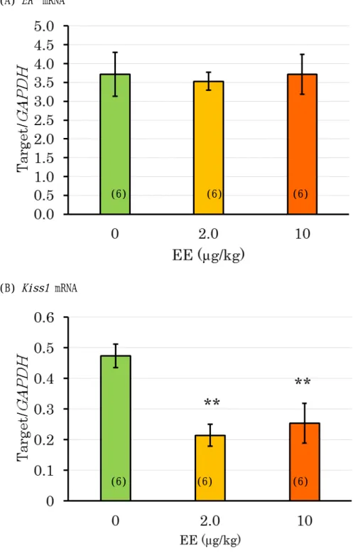
6. EE反復経口投与後の視床下部における
遺伝子発現(実験2)
図 3 に示すように、EE 2 µg/kg/day 群では 視床下部における
ERα mRNA量が対照群と比 較して有意な低値を示したが、上部領域では 群間に差は認められなかった。
図 4 に示すように、視床下部における遺伝 子発現量を対照群と
0.4 µg/kg/day以上の投与 群との間で比較したところ、 EE 投与群におけ
る
Kiss1mRNA 量が顕著に低下し、対照群と
の間に有意差が認められた。ERα mRNA につ いても、対照群と比較して、2 µg/kg/day 群で は有意な低値を示し、0.4 µg/kg/day 群でも低 下の傾向が認められた。 Kndy ニューロンでキ スペプチンとともに GnRH パルスの制御に関 わると考えられている NKB および Dyn なら びにそれらをコードする遺伝子については、
対照群と EE 投与群との間に有意差は認めら れなかった。しかし、EE の用量に依存して NKB 受容体をコードする Tacr3 の発現が低下 し、 DYN 受容体をコードする
Oprk1の発現が 増加した。
キスペプチン受容体をコードする
GPR54および EE が結合する可能性のある
ERβにつ いては遺伝子発現量に群間で著差は認められ なかった(図 5) 。
図 6 に示すように、
0.08 µg/kg/day以下の投 与群については、対照群が1例であったため 対照群との間で比較はできなかった。用量間 で比較しても
ERαおよび
Kiss1のいずれも
0.016 µg/kg/day群と
0.08 µg/kg/day群との間で 有意差は認められなかった。
7. EE単回経口投与後の視床下部における
遺伝子発現(実験2)
図 7 には 1 日齢における 2 あるいは 10
µg/kg
の EE 単回投与翌日の視床下部における
ERα
および
Kiss1発現量を示した。ERα につ いては対照群と EE 投与群との間で差異は認 められなかったが、
Kiss1は両投与群ともに対 照群と比べて有意な低値を示した。しかし、
用量間で差は認められなかった。
図 8 には 5 日齢における
20 µg/kgの EE 単 回投与翌日の視床下部における
ERαおよび
Kiss1発現量を示したが、ERα および
Kiss1と もに対照群と比較して有意な低値を示した。
8. EE単回経口投与後の視床下部における
遺伝子発現(実験2)
図 9 および 10 に代表例を示すように、
ERαmRNA については、 EE 反復経口投与した動物 でも対照群と同様に AVPV および ARCのいず れにおいても局在が認められ、その程度にも 明瞭な差は認められなかった。一方、Kiss1 mRNA については、EE 反復投与動物の ARC で顕著な発現低下が観察された(図 11) 。
D.考察
平成 26 年度のまでの研究から、新生期にお
ける EE の反復経口投与による遅発影響の閾
値は
0.016 µg/kg/day付近にあると推定されて
いる。これは
0.08 µg/kg/day群において、性周
期は回帰したものの、卵巣に嚢胞状卵胞を有
する動物が増加したことによるものである。
嚢胞状卵胞は加齢に伴い観察される変化であ り、0.08 µg/kg/day 投与では 26 週齢で対照群 との間に有意差が認められている(平成 25 年 度分担研究報告書)ことから、その生殖毒性 学的意義を検討するために、通常の生殖毒性 試験で評価される週齢で生殖能力を検討した。
その結果、全例の受胎能が確認され、胎児の 生存にも影響は認められなかった。しかし、
投与動物の妊娠後期における体重増加は有意 に抑制されていた。この時期は、胎児体重が 母体重に反映される時期であるが、 EE 投与群 の胎児体重は対照群と比べてむしろ有意に増 加していた。リッターサイズに差は認められ なかったことから、 EE 投与群では妊娠後期に おける母動物自身の体重増加が抑制されてい ることが示唆された。
胎児については外表および骨格に奇形や変 異の増加は認められなかったが、 EE 投与群で は対照群と比べて骨化が促進されていた。体 重変化を考慮すると、 EE 投与群では胎児の発 育が促進されていたものと考えられる。
新生児期に EE 投与を受けた動物が妊娠す ると妊娠末期における母体重は抑制されるに もかかわらず、胎児の発育が促進される理由 は明らかではない。用量反応性を考慮した検 討が必要とされるが、これまで検討されなか った糖質や脂質代謝などにおける遅発影響も 否定できない。
今年度は、新生児期の EE 投与による初発影 響を検討した。その結果、最も顕著な影響が
Kiss1発 現 に 認 め ら れ た 。 す な わ ち 、 0.4
μg/kg/day
以上の反復投与あるいは1日齢にお
ける 2 μg/kg 以上の単回投与あるいは 5 日齢に おける 20 μg/kg 投与によって、
Kiss1の発現低 下が認められた。 ISH でも ARC における
Kiss1発現に明瞭な低下が確認されている。この日
齢では AVPV に
Kiss1は発現していないこと
から、視床下部の定量解析で認められた
Kiss1発現の低下は、ARC における
Kiss1の発現低 下を反映したものと考えられる。さらに、
Kiss1
の発現低下が認められた群はいずれも
性周期の早期回帰停止が認められている。一 方、嚢胞状卵胞の形成は認められるが、性周 期の回帰は維持されている 0.08
μg/kg/day群
では
Kiss1 mRNAに影響が認められていない。
これらのことを考慮すると、ARC における
Kiss1
の発現低下は遅発影響の中でも性周期
の早期回帰停止に深く関与している可能性が 示唆される。
ARC における
Kiss1の発現が EE 投与によっ て低下する理由は本研究からは明らかではな い。EE はまず ER を介して生体に影響を及ぼ
すが、
Kiss1の発現低下が認められた 1 日齢で
の 10 μg/kg 単回投与では
ERαの発現量に影響 は認められなかったことから、ARC における
Kiss1
の発現低下は
ERαの発現低下に起因す
るものではないと考えられる。
脳の様々な部位に
ERαは発現するが、前交 連から上部の組織ではこのような変化は認め られなかったことから、 AVPV および ARC を 含む視床下部のみの変化であると推測される。
ERα
はこの日齢でも AVPV および ARC の両神 経核に発現していることが本研究でも確認さ れているが、ISH では両神経核ともに
ERαの 発現に EE 投与の影響は認められなかった。
従って、これらの神経核以外の部位での EE 投与による発現低下が疑われる。
ARC でキスペプチンとともに GnRH パルス 状分泌を促進する NKB の受容体遺伝子
Tacr3の発現が EE 投与群で低下の傾向を示し、パ ルス状分泌を抑制する DYN の受容体遺伝子
Oprk1
の発現が増加の傾向を示していた。
Kiss1
の発現低下のような顕著な変化ではな
いが、 Kndy ニューロンで作動する3つの分子 がいずれも GnRH パルスを抑制する方向に変 化していることは、遅発影響に繋がる変化と して注目すべきである。
前述のように
Kiss1の発現低下は ARC にお
ける Kiss1 の発現低下を反映すると考えられ
るが、定量解析では
Kiss1発現の低下に用量 反応関係が認められなかった。 この点は
Oprk1あるいは
Tacr3の変化とは異なっており、
GnRH パルスを抑制する方向の変化ではある
が、
Kiss1発現低下とは異なる機序の存在が伺
われる。
ERα
はエストロゲンによって誘導されるこ とが知られているが、EE 反復投与群では 0.4
μg/kg/day
以上の投与群で最終投与翌日の視床
下部における
ERα発現量が低下した。1 日齢
の単回投与では 10
μg/kgでも発現量が増減す
ることはなかったので、 EE を反復投与するこ
とで視床下部の
ERαが低下したものと推察さ れる。
以上のように、今年度の研究から、遅発影 響の最小影響量(0.08
μg/kg/day)は生殖能力評価では影響は認められないことが明らかに なったが、妊娠末期の生理状態についてはさ らに検討を要することが示唆された。また、
投与した EE は、 まず、 Kndy ニューロンの
kiss1遺伝子発現を低下させ、GnRH 分泌制御を変 化させることで、その後の視床下部/下垂体
/性腺軸の正常な発達を妨げ、遅発影響をも たらすことが示唆された。
F.研究発表 1. 論文発表
1) Shirota M, Kawashima J, Ogawa Y, Kamiie J, Yasuno K, Shirota K, Yoshida M. Delayed effects of single neonatal subcutaneous exposure of low-dose 17α-ethynylestradiol on reproductive function in female rats. Journal of Toxicological Sciences 37, 681-689 (2012)
2) Shirota M, Kawashima J, Nakamura T, Ogawa Y, Kamiie J, Shirota K. Vascular Hamartoma in the Uterus of a Female Sprague-Dawley Rat with an Episode of Vaginal Bleeding. Toxicologic Pathology 41, 1011-1015 (2013).
3) Shiorta M, Kawashima J, Nakamura T, Kamiie J, Shirota K, Yoshida M. Dose-dependent acceleration in the delayed effects of neonatal oral exposure to low-dose 17α-ethynylestradiol on reproductive functions in female Sprague–Dawley rats. Journal of Toxicological Sciences 40, 727-738 (2015)
2. 学会発表
1) 田 中 恵 他 「新 生 児 期 エ チ ニ ル エ ス トラジ オール(EE)曝露による遅発影響に関わる 初 発 影 響 の 探 索—視 床 下 部 に お け る エ ス ト ロ ジ ェ ン 受 容 体 (ER) 及 び Kisspeptin
(KP) シ グ ナ ル 伝 達 分 子 の 遺 伝 子 発 現 解 析」(第32回日本毒性病理学会、2016年1 月、徳島市)
2) 代田 眞理子、吉田 緑「幼若動物を用いた毒 性評価において認識すべき発達期の繁殖生物 学の特徴」(第42 回日本毒性学会シンポジウ ム、2015年6月、金沢市)
3) 田中 恵他「嚢胞状卵胞形成における新生期エ チニルエストラジオール経口曝露量と子宮肥 大試験の検出感度」(第 42 回日本毒性学会、
2015年6月、金沢市)
4) Shirota, M., et al. Gonadotropin-
independent follicle development in the Kiss1-/- female rats. (3rd World Congress on Reproductive Biology, August 2014, Edinburgh, UK)
5) 代田 眞理子「リプロダクティブヘルスからみ た遅発影響 −遅発影響検出のための実践的 指標の探索」(第 41 回日本毒性学会シンポジ ウム、2014年7月、神戸市)
6) 代田 眞理子「ラット周生期エストロゲン活性 物質曝露による遅発影響—毒性学的視点での 解析」(第106回日本繁殖生物学会大会シンポ ジウム、2013年9月、府中市)
7) 川嶋 潤他「新生ラットへのエチニルエストラ ジオール曝露が幼若期の卵巣における卵胞発 育関連遺伝子の発現に及ぼす影響」(第40回 日本毒性学会、2013年6月、千葉市)
G.知的財産権の出願・登録状況 (予定を含む。)
1. 特許取得 該当無し 2. 実用新案登録 該当無し 3. その他
無し 参考文献
Aatsinki JT, Pietilä EM, Lakkakorpi JT, Rajaniemi HJ.
Expression of the LH/CG receptor gene in rat ovarian tissue is regulated by an extensive alternative splicing of the primary transcript. Molecular Cellular Endocrinology 84,127-135 (1992)
Akieda-Asai S, Ohta R, Shirota M, Jaroenporn S, Watanabe G, Taya K. Endocrinological differences between Hatano high- and low-avoidance rats during early two-way avoidance acquisition. Experimental Animals 60, 509-516 (2011)
Asai S, Ohta R, Shirota M, Sato M, Watanabe G, Taya K. Reproductive endocrinology in Hatano high and low-avoidance rats during the estrous cycle. Endocrine 18, 161–166 (2002)
Ayyanan A, Laribi O, Schuepbach-Mallepell S, Schrick C, Gutierrez M, Tanos T, Lefebvre G, Rougemont J, Yalcin-Ozuysal O, Brisken C. Perinatal exposure to bisphenol A increases adult mammary gland progesterone response and cell number. Molecular Endocrinology 25, 1915-1923 (2011)
Bandiera S and Dworschak C. Effects of Testosterone and Estrogen on Hepatic Levels of Cytochromes P450 2C7 and P450 2Cll in the Rat. Archives of Biochemistry and Biophysics 296, 286-295 (1992)
Banu SK, Samuel JB, Arosh JA, Burghardt RC, Aruldhas MM. Lactational exposure to hexavalent chromium delays puberty by impairing ovarian development, steroidogenesis and pituitary hormone synthesis in developing Wistar rats. Toxicology and Applied Pharmacology 232,180-189 (2008)
Barkhem T, Carlsson B, Nilsson Y, Enmark E, Gustafsson J, Nilsson S. Differential response of estrogen receptor α and estrogen receptor β to partial estrogen agonists/antagonists. Molecular Pharmacology 54,105–112 (1998)
Barraclough CA. Production of anovulatory, sterile rats by single injections of testosterone propionate.
Endocrinology 68, 62-66 (1962)
Betancourt AM, Eltoum IA, Desmond RA, Russo J, Lamartiniere CA. In utero exposure to bisphenol A shifts the window of susceptibility for mammary carcinogenesis in the rat. Environmental Health Perspectives 118, 1614-1619 (2010)
Beale KE, Kinsey-Jones JS, Gardiner JV, Harrison EK, Thompson EL, Hu MH, Sleeth ML, Sam AH, Greenwood HC, McGavigan AK, Dhillo WS, Mora JM, Li XF, Franks S, Bloom SR, O’Byrne KT, Murphy KG.
The physiological role of arcuate kisspeptin neurons in the control of reproductive function in female rats.
Endocrinology 155, 1091–1098 (2014)
Becú-Villalobos D, González Iglesias A, Díaz-Torga G, Hockl P, Libertun C. Brain sexual differentiation and gonadotropins secretion in the rat. Cellular and Molecular Neurobiology 17, 699-715 (1997)
Biles JE, McNeal TP, Begley TH, Hollifield HC.
Determination of bisphenol-A in reusable polycarbonate food-contact plastics and migration to food simulating liquids. J Agriculture and Food Chemistry 45,3541–3544 (1997)
Blair RM, Fang H, Branham WS, Hass BS, Dial SL, Moland CL, Tong W, Shi L, Perkins R, Sheehan DM.
The estrogen receptor relative binding affinities of 188 natural and xenochemicals: structural diversity of ligands. Toxicological Sciences 54, 138-153 (2000)
Borgert CJ, LaKind JS, Witorsch RJ. A critical review
of methods for comparing estrogenic activity of endogenous and exogenous chemicals in human milk and infant formula. Environmental Health Perspectives 111, 1020-1036 (2003)
Boutin JM, Jolicoeur C, Okamura H, Gagnon J, Edery M, Shirota M, Banville D, Dusanter-Fourt I, Djiane J, Kelly PA. Cloning and expression of the rat prolactin receptor, a member of the growth hormone/prolactin receptor gene family.
Cell 53, 69-77 (1988)
Boutin JM, Edery M, Shirota M, Jolicoeur C, Lesueur L, Ali S, Gould D, Djiane J, Kelly PA.
Identification of a cDNA encoding a long form of prolactin receptor in human hepatoma and breast cancer cells. Molecular Endocrinology 3, 1455-1461 (1989)
Branham WS, Dial SL, Moland CL, Hass BS, Blair RM, Fang H, Shi L, Tong W, Perkins RG, Sheehan DM. Phytoestrogens and mycoestrogens bind to the rat uterine estrogen receptor. Journal of Nutrition 132, 658-664 (2002)
Brotons JA, Olea-Serrano MF, Villalobos M, Pedraza V, Olea N. Xenoestrogens released from lacquer coatings in food cans. Environment and Health Perspectives 103, 608-612 (1995)
Byers M, Kuiper GG, Gustafsson JA, Park-Sarge OK.
Estrogen receptor-beta mRNA expression in rat ovary:
down-regulation by gonadotropins. Molecular Endocrinology 11, 172-182 (1997)
Bukovsky A, Ayala ME, Dominguez R, Keenan JA, Wimalasena J, McKenzie PP, Caudle MR. Postnatal androgenization induces premature aging of rat ovaries.
Steroids 65, 190-205 (2000)
Cao J, Joyner L, Mickens JA, Leyrer SM, Patisau HB.
Sex-specific Esr2 mRNA expression in the rat hypothalamus and amygdala is altered by neonatal bisphenol A exposure. Reproduction 147, 537–554 (2014)
Chakraborty TR, Gore AC. Aging-Related Changes in Ovarian Hormones, Their Receptors, and Neuroendocrine Function. Experimental Biology and Medicine 229, 977–987 (2004)
Chang L-L, Wun W-S A, Paulus S. Wang PS. In utero and neonate exposure to nonylphenol develops
hyperadrenalism and metabolic syndrome later in life. I.
First generation rats (F1). Toxicology 301, 40-49 (2012) Chen Y, Jefferson WN, Newbold RR, Padilla-Banks E, Pepling ME. Estradiol, Progesterone, and Genistein Inhibit Oocyte Nest Breakdown and Primordial Follicle Assembly in the Neonatal Mouse Ovary in Vitro and in Vivo. Endocrinology 148, 3580–3590 (2007)
Chung WCJ, Anthony PA. Gender differences in neurodevelopment and epigenetics. Pflugers Archives European Journal of Physiology 465, 573–584 (2013) Cimafranca MA, Davila J, Ekman GC, Andrews RN, Neese SL, Peretz J, Woodling KA, Helferich WG, Sarkar J, Flaws JA, Schantz SL, Doerge DR, Cooke PS.
Acute and chronic effects of oral genistein administration in neonatal mice. Biology of Reproduction 83, 114-121 (2010)
Clarkson J. Effects of estradiol on kisspeptin neurons during puberty. Frontiers in Neuroendocrinology 34, 120-131 (2013)
d'Anglemont de Tassigny X, Fagg LA, Dixon JP, Day K, Leitch HG, Hendrick AG, Zahn D, Franceschini I, Caraty A, Carlton MB, Aparicio SA, Colledge WH.
Hypogonadotropic hypogonadism in mice lacking a functional Kiss1 gene. Proceeding of National Academy Scicience U S A. 104, 10714-10719 (2007)
Downsa JL, Wise PM. The role of the brain in female reproductive aging. Molecular and Cellular Endocrinology 299, 32-38 (2009)
Dufau ML, Tsai-Morris CH, Hu ZZ, Buczko E.
Structure and regulation of the luteinizing hormone receptor gene. Journal of Steroid Biochemistry and Molecular Biology 53, 283-291 (1995)
Durlinger AL, Gruijters MJ, Kramer P, Karels B, Ingraham HA, Nachtigal MW, Uilenbroek JT, Grootegoed JA, Themmen AP. Anti-Müllerian hormone inhibits initiation of primordial follicle growth in the mouse ovary. Endocrinology 143, 1076-1084 (2002)
Edson MA, Nagaraja AK, Matzuk MM. The mammalian ovary from genesis to revelation.
Endocrine Reviews 30, 624-712 (2009)
Fernández M, Bianchi M, Lux-Lantos V, Libertun C.
Neonatal exposure to bisphenol a alters reproductive parameters and gonadotropin releasing hormone
signaling in female rats. Environment and Health Perspectives 117, 757-762 (2009)
Frye CA, Bo E, Calamandrei G, Calzà L, Dessì-Fulgheri F, Fernández M, Fusani L, Kah O, Kajta M, Le Page Y, Patisaul HB, Venerosi A, Wojtowicz AK Panzica GC. Endocrine disrupters- a review of some sources, effects, and mechanisms of actions on behaviour and neuroendocrine systems. Journal of Neuroendocrinology 24, 144-159 (2012)
Goldenberg RL, Vaitukaitis JL, Ross GT. Estrogen and follicle stimulation hormone interactions on follicle growth in rats. Endocrinology 90, 1492-1498 (1972)
González-Martínez D, De Mees C, Douhard Q, Szpirer C, Bakker J. Absence of gonadotropin-releasing hormone 1 and Kiss1 activation in alpha-fetoprotein knockout mice: prenatal estrogens defeminize the potential to show preovulatory luteinizing hormone surges. Endocrinology 149, 2333-2340 (2008)
Gore AC, Martien KM, Gagnidze K, Pfaff D.
Implications of prenatal steroid perturbations for neurodevelopment, behavior, and Autism. Endocrine Reviews 35, 961-991(2014)
Gore AC, Walker DM, Zama AM, Armenti AE, Uzumcu M. Early life exposure to endocrine-disrupting chemicals causes lifelong molecular reprogramming of the hypothalamus and premature reproductive aging.
Molecular Endocrinology 25, 2157-2168 (2011) Graham R. Robertson, Geoffrey C. Farrell, Liddle C.
Sexually Dimorphic Expression of Rat CYP3A9 and CYP3A18 Genes Is Regulated by Growth Hormone.
Biochemical and Biophysical Research Communications 242, 57–60 (1998)
Greenstein BD. Effects of rat alpha-fetoprotein administration on estradiol free fraction, the onset of puberty, and neural and uterine nuclear estrogen receptors. Endocrinology 130, 3184-3190 (1992)
Kim H, Nakajima T, Hayashi S, Chambon P, Watanabe H, Iguchi T, Sato T. Effects of diethylstilbestrol on programmed oocyte death and induction of polyovular follicles in veonatal mouse ovaries. Biology of Reproduction 81, 1002–1009 (2009)
Gorski RA. Influence of age on the response to paranatal administration of a low dose of androgen.
Endocrinology 82, 1001-1004 (1968)
Herath CB, Yamashita M, Watanabe G, Jin W, Tangtrongsup S, Kojima A, Groome NP, Suzuki AK, Taya K. Regulation of follicle-stimulating hormone secretion by estradiol and dimeric inhibins in the infantile female rat. Biology of Reproduction 65,1623-1633 (2001)
Hirshield AH. Overview of Ovarian Follicular Development: Considerations for the Toxicologist.
Environmental and Molecular Mutagenesis 29,10-15 (1997)
Homma T, Sakakibara M, Yamada S, Kinoshita M, Iwata K, Tomikawa J, Kanazawa T, Matsui H, Takatsu Y, Ohtaki T, Matsumoto H, Uenoyama Y, Maeda K, Tsukamura H. Significance of neonatal testicular sex steroids to defeminize anteroventral periventricular kisspeptin neurons and the GnRH/LH surge system in male rats. Biology of Reproduction 81, 1216-1225 (2009)
Hong H, Branham WS, Dial SL, Moland CL, Fang H, Shen J, Perkins R, Sheehan D, Tong W. Rat α-Fetoprotein binding affinities of a large set of structurally diverse chemicals elucidated the relationships between structures and binding affinities.
Chemical Research of Toxicology 25, 2553-2566 (2012) Huang EJ, Manova K, Packer AI, Sanchez S, Bachvarova RF, Besmer P. The murine steel panda mutation affects kit ligand expression and growth of early ovarian follicles. Developmental Biology 157, 100-109 (1993)
Hutter HS, Gibson MJ. Effect of neonatal androgenization on positive feedback in female mice.
Biology of Reproduction 38, 636-638 (1988)
Horie K, Takakura K, Taii S, Narimoto K, Noda Y, Nishikawa S, Nakayama H, Fujita J, Mori T. The expression of c-kit protein during oogenesis and early embryonic development. Biology of Reproduction 45, 547-552 (1991)
Ichimura R, Takahashi M, Morikawa T, Inoue K, Maeda J, Usuda K, Yokosuka M, Watanabe G, Yoshida M. Prior attenuation of KiSS1/GPR54 signaling in the anteroventralperiventricular nucleus is a trigger for the delayed effect induced by neonatal exposure to 17alpha-ethynylestradiol in female rats. Reproductive Toxicolology 51, 145-156 (2015)
Ikeda Y, Nagai A, Ikeda M, AND Hayashi S. Increased Expression of Mullerian-Inhibiting Substance
Correlates with Inhibition of Follicular Growth in the Developing Ovary of Rats Treated with E2 Benzoate.
Endocrinology 143, 304–312 (2002)
Ikeda Y, Tanaka H, and Esaki M. Effects of Gestational Diethylstilbestrol Treatment on Male and Female Gonads during Early Embryonic Development.
Endocrinology 149, 3970-3979 (2008)
Jager W, Correia MA, Bornheim LM, Mahnke A, WALTER G. Hanstein WG, XUE L, and Benet LZ.
Ethynylestradiol-mediated induction of hepatic CYP3A9 in Female rats: implication for cyclosporine metabolism. Drug Metabolism and Disposition, 27, 1505–1511 (1999)
Jaroenporn S, Horii Y, Asai S, Wang KM, Nagaoka K, Ohta R, Shirota M, Watanabe G, Taya K. Endocrine mechanisms responsible for different follicular development during the estrous cycle in Hatano high- and low-avoidance rats. Journal of Reproduction and Development 57, 690-699 (2012)
Jefferson W, Newbold R, Padilla-Banks E, and Pepling M. Neonatal Genistein Treatment Alters Ovarian Differentiation in the Mouse: Inhibition of Oocyte Nest Breakdown and Increased Oocyte Survival. Biology of Reproduction 74, 161–168 (2006)
Jefferson WN, Patisaul HB Williams CJ. Reproductive consequences of developmental phytoestrogen exposure.
Reproduction 143, 247-260 (2012)
Hu J, Du G, Zhang W, Huang H, Chen D, Wu D, Wang X. Short-term neonatal/prepubertal exposure of dibutylphthalate (DBP) advanced pubertal timing and affected hypothalamic kisspeptin/GPR54 expression differently in female rats. Toxicology 314, 65–75 (2013)
Kanno J, Onyon L, Haseman J, Fenner-Crisp P, Ashby J, Owens W. The OECD Program to Validate the Rat Uterotrophic Bioassay to Screen Compounds for in Vivo Estrogenic Responses: Phase 1. Environmental Health Perspectives 109, 785–794 (2001)
Kelly PA, Boutin JM, Jolicoeur C, Okamura H, Shirota M, Edery M, Dusanter-Fourt I, Djiane J. Purification, cloning, and expression of the prolactin receptor.
Biology of Reproduction 40, 27-32 (1989)
Kenny HA, Woodruff TK. Follicle size class contributes to distinct secretion patterns of inhibin isoforms during the rat estrous cycle. Endocrinology 147, 51-60 (2006)
Kezele P, Skinner MK. Regulation of Ovarian Primordial Follicle Assembly and Development by Estrogen and Progesterone: Endocrine Model of Follicle Assembly. Endocrinology 144, 3329–3337 (2003)
Kimura F, Funabashi T. Two subgroups of gonadotropin releasing hormone neurons control gonadotropin secretion in rats. News of Physiological Sciences 13,225-231 (1998)
Kipp JL, Kilen SM, Bristol-Gould S, Woodruff TK, Mayo KE. Neonatal exposure to estrogens suppresses activin expression and signaling in the mouse ovary.
Endocrinology 148:1968-1976 (2007)
Kuiper GG, Lemmen JG, Carlsson B, Corton JC, Safe SH, van der Saag PT, van der Burg B, Gustafsson JA.
Interaction of estrogenic chemicals and phytoestrogens with estrogen receptor beta. Endocrinology 139, 4252-4263 (1998)
Kurian JR, Olesen KM, Auger AP. Sex differences in epigenetic regulation of the estrogen receptor-promoter within the developing preoptic area. Endocrinology 151, 2297–2305 (2010)
Lei L, Shiying J, Kelly EM, Woodruff TK. The Interactions Between the Stimulatory Effect of Follicle-Stimulating Hormone and the Inhibitory Effect of Estrogen on Mouse Primordial Folliculogenesis.
Biology of Reproduction 82, 13–22 (2010)
Mazaud Guittot S, Guigon CJ, Coudouel N, Magre S.
Consequences of fetal irradiation on follicle histogenesis and early follicle development in rat ovaries. Biology of Reproduction 75, 749–759 (2006)
McCarthy MM. Estradiol and the developing brain.
Physiological Reviews 88, 91-124 (2008)
McGee EA, Smith R, Spears N, Nachtigal MW, Ingraham H, Hsueh AJ. Müllerian inhibitory substance induces growth of rat preantral ovarian follicles.
Biology of Reproduction 64, 293-298 (2001)
McLachlan JA, Tilghman SL, Burow ME, Bratton MR.
Environmental signaling and reproduction: A comparative biological and chemical perspective.
Molecular and Cellular Endocrinology 354, 60-62 (2012)
Melinda E, Wilson R, Handa J. Ontogeny of Gene expression in the gonadotroph of the developing female rat. Biology of Reproduction 56, 563-568 (1997)
Mena MA, Arriaza CA, Tchernitchin AN. Early postnatal androgenization imprints selective changes in the action of estrogens in the rat uterus. Biology of Reproduction 46, 1080-1085 (1992)
Mineshige T, Kamiie J, Sugahara G, Yasuno K, Aihara N, Kawarai S, Yamagishi K, Shirota M, Shirota K.
Expression of periostin in normal, atopic, and nonatopic chronically inflamed canine skin. Veterinary Pathology 52, 1118-1126 (2015)
Mogi K, Takanashi H, Nagasawa M, Kikusui T. Sex differences in spatiotemporal expression of AR, ERα, and ERβ mRNA in the perinatal mouse brain.
Neuroscience Letters 584, 88–92 (2015)
Montano MM, Welshons WV, vom Saal FS. Free estradiol in serum and brain uptake of estradiol during fetal and neonatal sexual differentiation in female rats.
Biology of Reproduction 53, 1198-207 (1995)
Moral R, Santucci-Pereira J, Wang R, Russo IH, Lamartiniere CA, Russo J. In utero exposure to butyl benzyl phthalate induces modifications in the morphology and the gene expression profile of the mammary gland: an experimental study in rats.
Environmental Health 10, 5 (2011)
Morohashi KI, Omura T. Ad4BP/SF-1, a transcription factor essential for the transcription of steroidogenic cytochrome P450 genes and for the establishment of the reproductive function. FASEB Journal 10, 1569-1577 (1996)
Murakami T, Sato A, Inatani M, Sakurai H, Yumoto R, Nagai J, Takano M. Effect of neonatal exposure of 17beta-estradiol and tamoxifen on hepatic CYP3A activity at developmental periods in rats. Drug Metabolism and Pharmacokinetics 19, 96-102 (2004) Murray AA, Gosden RG, Allison V, Spears N. Effect of androgens on the development of mouse follicles growing in vitro. J Reprod Fertility 113, 27-33 (1998)
Nagai A, Ikeda Y, Aso T, Eto K, Ikeda MA. Exposure of neonatal rats to diethylstilbestrol affects the expression of genes involved in ovarian differentiation.
Journal of Medical and Dental Sciences 50, 35-40 (2003)
Nagaraja AK, Middlebrook BS, Rajanahally S, Myers M, Li Q, Matzuk MM, Pangas SA. Defective gonadotropin-dependent ovarian folliculogenesis and granulosa cell gene expression in inhibin-deficient mice.
Endocrinology 151, 4994-5006 (2010)
Navarro VM, Sánchez-Garrido MA, Castellano JM, Roa J, García-Galiano D, Pineda R, Aguilar E, Pinilla L, Tena-Sempere M. Persistent impairment of hypothalamic KiSS-1 system after exposures to estrogenic compounds at critical periods of brain sex differentiation. Endocrinology 150, 2359-2567 (2009)
Nilsson S, Mäkelä S, Treuter E, Tujague M, Thomsen J, Andersson G, Enmark E, Pettersson K, Warner M, Gustafsson JA. Mechanisms of estrogen action.
Physiological Reviews 81, 1535-1565 (2001)
Nozawa K, Nagaoka K, Zhang H, Usuda K, Okazaki S, Taya K, Yoshida M, Watanabe G. Neonatal exposure to 17α-ethynyl estradiol affects ovarian gene expression and disrupts reproductive cycles in female rats.
Reproductive Toxicology 46, 77–84 (2014)
Ohta R, Ohmukai H, Marumo H, Shindo T., Nagata, T Ono H. Delayed reproductive dysfunction in female rats induced by early life exposure to low-dose diethylstilbestrol. Reproductive Toxicology 34, 323-330 (2012)
Ohta R, Shirota M, Kanazawa Y, Shindo T, Furuya M, Seki T, Ono H, Kojima K, Asai S, Watanabe G, Taya K.
Effects of transmaternal exposure to genistein in Hatano high- and low-avoidance rats. Experimental Animals 58, 471-479 (2009)
Ojeda SR, Advis JP, Andrews WW. Neuroendocrine control of the onset of puberty in the rat. Federal Proceedings 39, 2365-2371 (1980)
Olea N, Pulgar R, Perez P, Olea-Serrano F, Rivas A, Novillo-Fertrell A, Pedraza V, Soto AM, Sonnenschein C. Estrogenicity of resin-based composites and sealants used in dentistry. Environment and Health Perspectives 104, 298–305 (1996)
Orisaka M, Hattori K, Fukuda S, Mizutani T, Miyamoto K, Sato T, Tsang BK, Kotsuji F, Yoshida Y.
Dysregulation of ovarian follicular development in female rat: LH decreases FSH sensitivity during preantral-early antral transition. Endocrinology 154, 2870-2880 (2013)
Osterlund M, Kuiper GG, Gustafsson JA, Hurd YL.
Differential distribution and regulation of estrogen receptor-alpha and -beta mRNA within the female rat brain. Molecular Brain Research 54, 175-180 (1998)
Pedersen T, Peters H. Proposal hor a classification of oocytes and follicles in the mouse ovary. Journal of Reproduction and Fertility 17, 555-557 (1968)
Pineda R, Garcia-Galiano D, Roseweir A, Romero M, Sanchez-Garrido MA, Ruiz-Pino F, Morgan K, Pinilla L, Millar RP, Tena-Sempere M. Critical roles of kisspeptins in female puberty and preovulatory gonadotropin surges as revealed by a novel antagonist.
Endocrinology 151, 722-730 (2010)
Pinilla L, Castellano JM, Romero M, Tena-Sempere M, Gaytán F, Aguilar E. Delayed puberty in spontaneously hypertensive rats involves a primary ovarian failure independent of the hypothalamic KiSS-1/GPR54/
GnRH system. Endocrinology 150, 2889-2897 (2009)
Ramirez MC, Luque GM, Ornstein AM, Becu-Villalobos D. Differential neonatal testosterone imprinting of GH-dependent liver proteins and genes in female mice. Journal of Endocrinology 207, 301-308 (2010)
Rasier G, Parent AS, Gérard A, Lebrethon MC, Bourguignon JP. Early maturation of gonadotropin-releasing hormone secretion and sexual precocity after exposure of infant female rats to estradiol or dichlorodiphenyltrichloroethane. Biology of Reproduction 77, 734–742 (2007)
Richards JS. Estradiol receptor content in rat granulosa cells during follicular development: modification by estradiol and gonadotropins. Endocrinology 97, 1174-1184 (1975)
Rubin BS, Murray MK, Damassa DA, King JC, Soto AM. Perinatal exposure to low doses of bisphenol A affects body weight, patterns of estrous cyclicity, and plasma LH levels. Environment and Health Perspectives 109, 675-680 (2001)
Ryan BC, Hotchkiss AK, Crofton KM, Gray LE Jr. In utero and lactational exposure to bisphenol A, in contrast to ethinyl estradiol, does not alter sexually dimorphic behavior, puberty, fertility, and anatomy of female LE rats. Toxicological Sciences 114, 133-148 (2010)
Sandhoff TW, Hales DB, Hales KH, McLean MP.
Transcriptional regulation of the rat steroidogenic acute regulatory protein gene by steroidogenic factor 1.
Endocrinology. 1998 139:4820-4831.
Sakuma Y. Gonadal steroid action and brain sex differentiation in the rat. Journal of Neuroendocrinology 21, 410-414 (2009)
Sakurada Y, Sawai M, Inoue K, Shirota M, Shirota K.
Comparison of aryl hydrocarbon receptor gene expression in laser dissected granulosa cell layers of immature rat ovaries. Journal of Veterinary Medical Science 73, 923-926 (2011)
Schindler R, Nilsson E, Skinner MK. Induction of ovarian primordial follicle assembly by connective tissue growth factor CTGF. PLoS ONE 5, e12979 (2010)
Schwarz JM, McCarthy MM. The role of neonatal NMDA receptor activation in defeminization and masculinization of sex behavior in the rat. Hormones and Behavior 54, 662-668 (2008)
Seminara SB, Messager S, Chatzidaki EE, Thresher RR, Acierno JS Jr, Shagoury JK, Bo-Abbas Y, Kuohung W, Schwinof KM, Hendrick AG, Zahn D, Dixon J, Kaiser UB, Slaugenhaupt SA, Gusella JF, O'Rahilly S, Carlton MB, Crowley WF Jr, Aparicio SA, Colledge WH. The GPR54 gene as a regulator of puberty. New England Journal of Medicine 349,1614-1627 (2003)
Shiorta M, Kawashima J, Nakamura T, Kamiie J, Shirota K, Yoshida M. Dose-dependent acceleration in the delayed effects of neonatal oral exposure to low-dose 17α-ethynylestradiol on reproductive functions in female Sprague–Dawley rats. Journal of Toxicological Sciences 40, 727-738 (2015)
Shirota M, Kawashima J, Nakamura T, Ogawa Y, Kamiie J, Yasuno K, Shirota K, Yoshida M. Delayed effects of single neonatal subcutaneous exposure of low-dose 17α-ethynylestradiol on reproductive function in female rats. Journal of Toxicological Science 37, 681-690 (2012)
Shirota M, Soda S, Katoh C, Asai S, Sato M, Ohta R, Watanabe G, Taya K, Shirota K. Effects of reduction of the number of primordial follicles on follicular development to achieve puberty in female rats.
Reproduction 125, 85-94 (2003)
Shirota M, Kurohmaru M, Hayashi Y, Shirota K, Kelly PA. Detection of in situ localization of long form
prolactin receptor messenger RNA in lactating rats by biotin-labeled riboprobe. Endocrine Journal 42, 69-76 (1995)
Shirota M, Banville D, Ali S, Jolicoeur C, Boutin JM, Edery M, Djiane J, Kelly PA. Expression of two forms of prolactin receptor in rat ovary and liver. Molecular Endocrinology 4, 1136-1143 (1990)
Soga T, Kitahashi T, Clarke IJ, Parhar IS.
Gonadotropin-inhibitory hormone promoter-driven enhanced green fluorescent protein expression decreases during aging in female rats Endocrinology 155, 1944–1955 (2014)
Sokka T, Huhtaniemi I. Ontogeny of gonadotrophin receptors and gonadotrophin -stimulated cyclic AMP production in the neonatal rat ovary. Journal of Endocrinology 127, 297-303 (1990)
Sokka T, Hämäläinen T, Huhtaniemi L. Functional LH receptor appears in the neonatal rat ovary after changes in the alternative splicing pattern of the LH receptor mRNA. Endocrinology 130, 1738-1740 (1992)
Sokka TA, Huhtaniemi IT. Functional maturation of the pituitary-gonadal axis in the neonatal female rat.
Biology of Reproduction 52, 1404-1409 (1995)
Sokka TA, Hämäläinen TM, Kaipia A, Warren DW, Huhtaniemi IT. Development of luteinizing hormone action in the perinatal rat ovary. Biology of Reproduction 55, 663-670 (1996)
Shirwalkar H, Modi DN, Maitra A. Exposure of adult rats to estradiol valerate induces ovarian cyst with early senescence of follicles. Molecular Cellular Endocrinology 272, 22-37 (2007)
Takahashi M, Inoue K, Morikawa T, Matsuo S, Hayashi S, Tamura K, Watanabe G, Taya K, Yoshida M. Delayed effects of neonatal exposure to 17alpha-ethynylestradiol on the estrous cycle and uterine carcinogenesis in Wistar Hannover GALAS rats.
Reproductive Toxicology 40, 16-23 (2013)
Takumi K, Iijima N, Iwata K, Higo S, Ozawa H. The effects of gonadal steroid manipulation on the expression of Kiss1 mRNA in rat arcuate nucleus during postnatal development. Journal of Physiological Sciences 62, 453–460 (2012)
Tena-Sempere M. Kisspeptin⁄GPR54 system as potential target for endocrine disruption of reproductive
development and function. International Journal of Andrology 33, 360–368 (2010)
Thomas FH, Vanderhyden BC. Oocyte-granulosa cell interactions during mouse follicular development:
regulation of kit ligand expression and its role in oocyte growth. Reproductive Biology and Endocrinology 4, 19 (2006)
Tingen C, Kim A, and Woodruff TK. The primordial pool of follicles and nest breakdown in mammalian ovaries. Molecular Human Reproduction 15, 795–803 (2009)
Topper VY, Walker DM, Gore AC. Sexually dimorphic effects of gestational endocrine-disrupting chemicals on microRNA expression in the developing rat hypothalamus. Molecular and Cellular Endocrinology 414, 42-52 (2015)
Toran-Allerand CD. Gonadal hormones and brain development: implications for the genesis of sexual differentiation. Annual New York Academy of Sciences 435, 101-111 (1984)
Tsai-Morris CH, Buczko E, Wang W, Xie XZ, Dufau ML. Structural organization of the rat luteinizing hormone (LH) receptor gene. Journal of Biological Chemistry 266, 1355-11359 (1991)
Umekita Y, Souda M, Hatanaka K, Hamada T, Yoshioka T, Kawaguchi H, animoto A. Gene expression profile of terminal end buds in rat mammary glands exposed to diethylstilbestrol in neonatal period.
Toxicology Letter 205, 15-25 (2011)
Usuda K, Nagaoka K, Nozawa K, Zhang H, Taya K, Yoshida M, Watanabe G. Neonatal exposure to 17α-ethinyl estradiol affects kisspeptin expression and LH surge level in female rats. Journal of Veterinary Medical Sciences 76, 1105-1110 (2014)
Uzumcu M, Kuhn PE, Marano JE, Armenti AE, Passantino L. Early postnatal methoxychlor exposure inhibits folliculogenesis and stimulates anti-Mullerian hormone production in the rat ovary. Journal of Endocrinology 191, 549–558 (2006)
Vannier B, Raynaud JP. Long-term effects of prenatal oestrogen treatment on genital morphology and reproductive function in the rat. Journal of Reproduction and Fertility 59, 43-49 (1980)
Varayoud J, Ramos JG, Bosquiazzo VL, Muñoz-de-Toro M, Luque EH. Developmental exposure to Bisphenol A impairs the uterine response to ovarian steroids in the adult. Endocrinology 149, 5848-5860 (2008)
Vitt UA, McGee EA, Hayashi M, Hsueh AJ. In vivo treatment with GDF-9 stimulates primordial and primary follicle progression and theca cell marker CYP17 in ovaries of immature rats. Endocrinology 141, 3814-3820 (2000)
Visser JA, de Jong FH, Laven JS, Themmen AP.
Anti-Müllerian hormone: a new marker for ovarian function. Reproduction 131,1-9 (2006)
Walker DM, Kirson D, Perez LF, Gore AC. Molecular profiling of postnatal development of the hypothalamus in female and male rats. Biology of Reproduction 129, 1-12 (2012)
Wang C and Roy SK. Development of Primordial Follicles in the Hamster: Role of Estradiol-17β.
Endocrinology 148, 1707–1716 (2007)
Watanabe C, Kuwagata M, Yoshimura S, Azegami J, Kojima K, Ono H, Nagao T. An improved technique for repeated gavage administration to rat neonates.
Congenital Anomalies (Kyoto) 43, 177-179 (2003)
Wu J, Nayudu PL, Kiesel PS, Michelmann HW.
Luteinizing hormone has a stage-limited effect on preantral follicle development in vitro. Biology of Reproduction 63, 320-327 (2000)
Yamasaki K, Takeyoshi M, Sawaki M, Imatanaka N, Shinoda K, Takatsuki M. Immature rat uterotrophic assay of 18 chemicals and Hershberger assay of 30 chemicals. Toxicology 183, 93-115 (2003)
Yasuno K, Sakashita H, Araki S, Kobayashi R, Sakurada Y, Shirota M, Kamiie J, Shirota K. Cutaneous hybrid cyst in a Sprague-Dawley rats.
Journal of Toxicologic Pathology 25, 175-178 (2012) Yeh J, Kim B. Increasing blunting of inhibin responses to dynamic ovarian challenge is associated with reproductive aging in the rat. Reproductive Sciences 14, 10-19 (2007)
Yoshida H, Takakura N, Kataoka H, Kunisada T, Okamura H, Nishikawa SI. Stepwise requirement of c-kit tyrosine kinase in mouse ovarian follicle development. Developmental Biology 184, 122-137
(1997)
Yoshida M, Takahashi M, Inoue K, Hayashi S, Maekawa A, Nishikawa A. Delayed adverse effects of neonatal exposure to diethylstilbestrol and their dose dependency in female rats. Toxicologic Pathology 39, 823-834 (2011)
Yoshida M, Watanabe G, Shirota M, Maekawa A, Taya K. Reduction of primordial follicles caused by maternal treatment with busulfan promotes endometrial adenocarcinoma development in donryu rats. Journal of Reproduction and Development 51, 707-714 (2005) Zhang FP, Poutanen M, Wilbertz J, Huhtaniemi I.
Normal prenatal but arrested postnatal sexual development of luteinizing hormone receptor knockout (LuRKO) mice. Molecular Endocrinology 15, 172-183 (2001)
Zhuang XL, Fu YC, Xu JJ, Kong XX, Chen ZG, Luo LL. Effects of genistein on ovarian follicular development and ovarian life span in rats. Fitoterapia.
81, 998-1002 (2010)