145
Sex differences in the influence of
elevated serum uric acid levels for cardiovascular risk in the general population with normal renal function
Yuki M
atsuura1), Fumitaka t
anaka1), toshie s
egawa 2),
kozo t
anno 3), kiyomi s
akata 3), Masaki o
hsawa 4), shinichi o
MaMa 5), kuniaki o
gasawara 6), koichi a
sahi1)and Motoyuki n
akaMura 7)1) Division of Nephrology and Hypertension, Department of Internal Medicine, School of Medicine, Iwate Medical University, Yahaba, Japan
2) Division of Diabetes, Metabolism and Endocrinology, Department of Internal Medicine,
School of Medicine, Iwate Medical University, Yahaba, Japan
3) Department of Hygiene and Preventive Medicine, School of Medicine, Iwate Medical University, Yahaba, Japan
4) Department of Internal Medicine, Morioka Tsunagi Onsen Hospital
5) Emergency and Critical Care Center, School of Medicine, Iwate Medical University, Yahaba, Japan
6) Department of Neurosurgery, School of Medicine, Iwate Medical University, Yahaba, Japan
7) Professor Emeritus, School of Medicine, Iwate Medical University, Yahaba, Japan
(Received on January 20, 2020 & Accepted on February 13, 2020)
Key words: serum uric acid, chronic kidney disease, cardiovascular disease, risk factor, women Corresponding author: Yuki Matsuura
[email protected] The role of serum uric acid (SUA) as an independent predictor of cardiovascular events (CVEs) is uncertain, as previous studies may not have sufficiently accounted for risk factors that are strongly related to the SUA level, such as chronic kidney disease (CKD). We examined the association between the SUA level and the incidence of CVEs in each gender without CKD. Baseline data, including the SUA level, estimated glomerular filtration rate, and urinary albumin/creatinine ratio were determined in participants from a community-based population. After the exclusion of CKD, the subjects were stratified into sex-specific quartiles of SUA (n
= 15,036). A Cox regression analysis was performed to examine the sex-specific relationship between the baseline SUA level and the onset of CVEs. During a mean follow-up period of 8 . 8 years, there were 611 CVEs ( 304 in men, 307 in women). After adjusting for traditional risk factors, the hazard ratio for the onset of CVEs did not differ among the quartiles in men. In contrast, in women, a significant trend was observed. In the Japanese general population without CKD, moderately elevated SUA level is considered an independent risk factor for the onset of CVEs in women but not in men.
Abstract
Yuki Matsuura, et al.
146
I
. IntroductionThe significance of an elevated serum uric acid (SUA) level as a risk factor for the onset of cardiovascular events (CVEs) remains controversial. Several studies have suggested that an elevated SUA level was a risk factor for stroke
1-4)and coronary heart disease
3-6). However, other studies have suggested that the SUA level was not related to the onset of coronary heart disease
7). Some studies have shown that the association between the SUA level and CVEs had gender differences
2, 5, 6), while others have found no such differences
1, 3, 4). Given these conflicting findings, whether or not increased levels of SUA are a direct risk factor for the incidence of CVEs in the general population remains unclear.
Chronic kidney disease (CKD) is an independent risk factor for CVEs
8). Since the SUA level is affected by renal function, the SUA level increases when renal dysfunction occurs. This suggests that the association between an elevated SUA level and CVEs may be a secondary phenomenon due to chronic renal dysfunction rather than an elevated SUA level per se. However, few studies have examined whether or not an elevated SUA level is related to the onset of CVEs independently observed in subjects without renal function impairment.
Therefore, in the present study, we evaluated the association between the SUA level and the incidence of CVEs in the Japanese general population without CKD.
II
. Materials and methods1.Study participants
This study [Iwate-KENCO (Kenpoku Cohort) Study] was a prospective community-
based cohort study examining CVEs in Iwate Prefecture in northern Honshu, Japan. From April 2002 to January 2005, 26,469 participants in Ninohe, Kuji, and Miyako consented to participate in the study. All participants provided their written informed consent.
Baseline survey items were the SUA level, age, sex, body mass index (BMI), blood pressure, blood sugar, hemoglobin A1c (HbA1c), serum creatinine, estimated glomerular filtration rate (eGFR), urinary albumin/creatinine ratio (UACR), total cholesterol, and electrocardiogram findings (existence of atrial fibrillation). The SUA level was measured by the Uricase-Peroxidase method using the Shino-Test Quick Auto neo UA II with a Hitachi 7700 Automatic Analyzer (Hitachi High-Technologies Corporation, Tokyo, Japan). We also confirmed the medical history, lifestyle (drinking and current smoking), and prescribed oral medications (antihypertensive, antihypercholesteremic, antidiabetic, and antihyperuricemic agents) in the pre-planned structural questionnaire.
The study protocol was approved by the Ethics Committee of Iwate Medical University School of Medicine (IRB approval number H14-13) and conducted in accordance with the principles contained in the Declaration of Helsinki.
The blood pressure was measured twice, and we used the mean value. Hypertension was defined as systolic blood pressure ≧ 140 mmHg and/or diastolic blood pressure
≧ 90 mmHg, or undergoing treatment with
antihypertensive drugs. Obesity was defined
as a BMI ≧ 25 kg/m
2. BMI was calculated
by dividing the weight (in kilograms) by the
square of the height (in meters). Diabetes
mellitus (DM) was defined as an HbA1c [National Glycohemoglobin Standardization Program (NGSP)] ≧ 6.5%, fasting blood glucose ≧ 126 mg/dL, casual blood glucose
≧ 200 mg/dL, or undergoing treatment with antidiabetic drugs including insulin. HbA1c (NGSP) was calculated with the following formula: HbA1c (NGSP) (%) = 1.02 × HbA1c [Japan Diabetes Society (JDS)] (%) + 0.25 (%)
9)
. Hypercholesterolemia was defined as total cholesterol ≧ 240 mg/dL or undergoing treatment with antihypercholesteremic drugs.
CKD was defined as an eGFR < 60 mL/
min/1.73 m
2or UACR ≧ 30 mg/gCr. eGFR was calculated using the formula devised by the Chronic Kidney Disease Epidemiology Collaboration (CKD-EPI)
10).
Subjects who had evidence of CKD (n = 8,171) were excluded in order to eliminate the influence of renal dysfunction on the SUA level. We also excluded subjects without
SUA data or who had a low SUA (< 2.0 mg/
dL; n = 121), as hypouricemia is known to be a significant risk factor for CVEs
11)and acute kidney injury, and those who had taken antihyperuricemic drugs at baseline (n = 1,881).
Furthermore, subjects who were < 40 or ≧ 90 years of age or had a history of CVEs (n
= 1,349) were excluded. Finally, we excluded subjects without adjustment factors (n = 32).
A total of 15,036 participants were ultimately enrolled in the present study (Fig. 1).
2.Endpoint
The endpoint was determined as the composite of CVEs [stroke, myocardial infarction (MI), and sudden cardiac death].
Diagnosis of stroke was based on the criteria established for the Monitoring System for Cardiovascular Disease commissioned by the Ministry of Health and Welfare
12). These criteria correspond with those published by the World Health Organization
13). Diagnosis
Fig. 1. The flowchart of the subject selection.
AMI, acute myocardial infarction; CKD, chronic kidney disease; SUA, serum uric acid.
of MI was defined by the multinational monitoring of trends and determinants in cardiovascular disease (MONICA) criteria (type II definition)
14). Registration was initially performed by attending physicians at all the general public hospitals located in the present study area. Furthermore, to ensure the complete capture of all registrations, physicians or trained research nurses visited those hospitals and reviewed the medical charts and/or discharge summaries. The onset of any type of CVEs was followed from the baseline study to December 2012. Subjects
who developed CVEs multiple times were analyzed at the first time.
3.Statistical analyses
Continuous variables are expressed as the mean ± standard deviation. Comparisons among sex-specific quartiles were conducted using a one-way analysis of variance or chi- squared tests. The incidence rates per 1,000 person-years were calculated as the observed number of CVEs cases divided by the area’
s population. The Cox proportional hazards model was used to analyze the hazard ratio of the SUA level quartile for the onset of
148 Yuki Matsuura, et al.
Table 1.Baseline characteristics of participants by gender total
Men
Uric acid (mg/dL) No. of participants Age (years) Hypertension (%) Obesity (%)
Diabetes mellitus (%) Hypercholesterolemia (%) Atrial fibrillation (%) Alcohol intake (%) Smoking (%) Hyperuricemia (%) eGFR (ml/min/1.73m2) Women
Uric acid (mg/dL) No. of participants Age (years) Hypertension (%) Obesity (%)
Diabetes mellitus (%) Hypercholesterolemia (%) Atrial fibrillation (%) Alcohol intake (%) Smoking (%) Hyperuricemia (%) eGFR (ml/min/1.73m2)
q1 q2 q3
2.1-12.0 5,038 63.2 ± 10.0
37.6 30.4 6.8 8.9 1.6 69.6 32.1 15.2 77.6 ± 7.9
2.0-10.3 9,998 60.6 ± 9.6
31.1 33.2 3.9 19.4 0.30 19.9 2.4 0.9 79.3 ± 7.8
2.1-4.8 1,318 65.3 ± 9.5
34.5 23.3 8.3 7.2 1.0 62.9 30.0 - 78.2 ± 7.6
2.0-3.7 2,632 59.1 ± 10.1
22.9 23.9 3.8 14.7 0.04 18.0 2.7
- 82.0 ± 7.5
4.9-5.6 1,265 63.1 ± 10.2
33.5 26.2 7.4 8.0 1.6 67.9 30.6 - 78.4 ± 8.0
3.8-4.3 2,504 60.3 ± 9.8
29.2 28.2 3.1 18.5 0.20 20.4 2.4
- 80.0 ± 7.4
5.7-6.4 1,156 63.2 ± 9.9
39.2 32.3 7.4 8.0 2.2 70.2 33.1 - 77.4 ± 7.8
4.4-4.9 2,235 61.0 ± 9.4
31.6 34.9 4.0 21.9 0.40 19.6 2.0
- 78.4 ± 7.5
6.5-12.0 1,299 61.1 ± 10.1
43.1 40.0 4.5 12.1
1.6 77.4 34.8 58.8 76.5 ± 8.0
5.0-10.3 2,627 62.2 ± 9.0
40.5 45.6 4.8 22.8 0.57 21.5 2.4 3.5 76.5 ± 7.7
< 0.001
< 0.001
< 0.001 0.001
< 0.001 0.135
< 0.001 0.030
<0.001
< 0.001
< 0.001
< 0.001 0.016
< 0.001 0.003 0.010 0.422
<0.001 q4 p-value(*)
* among quartiles
q, quartile; eGFR, estimated glomerular filtration rate.
CVEs after adjusting for several established cardiovascular risk factors (age, BMI, hypertension, DM, hypercholesterolemia, atrial fibrillation, and current smoking) and to examine the interaction between SUA quartile and sex in the association of the SUA level with CVEs. Analyses were performed using IBM SPSS Statistics for Windows. p values <
0.05 were considered statistically significant.
III
. ResultsThe baseline characteristics of participants are presented in Table 1. There were a total of 15,036 participants, including 5,038 (33.5%) men and 9,998 (66.5%) women. The mean age was 63.2 ± 10.0 years in men and 60.6
± 9.6 years in women. The prevalence of hyperuricemia (baseline SUA level ≧ 7.0 mg/
dL) was 15% in men and 1% in women.
The median SUA level at baseline was 5.6 mg/dL (interquartile range 4.8-6.5 mg/dL) in men and 4.3 mg/dL (interquartile range 3.7-
5.0 mg/dL) in women. The SUA level was significantly higher in men than in women (p
< 0.001).
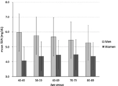
Figure 2 shows the mean SUA level by 10- year age groups in both sexes. The mean SUA level in women increased with age ( p for trend < 0.001), whereas the mean SUA level in men decreased with age (p for trend < 0.001).
The baseline characteristics of participants according to sex-specific quartiles of SUA level are presented in Table 1. Compared with quartile 1, the higher quartiles in men were more likely to have hypertension, obesity, hypercholesterolemia, alcohol intake, a younger age, less-frequent DM and a lower eGFR. In contrast, the higher quartiles in women were likely to be older and to have DM more frequently.
During a mean follow-up period of 8.8 years, we confirmed 611 CVEs. There were 304 CVEs in men (quartile 1 = 91, quartile 2 = 64, quartile 3 = 76, quartile 4 = 73) and
Fig. 2. The mean SUA level by 10-year age groups in women and men.
The level increased with age in women (p for trend < 0.001), whereas the level in men decreased with age (p for trend < 0.001).
SUA, serum uric acid.
Yuki Matsuura, et al.
150
307 in women (quartile 1 = 52, quartile 2 = 72, quartile 3 = 80, quartile 4 = 103). The CVEs were 248 strokes and 59 cases of MI/
sudden cardiac death in men and 280 strokes and 30 cases of MI/sudden cardiac death in women. The incidence rates of CVEs per 1,000 person-years in men were quartile 1 = 8.1, quartile 2 = 5.8, quartile 3 = 7.6, and quartile 4 = 6.5, with no significant trends (p = 0.409).
In contrast, in women, the incidence rates of CVEs per 1000 person-years were quartile 1 = 2.2, quartile 2 = 3.2, quartile 3 = 4.0 and quartile 4 = 4.5, with a significant positive relationship between the elevation of SUA levels and the incidence rate of CVEs (p <
0.001).
The sex-specific Cox proportional hazards model was used to analyze the hazard ratio of the SUA quartile for the onset of CVEs adjusted by several established cardiovascular risk factors (age, BMI, hypertension, DM, hypercholesterolemia, atrial fibrillation, and
current smoking) (Fig. 3). There was no significant association between them in men [quartile 1 = reference, quartile 2 = 0.80 (95%
confidence interval {CI}: 0.58-1.10), quartile 3 = 1.01 (95% CI: 0.75-1.38), quartile 4 = 0.99 (95%
CI: 0.72-1.36); p = 0.447]. However, in women, as the SUA level increased, the hazard ratio of incident CVEs increased [quartile 1 = reference, quartile 2 = 1.28 (95% CI: 0.89-1.83), quartile 3 = 1.58 (95% CI: 1.11-2.24), quartile 4 = 1.58 (95% CI: 1.12-2.22); p = 0.035]. The interaction between the SUA quartiles and the onset of CVEs was observed by gender (p for interaction = 0.047).
IV
. DiscussionThe present study showed that, in the Japanese general population without CKD, mildly elevated SUA level is an independent risk factor for the onset of CVEs in women but not in men.
Previous studies have shown that an
Fig. 3. The hazard ratio of the SUA quartile for the onset of CVEs adjusted by cardiovascular factors (age, BMI, hypertension, DM, hypercholesterolemia, atrial fibrillation, and current smoking).
There was no significant association between them in men (p = 0.447). However, in women, as the SUA level increased, the hazard ratio increased (p = 0.035). The interaction between the SUA quartile and the onset of CVEs was observed by gender (p for interaction = 0.047).
CI, confidence interval; q, quartile
increased SUA level is a risk factor for CVEs
1-6). Several mechanisms have been reported by which elevated SUA levels induce CVEs. First, an elevated SUA level has been reported to impair endothelial function by inducing intracellular oxidative stress and inflammation through the activation of the local renin-angiotensin system and the pro-oxidant effect of uric acid
15). Second, an elevated SUA level might accelerate renal vascular inflammation
16), which might activate the renin-angiotensin system and induce hypertension. Third, an elevated SUA level induces the proliferation of vascular smooth muscle cells
17, 18)and the development of glomerular hypertension and
CKD
19). Fourth, an elevated SUA level has
been reported to induce insulin resistance, as uric acid might exert cytotoxic effects against pancreatic beta cells
20), thereby increasing the incidence of DM. Fifth, an elevated SUA level has been suggested to be an important pathophysiological factor inducing incident atrial fibrillation through its inflammatory and oxidative natures with the activation of pro- inflammatory cytokines in atrial myocytes
21). Any of these potential pathophysiological natures of elevated SUA levels might be associated with CVEs.
However, the present results suggest that an elevated SUA level is an independent risk factor for the onset of CVEs in women without CKD. This association was obscure in men. Although the detailed mechanisms underlying the gender differences in the relationship between SUA levels and the onset of CVEs were unclear based on the present findings, several cohort studies have described a close relationship between SUA levels and
atherosclerotic biomarkers in women
22-29). In two unconnected cross-sectional studies of a healthy general population, Choi et al.
22)and Fang et al.
23)reported that an elevated SUA level above the highest quartile was significantly associated with a risk of elevated arterial stiffness parameters, such as the brachial-ankle pulse wave velocity in women. Similarly, Park et al. showed that the odds ratio of hyperuricemia in the high pulse pressure group was significantly greater than in the normal pulse pressure group in apparently healthy women
24). In addition, numerous cross-sectional
25-27)and longitudinal
20, 28-30)cohort studies have shown that an elevated SUA level is closely associated with the components of metabolic syndrome and the future occurrence of metabolic syndrome, especially in women.
Furthermore, a large cohort study in a healthy Korean population recently showed that, during 5.4 years of follow-up, the highest SUA quartile group showed a 4.3-fold higher hazard ratio of new onset of atrial fibrillation in women than in men
31).
These previous epidemiological studies might accord with the sex-linked differences in the influence of elevated SUA levels for cardiovascular risk in the present population.
The exact biological mechanisms underlying
the present female-dominant relationship
between SUA levels and cardiovascular risks
could not be determined from epidemiological
study. However, in the present study, although
the mean SUA levels in men gradually
decreased with age, these values gradually
and significantly increased in women from 50
years of age (in menopause) (Fig. 2). Therefore,
the association between SUA levels and CVEs
152 Yuki Matsuura, et al.
in women might be related to the decrease in estrogen levels. Mammalian kidneys are known to be affected by sex hormones, including their secretory and reabsorptive functions. Indeed, estrogen increases the renal clearance of uric acid by inhibiting urate transporter-1, which reabsorbs uric acid
32). As estrogen decreases with age, urinal excretion of uric acid might decrease in women. Given that the uric acid reserve capacity in the body is half in women compared with men (600 mg versus 1200 mg)
33), circulating levels of uric acid might be abruptly increased after menopause. This different metabolic nature of uric acid with aging between genders might be one of reasons for the sex differences in the influence of elevated SUA levels and CVEs.
Several previous studies have shown that CVE risk is associated with an elevated SUA level in both men and women
1, 3, 4). One reason the results of this study differ from those of previous studies might be our exclusion of subjects with CKD, a condition which might increase the risk of CVE as well as the SUA level. Indeed, since there are few general population studies that exclude CKD patients, it cannot be denied that CKD secondarily increases the SUA level and thus affects the onset of CVEs. The exclusion of CKD patients from this study might therefore have resulted in our observation of a more accurate association between elevated SUA levels and incident CVEs. Moreover, this study has several strengths. CVEs were captured prospectively according to previously determined standard epidemiological criteria and confirmed by research staff on medical
chart reviews. This study also had a large general population with data obtained on their SUA, eGFR, and UACR values.
However, there are several limitations associated with this study that need to be considered. First, the eGFR and UACR were measured only once (at the baseline in this study). The prevalence of CKD may therefore have been overestimated, meaning that the results of this study were underestimated.
Second, although we excluded subjects taking antihyperuricemic drugs at the start of the study, we could not completely exclude subjects who started taking antihyperuricemic drugs during the follow-up period. Third, some substances involved in uric acid metabolism other than uric acid, especially xanthine oxidase, have an oxidizing action. The present study did not measure these substances.
Finally, although the present study has shown that mildly elevated SUA is a cardiovascular r i s k f a c t o r i n w o m e n , w h e t h e r a n y interventions to decrease SUA are effective to reduce the incidence of CVEs is unclear in this study.
In conclusion, in the Japanese general population without CKD, moderately elevated SUA level is considered to be an independent risk factor for the onset of CVEs in women but not in men.
Conflict of Interest: The authors have no conflict of interest to declare.
1) Kamei K, Konta T, Hirayama A, et al.:
Associations between serum uric acid levels and the incidence of nonfatal stroke: a nationwide community-based cohort study. Clin Exp Nephrol 21, 497-503, 2017.
2) Storhaug HM, Norvik JV, Toft I, et al.: Uric acid is a risk factor for ischemic stroke and all- cause mortality in the general population: a gender specific analysis from The Tromsø Study.
BMC Cardiovasc Disord 13, 115, 2013.
3) Holme I, Aastveit AH, Hammar N, et al.: Uric acid and risk of myocardial infarction, stroke and congestive heart failure in 417,734 men and women in the Apolipoprotein MOrtality RISk study (AMORIS). J Intern Med 266, 558-570, 2009.
4) Bos MJ, Koudstaal PJ, Hofman A, et al.: Uric acid is a risk factor for myocardial infarction and stroke: the Rotterdam study. Stroke 37, 1503- 1507, 2006.
5) Braga F, Pasqualetti S, Ferraro S, et al.:
Hyperuricemia as risk factor for coronary heart disease incidence and mortality in the general population: a systematic review and meta- analysis. Clin Chem Lab Med 54, 7-15, 2016.
6) Tuttle KR, Short RA and Johnson RJ: Sex differences in uric acid and risk factors for coronary artery disease. Am J Cardiol 87, 1411- 1414, 2001.
7) Culleton BF, Larson MG, Kannel WB, et al.:
Serum uric acid and risk for cardiovascular disease and death: the Framingham Heart Study.
Ann Intern Med 131, 7-13, 1999.
8) Go AS, Chertow GM, Fan D, et al.: Chronic k i d n e y d i s e a s e a n d t h e r i s k s o f d e a t h , cardiovascular events, and hospitalization. N Engl J Med 351, 1296-1305, 2004.
9) Committee of the Japan Diabetes Society on the Diagnostic Criteria of Diabetes Mellitus: Report of the committee on the classification and diagnostic criteria of diabetes mellitus. J Diabetes Investig 1, 212-228, 2010.
10) Horio M, Imai E, Yasuda Y, et al.: Modification of the CKD epidemiology collaboration (CKD- EPI) equation for Japanese: accuracy and use for population estimates. Am J Kidney Dis 56, 32-38, 2010.
11) Essex MN, Hopps M, Bienen EJ, et al.:
Evaluation of the relationship between serum uric acid levels and cardiovascular events in
patients with gout: a retrospective analysis using electronic medical record data. J Clin Rheumatol 23, 160-166, 2017.
12) Study Project of Monitoring System for Cardiovascular Disease Commissioned by the Ministry of Health and Welfare: Manual for the registry and follow-up of stroke. National Cardiovascular Center, Osaka, Japan, 1998.
13) World Health Organization MONICA Project:
Event registration data component, MONICA manual version 1.1. Document for meeting of MONICA principal investigators. World Health Organization, Geneva, 1986.
14) Tunstall-Pedoe H, Kuulasmaa K, Amouyel P, et al.: Myocardial infarction and coronary deaths in the World Health Organization MONICA Project:
Registration procedures, event rates, and case- fatality rates in 38 populations from 21 countries in four continents. Circulation 90, 583–612, 1994.
15) Maruhashi T, Nakashima A, Soga J, et al.:
Hyperuricemia is independently associated with endothelial dysfunction in postmenopausal women but not in premenopausal women. BMJ Open 3, e003659, 2013.
16) Sundström J, Sullivan L, D'Agostino RB, et al.:
Relations of serum uric acid to longitudinal blood pressure tracking and hypertension incidence.
Hypertension 45, 28-33, 2005.
17) Cao X, Wu L and Chen Z: The association between elevated serum uric acid level and an increased risk of renal function decline in a health checkup cohort in China. Int Urol Nephrol 50, 517-525, 2018.
18) Tsai CW, Lin SY, Kuo CC, et al.: Serum uric acid and progression of kidney disease: a longitudinal analysis and mini-review. PLoS One 12, e0170393, 2017.
19) Kamei K, Konta T, Hirayama A, et al.: A slight increase within the normal range of serum uric acid and the decline in renal function: associations in a community-based population. Nephrol Dial Transplant 29, 2286-2292, 2014.
20) Lin KC, Tsai ST, Lin HY, et al.: Different progressions of hyperglycemia and diabetes among hyperuricemic men and women in the kinmen study. J Rheumatol 31, 1159-1165, 2004.
21) Zhang CH, Huang DS, Shen D, et al.: Association between serum uric acid levels and atrial fibrillation risk. Cell Physiol Biochem 38, 1589-1595, 2016.
References
154 Yuki Matsuura, et al.
22) C h o i H Y , K i m S H , C h o i A R , e t a l .: Hyperuricemia and risk of increased arterial stiffness in healthy women based on health screening in Korean population. PLoS One 12, e0180406, 2017.
23) Fang JI, Wu JS, Yang YC, et al.: High uric acid level associated with increased arterial stiffness in apparently healthy women. Atherosclerosis 236, 389-393, 2014.
24) Park CE, Sung HH, Jung EY, et al.: Gender difference in the relationship between uric acid and pulse pressure among Korean adults. Clin Exp Hypertens 41, 499-504, 2019.
25) Chiou WK, Wang MH, Huang DH, et al.: The relationship between serum uric acid level and metabolic syndrome: differences by sex and age in Taiwanese. J Epidemiol 20, 219-224, 2010.
26) Zhang Q, Lou S, Meng Z, et al.: Gender and age impacts on the correlations between hyperuricemia and metabolic syndrome in Chinese. Clin Rheumatol 30, 777-787, 2011.
27) Yang T, Chu CH, Bai CH, et al.: Uric acid level as a risk marker for metabolic syndrome: a Chinese cohort study. Atherosclerosis 220, 525- 531, 2012.
28) Liu M, He Y, Jiang B, et al.: Association between serum uric acid level and metabolic syndrome and its sex difference in a Chinese community elderly population. Int J Endocrinol 2014, doi: 10.1155/2014/754678, 2014.
29) Yadav D, Lee ES, Kim HM, et al.: Prospective study of serum uric acid levels and incident metabolic syndrome in a Korean rural cohort.
Atherosclerosis 241, 271-277, 2015.
30) Cicero AFG, Fogacci F, Giovannini M, et al.:
Serum uric acid predicts incident metabolic syndrome in the elderly in an analysis of the Brisighella Heart Study. Sci Rep 8, 11529, 2018.
31) Kwon CH, Lee SH, Lee JY, et al.: Uric Acid and Risk of Atrial Fibrillation in the Korean General Population. Circ J 82, 2728-2735, 2018.
32) Ljubojevic M, Herak-Kramberger CM, Hagos Y, et al.: Rat renal cortical OAT1 and OAT3 exhibit gender differences determined by both androgen stimulation and estrogen inhibition.
Am J Physiol Renal Physiol 287, F124-138, 2004.
33) Burtis CA, Ashwood ER and Bruns DE: Tietz textbook of clinical chemistry and molecular disgnostics 5th ed, pp.686-691, Saunders, Philadelphia, 2012.
155
腎機能正常な一般住民における血清尿酸値と 心血管疾患発症の関連,性差について
松浦佑樹
1),田中文隆
1),瀬川利恵
2),
丹野高三
3),坂田清美
3),大澤正樹
4),大間々真一
5), 小笠原邦昭
6),旭 浩一
1),中村元行
7)1)岩手医科大学医学部,内科学講座腎・高血圧内科分野
2)岩手医科大学医学部,内科学講座糖尿病・代謝・内分泌内科分野
3)岩手医科大学医学部,衛生学公衆衛生学講座
4)特定医療法人盛岡つなぎ温泉病院
5)岩手医科大学医学部,救急・災害・総合医学講座救急医学分野
6)岩手医科大学医学部,脳神経外科学講座
7)岩手医科大学医学部,名誉教授
(Received on January 20, 2020 & Accepted on February 13, 2020)
血清尿酸値が心血管疾患発症の独立した危険因子で あるかについては議論が分かれている.また血清尿酸 値は慢性腎臓病があると二次的に上昇するため,血清 尿酸値自体の評価を行うためには慢性腎臓病を除外す る必要がある.そこで本研究は慢性腎臓病を除外した 一般住民 15,036 人(男性 5038 人,女性 9998 人)にお いて,血清尿酸値 4 分位と心血管疾患発症の関連,性 差について解析した.平均追跡期間は 8.8 年で,男性 で 304 件(脳卒中 248 件,心筋梗塞 / 突然死 59 件),
女性で 307 件(脳卒中 280 件,心筋梗塞 / 突然死 30 件)
の心血管疾患が認められた.確立された心血管リスク で調整してハザード比を解析したところ,男性では血 清尿酸値と心血管疾患発症との間に明らかな関連はな かったが,女性ではベースラインの血清尿酸値が高い ほど心血管疾患発症のハザード比が高かった.血清尿 酸値は女性でのみ心血管疾患発症の独立した危険因子 と考えられた.
要旨