ヒト剖検心における Bachmann 束の検討
1)昭和大学歯学部全身管理歯科学講座総合内科部門
2)昭和大学医学部内科学講座(循環器内科学部門)
3)昭和大学医学部臨床病理診断学講座
4)国立循環器病研究センター中央診療部門病理部
小川 玄洋1) 井 上 紳*1) 酒井 哲郎2)
小林 洋一2) 瀧本 雅文3) 松山 高明3,4)
抄録:Bachmann 束は冠静脈洞とともに洞調律時の心房間興奮伝搬に関わる主要な筋束である が,肉眼的な定義・分布は必ずしも明確でない.ヒト剖検心において左房天蓋部における Bachmann 束の分布を検討した.頻脈性不整脈を認めない非循環器疾患の剖検心 15 例,平均 年齢 61.4 歳(51 〜 79 歳,女性 6 例)について左房側の付着部位である天蓋部中央における Bachmann 束の筋束厚,幅を計測し周辺の組織性状を観察した.Bachmann 束の平均厚は 3.67 mm,平均幅 21.4 mm で,心重量と筋束の厚みは相関係数
−
0.56 で逆相関した.Bachmann 束は左右心耳を結ぶ主幹部と,左右心耳の静脈側および房室弁輪側に伸展する周辺部に分けら れた.主幹部は房室弁輪に平行に走行する筋束であるが,4 例が上下に二分していた.また,上大静脈筋袖が Bachmann 束表層に伸展している例もみられた.Bachmann 束は心重量と相 関して菲薄化するが,各種不整脈との関連ではより詳細な検討が必要であると考えられた.
キーワード:心房間興奮伝搬,Bachmann 束,心房リモデリング
ヒト心房間興奮伝搬について,近年の左房内電位 の解析から,洞調律時の心房間の興奮伝搬には Bachmann 束と冠静脈洞開口部の 2 つの主要な経路 があること1),主要な心房中隔組織である卵円孔周 囲は心房間の伝導にあまり関与していないこと2,3), 心房細動例では Bachmann 束の伝導に遅延がみら れること4,5),等が明らかになった.
左右心房間興奮伝搬について左房側の詳細なデー タが得られるようになったのは,Haïssaguerre ら により肺静脈開口部周辺の異所性興奮が心房細動の 誘因となることが報告されて以来,経心房中隔的カ テーテル操作による心房電位の計測が一般的になっ たためである6).これ以降,肺静脈隔離術の普及に 伴い 3D マッピングや CT integration など左房内電 位と解剖学的な位置関係が詳細に検討されるように なった7).しかし,Bachmann の報告が主に生理学 的研究であり詳細な心筋構築に関する記載がないこ とから8),左右心房筋の解剖学的連結については報 告者によってばらつきがある9‑11).
Bachmann 束の分布および性状を再検討するた め,主たる不整脈を認めなかったヒト剖検心につい て肉眼的および組織学的にその形態を検索した.
研 究 方 法
対象は剖検心 15 例.男性 9 人,女性 6 人.年齢 は 51 歳から 79 歳,平均 61.4 歳であった.表 1 に 死因を示す.全例頻脈性不整脈の既往を認めず,死 因は循環器疾患ではなかった.Bachmann 束は大動 脈基部背面で左右心房間を連結する筋束と定義し肉 眼的に検討した.ホルマリン固定後に両肺静脈間中 央の左心房天蓋部を心長軸方向に切開し組織切片を 作成した.同部位において左房壁厚,Bachmann 束 の厚みと幅を測定し,それぞれ年齢,心重量につい てピアソン相関係数を用いて検討した.計測後に Bachmann 束周辺の組織を観察した.
結 果
図 1a は心外膜および脂肪織を剥離したものだが,
原 著
*責任著者
Bachmann 束主幹部は弁輪に平行な等方性の心房筋 走行を示し左右の心耳を結んでいる.左右心房付着 部をより詳細に検討すると,左右心耳開口部の静脈 側と房室弁輪側にそれぞれ分かれて付着している.
図 1b は右房の展開図であるが,Bachmann 束(BB)
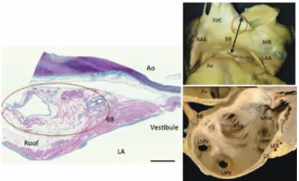
は分界稜心房中隔付着部深部で分界稜から分岐する 形で左房側へ伸展する.その走行と各部の命名につ いては Papes が詳細に描画しており,図 1 に示す12). 左房天蓋部の組織標本では短軸方向の筋束として 観察される.左房天蓋部中央においても 4 例で静脈 側と房室弁輪側に二分した例を認めた(図 2).し かし,主幹部が一本である 11 例も左心耳開口部で は静脈側と房室弁輪側に分岐する.それぞれ left posterior crest と left anterior crest と呼ばれ,同 様に分岐する右房側の right posterior crest,right anterior crest と対応する(図 1,3).
特に left posterior crest は左上肺静脈基部を走行 し,Marshall 筋束に接する(図 4).一方の left an-
terior crest は僧帽弁前庭部筋束に付着する(図 5).
Bachmann 束の周辺は心外膜脂肪織で覆われてい るが,1 例に上大静脈筋袖が伸展・付着したものが 認められた(図 6).
天蓋部Bachmann束を含む左房壁厚5.48
±
1.10 mm(平均
±
標準偏差),Bachmann 束厚 3.67±
0.84 mm,Bachmann 束幅 21.4
±
2.82 mm,一般左心房筋平均 厚 1.81±
0.60 mm であった(表 2).年齢は各心筋 厚,Bachmann 束幅と相関を認めなかったが,心重 量は左心房天蓋部の Bachmann 束最厚部の全心房 筋厚(相関係数−
0.55(P 値= 0.04)),Bachmann 束厚(相関係数−
0.56(P 値= 0.03))と逆相関し た(図 7).考 察
Bachmann 束は正確には interatrial bundle と呼 ぶべきであるが,その分布や頻度に関しては文献に より差がある.Papez は interatrial bundle の右房
表 1
死因 症例数
悪性疾患 10
炎症性疾患 2
脳卒中 1
その他 2
合計 15
図 1 Bachmann 束の走行と周囲の筋束(右図は文献 12 より引用)
図 2 左房天蓋部における Bachmann 束の組織形態
図 5 Left anterior crest と僧帽弁前庭部(vestibule)
図 3 左心耳と Bachmann 束の伸展(left anterior and posterior crest)
図 4 Left posterior crest と Marshall 筋束
図 6 Bachmann 束に伸展した上大静脈筋袖
側起始として,分界稜に相当する right posterior crest(terminal crest)と,右心耳三尖弁輪間(三 尖弁輪前庭部)の right anterior crest の 2 つを挙 げている12).両者は合流して左房天蓋部に付着した 後に左心耳開口部で再び 2 つの筋束に別れる.特に left posterior crest 部は左房側方峡部の心外膜側に 付着するが,同部位には Marshall 筋束(図 4,MB)
が付着し,肺静脈隔離において焼灼線が通るため重 要である13).Bachmann 束主幹部が天蓋部中央で二 分した症例を 4 例(26.7%)認めた(図 2).主幹部 は左右心耳で前後の crest として静脈側と房室弁輪 側に分かれるが,中央で主幹部が二分された例は,
この分岐のバリエーションと考えられた.
左右の房室弁輪近傍を結ぶ筋束はその厚みや幅,
分布が主幹部に比べて極めて薄く,分布も多様であ るが,ヒト剖検心を用いた報告で一部の症例には Bachmann 束を認めないとするものがある11).これ らは組織検索方法として大動脈基部で房室弁輪に平 行に走行する筋束のみを検討したためと思われる.
左右の anterior crest は心耳下部で大動脈弁基部の 湾曲に沿い房室弁輪前庭部(vestibule)に付着す るため,弁輪と平行に標本を作製した際に検索しや すい.しかし,洞調律の際の興奮伝搬の主たる伝搬 路と考えられる分界稜 /right posterior crest の走 行は図 1 に示すように,右房心内膜下に移り,筋走 行も房室弁輪に対して斜交するため標本上で追うの は困難となる.そのために見落とされた可能性が示 唆される.
Bachmann 束はその発生学的由来が必ずしも明ら か で な い.Lamers ら は Bachmann 束 は atrioven- tricular canal myocardium 由来を示唆し,van den Hoffらは心房中隔への sinoatrial ring tissue の伸展で あると述べている14,15).彼らによれば発生学的な本 来の心房筋は左右心耳の櫛状筋部分であり,平坦な 心筋構造をもつ Bachmann 束はより幼弱な洞房輪や 房室接合部心筋に近い性格を残すとする.Bachmann 束が分布する大動脈弁基部は胎生期には心内膜床組 織が隣接し,生下後はその名残として中心線維体や
表 2
平均±標準偏差 最少 最大
年齢(y.o.) 61.4±7.8 51 79 心重量(g) 349.6±42.4 260 420 Bachmann 束厚(mm) 3.67±2.82 2.6 5.7 Bachmann 束幅(mm) 21.4±2.82 18 28 左房天蓋部(roof)全体厚(mm) 5.28±1.10 3.7 8.5 左房天蓋部一般左房心筋厚(mm) 1.81±0.60 1.1 2.8
図 7 Bachmann 束の厚と年齢,心重量
fat pad など間質組織が入り込んでいる.Bachmann 束の後方に存在する二次中隔は,心外膜側の脂肪組 織が心房間溝として発育したものであり,その過程 で残存したものが Bachmann 束と考えられる.刺 激伝導系との関連では,Bachmann 束は洞結節が存 在する分解稜や房室結節が隣接する房室弁輪前庭部 に付着し,刺激伝導系組織との密接な関連が推察さ れる.
James らは電子顕微鏡による心筋細胞分類から Bachmann 束内に他部位の結節間伝導路と同様に結 節様細胞や移行細胞,Purkinje 様細胞を認めると した16).電子顕微鏡を用いた細胞形態の検討では,
Yamaguchi らも Bachmann 束の心筋細胞形態に一 般心房筋とは異なった伝導系細胞に近い所見を報告 している17).光学顕微鏡を用いた今回の検討では,
図 6 のように Bachmann 束が左房天蓋で上大静脈 筋袖の伸展部と接合しているものや,left posterior crest では Marshall 筋束と接しているものもみられ
(図 4),催不整脈性との関連が推察された.
全症例の死因が心疾患ではないため心臓超音波検 査などが施行されておらず,左心房拡大の有無は不 明であるが,平均心重量 349 g とやや大きく,心肥 大症例を含むと考えられた.左心房壁厚は年齢と共 に肥厚するとの報告があるが18),本研究の対象が平 均年齢 64 歳と高く,また前述のように心肥大症例を 少なからず含む影響が示唆された.一方,Bachmann 束厚と心重量は相関係数
−
0.56(P = 0.03)で有意 に逆相関し,心重量の増加と共に Bachmann 束は 菲薄化していた(図 7).左室肥大が生じると左室 拡張機能の低下から左房負荷が生じ,左心房拡大を 招くことで心房細動などの催不整脈性基質が生じる ことが報告されている19).Bachmann 束の構成細胞 が伝導系細胞の性格を持つとすると左房拡大の影響 も受け易いことが推察される.種々の心房不整脈の 症例における Bachmann 束についてはより詳細な 組織学的な検索が必要であると推察された.利益相反
本研究に関し開示すべき利益相反はない.
文 献
1) Markides V, Schilling RJ, Ho SY, . Charac- terization of left atrial activation in the intact human heart. . 2003;107:733‑739.
2) Lemery R, Soucie L, Martin B, . Human study of biatrial electrical coupling: determinants of endocardial septal activation and conduction over interatrial connections. 2004;
110:2083‑2089.
3) Sakamoto S, Nitta T, Ishii Y, . Interatrial electrical connections: the precise location and preferential conduction.
2005;16:1077‑1086.
4) Roithinger FX, Abou-Harb M, Pachinger O, . The effect of the atrial pacing site on the total atrial activation time.
2001;24:316‑322.
5) Jurkko R, Mantynen V, Lehto M, . Inter- atrial conduction in patients with paroxysmal atrial fibrillation and in healthy subjects.
2010;145:455‑460.
6) Haissaguerre M, Jais P, Shah DC, . Sponta- neous initiation of atrial fibrillation by ectopic beats originating in the pulmonary veins.
. 1998;339:659‑666.
7) Faletra FF, Nucifora G, Regoli F, . Anato- my of pulmonary veins by real-time 3D TEE:
implications for catheter-based pulmonary vein
ablation. . 2012;5:456‑
462.
8) Bachmann G. The inter-auricular time interval.
. 1916;41:309‑320.
9) Ho SY, Anderson RH, Sanchez-Quintana D.
Atrial structure and fibres: morphologic bases of atrial conduction. 2002;54:
325‑336.
10) Lemery R, Guiraudon G, Veinot JP. Anatomic description of Bachmann s bundle and its rela- tion to the atrial septum. . 2003;
91:1482‑1485.
11) Platonov PG, Mitrofanova L, Ivanov V, . Substrates for intra-atrial and interatrial con- duction in the atrial septum: anatomical study on 84 human hearts. . 2008;5:
1189‑1195.
12) Papez JW. Heart musculature of the atria.
. 1920;27:255‑277, 4pl.
13) Makino M, Inoue S, Matsuyama TA, . Di- verse myocardial extension and autonomic in- nervation on ligament of Marshall in humans.
2006;17:594‑599.
14) Lamers WH, Moorman AF. Cardiac septation:
a late contribution of the embryonic primary myocardium to heart morphogenesis.
2002;91:93‑103.
15) van den Hoff MJ, Kruithof BP, Moorman AF.
Making more heart muscle. 2004;26:
248‑261.
16) James TN. The connecting pathways between the sinus node and A-V node and between the right and the left atrium in the human heart.
. 1963;66:498‑508.
17) Yamaguchi T, Yi SQ, Tanaka S, . Ultra- structure and cytoarchitecture of Bachmann s bundle in the mammalian heart. . 2009;25:24‑31.
18) Pan NH, Tsao HM, Chang NC, . Aging di- lates atrium and pulmonary veins: implications for the genesis of atrial fibrillation.
2008;133:190‑196.
19) Hennersdorf MG, Schueller PO, Steiner S, . Prevalence of paroxysmal atrial fibrillation de- pending on the regression of left ventricular hy- pertrophy in arterial hypertension.
2007;30:535‑540.
HISTOLOGICAL EXAMINATION OF BACHMANN BUNDLE IN HEARTS FROM HUMAN AUTOPSIES
Genyo OGAWA1), Shin INOUE1), Tetsuo SAKAI2),
Yoichi KOBAYASHI2), Masafumi TAKIMOTO3) and Taka-aki MATSUYAMA3,4)
1)Division of General Medicine, Department of Perioperative Medicine, Showa University Dental Hospital
2)Division of Cardiology, Department of Medicine, Showa University School of Medicine
3)Department of Pathology, Showa University School of Medicine
4)Department of Pathology, National Cerebral and Cardiovascular Center
Abstract Bachmann s bundle is an important band of muscle that is, along with the coronary si- nus musculature, involved in interatrial conduction during sinus rhythm. Despite its electrophysiological significance in maintaining normal heart beats, however, the macroscopic features and distribution have not been clearly delineated. Via autopsy, this study examined the distribution of Bachmann s bundle in the roof of the left atrium. Dissected hearts were from 15 individuals without conspicuous cardiovascular disease or tachy-arrhythmia, who had a mean age of 61.4 years (51 79 years, 6 females). The thickness and width of Bachmann s bundle were measured in the center of the roof of the left atrium, which is where the bundle is attached. The histology of the surrounding tissue was also examined. The mean thickness of Bachmann s bundle was 3.67 mm and its mean width was 21.4 mm. The weight of the heart and thickness of the bundle were inversely correlated with a correlation coefficient of
−
0.56. Bach- mann s bundle was divided into the main trunk, that connects the atrial appendages, and extensions, which project into the area around the caval and left pulmonary veins or both atrioventricular annuli.The main trunk runs parallel to the atrioventricular annuli, but in 4 hearts it ran up and down indepen- dently. The myocardial sleeve of the superior vena cava was also found to extend into the surface layer of Bachmann s bundle in one heart. The thickness of Bachmann s bundle was inversely correlated with the weight of the heart. More detailed studies are needed to examine the bundle s association with vari- ous arrhythmias.
Key words: interatrial conduction, Bachmann s bundle, atrial remodeling
〔受付:4 月 24 日,受理:5 月 13 日,2015〕