九州大学学術情報リポジトリ
Kyushu University Institutional Repository
ペルオキシソーム形成因子(assembly factor-1)の同 定
塚本, 利朗
https://doi.org/10.11501/3070108
出版情報:Kyushu University, 1993, 博士(理学), 論文博士
Identification of peroxisome assembly factor-1
Toshiro TSUKAMOTO
CONTENTS
page Introduction . . . . . . . . 3
Chapter I
Isolation and characterization of Chinese hamster ovary
cell mutants defective in assembly of �eroxisomes ... . .... . . 5
Chapter II
eDNA cloning and characterization of peroxisome assembly
factor-1 . . . . . . . . . . . . . . . . . ... . . . . . ... . ..... . . . ..... ...... 50
Conclusion . . . . . . . . . . . . . . . . . . . . . . . . . . . . . . . . . . . . . . . . . . . . . . . . . . . . 83
Acknowledgment . . . . . . . . ... . . . . . ... . . . . ... . ... . . . . ... ........... 85
INTRODUCTION
Intracellular organelles present in eukaryotic cells have different sets of enzymes with specific functions. Peroxisome is a ubiquitous organelle found in a wide variety of eukaryotes, from yeast through human. It is bounded by a single membrane and carries many functions including respiration (based on H2o2- forming oxidases and catalase),
in ether
fatty acid /3 -oxidation, (plasmalogen) the initial reactions glycerolipid
biosynthesis, the glyoxylate cycle, transamination of amino acids, purine and polyamine catabolism, metabolism of cholesterol and phytanic acid, and synthesis of bile acids. To maintain intracellular structures and functions, newly synthesized proteins need to be sorted to their correct compartments. All peroxisomal proteins so far studied are synthesized on free polyribosomes.
imported into enzymes use an
Most of them are made at their final sizes and peroxisomes posttranslationally. Most matrix S KL tripeptide at the C-terminus as c i s -acting topogenic signal and the import of at least one enzyme, acyl-CoA oxidase, is ATP-dependent. Another targeting signal has also been identified in the N-terminal presequence of 3-ketoacyl-CoA thiolase. However, little is known of molecular mechanisms of the protein translocation system and the import receptors present in peroxisomal membrane. A dysfunction of any of these factors would cause defective biogenesis of peroxisomes. Indeed, several human autosomal recessive, peroxisome-deficient genetic disorders such as Zellweger syndrome and neonatal adrenoleukodystrophy have been reported. It is thought that defect of peroxisome assembly
or protein import into peroxisomes is the primary lesion in these diseases. These patients show characteristic dysmorphism, severe hypotonia, psychomotor retardation, and peroxisomal dysfunctions and rarely survive early childfood. Hepatocytes and fibroblasts of the patients lack peroxisomes; catalase is mislocalized in the cytosol, and many peroxisomal enzymes are absent or greatly decreased due to rapid degradation. Cell-fusion experiments using fibroblasts obtained from peroxisome-deficient patients revealed that at least eight complementation groups were present. This suggests that many factors are required for peroxisome biogenesis. To reveal molecular mechanism of peroxisome biogenesis and primary defects of peroxisome-deficient disorders, two peroxisome-deficient mutants were isolated from Chinese hamster ovary (CHO)
morphologically and biochemically.
restore peroxisome of the mutant mammalian expression vector, pcD2.
cells and characterized A eDNA clone that could using cell was isolated
Chapter I
Isolation and characterization of Chinese hamster ovary cell mutants defective in assembly of peroxisomes
Abbreviations
AOx, acyl-coenzyme A oxidase; CHO, Chinese hamster ovary;
DAB, 3,3'-diaminobenzidine tetrahydrochloride;
DHAP-ATase, dihydroxyacetonephosphate acyltransferase;
EMS, ethyl methanesu fonate; FCS, fetal calf serum;
HAT, hypoxanthine/aminopterin/thymidine; 70 IMP, 70-kD integral membrane protein; 22 IMP, 22-kD integral membrane protein;
MEM, minimum essential medium; NEM, N-ethylmaleimide;
Oua, ouabain; TG, 6-thioguanine.
SUMMARY
I made use of autoradiographic screening to isolate two Chinese hamster ovary (CHO) cell mutants deficient in peroxisomal dihydroxyacetonephosphate acyltransferase, a key enzyme for the biosynthesis of ether glycerolipids such as plasmalogens.
Morphological analys·s revealed no evidence of peroxisome in these mutants. Catalase was as active as in the normal cells but was not sedimentable. Pulse-chase radioLabeling experiments and cell-free translation of R A demonstrated that acyl-CoA oxidase, the first enzyme of peroxisomal e-oxidation system, was synthesized as the 75-kD form but was not converted to 53- and 22-kD mature components that were present in the wild-type CHO cells; rather, degradation was apparent. Peroxisomal thiolase was synthesized as in normal cells but remained as a larger, 44- kD precursor, whereas maturation to the 41-kD enzyme was detected in the wild-type cells. The peroxisomal 70-kD integral membrane protein was also equally synthesized, as in the wild-type cells, and was not degraded. These resu ts suggest that assembly of the peroxisomes is defective in the mutants, whereas the synthesis of peroxisomal proteins appears to be normal. Cell-fusion studies revealed that the two mutants are recessive to the wild-type CHO cells and belong to different complementation groups. Thus, these mutants presumably contain different lesions in gene ( s) encoding factor(s) required for peroxisome assembly.
INTRODUCTION
The peroxisome is a ubiquitous intracellular organelle present in almost all, if not all, eukaryotes. It is classically defined as a subcellular organelle containing catalase and at least one H2o2-producing oxidase ( 1). Peroxisome functions in the catabolism of a wide variety of substrates such as fatty acid, D-amino acid, L-a-hydroxy acids, uric acid and polyamine.
Several human genetic disorders with evidence of absence of peroxisome are linked to various biochemical dysfunctions.
Hence, the peroxisome plays crucial metabolic roles, including the catabolism of very long chain fatty acids by f3 -oxidation system, biosynthesis of ether-linked glycerolipids such as plasmalogens, metabolism of cholesterol and phytanic acid, and synthesis of bile acids (2, 3). Among these disorders, cerebro
hepato-renal syndrome ( Zellweger syndrome) is a typical, severe disease (4, 5).
Many lines of biochemical and morphological evidence are consistent with the idea that peroxisomes are formed by division
after posttranslational of preexisting peroxisomes
newly synthesized proteins (6, 7) . Peroxisomal
import of proteins, including membrane polypeptides, are
polyribosomes in the cytosol, mostly
synthesized on free at the final sizes.
Posttranslational import of several proteins into peroxisomes has been reproduced in vitro (8, 9, 10, 11). Targeting signal (s) in in v · vo and in vitro import have been noted for peroxisomal enzymes such as luciferase ( 12, 13) and acyl-CoA oxidase (AOx) of rat liver ( 11) as well as C. tropicalis ( 14). The topogenic
signal identified for several enzymes resides at the extreme COOH terminus and comprises the sequence -Ser-Lys-Leu-COOH (11, 13).
Zoeller and Raetz isolated ch·nese hamster ovary (CHO) cell mutants deficient in dihydroxyac tonephosphate acyltransferase (DHAP-ATase) ( 15), a peroxisomal key enzyme in the synthesis of ether-linked glycerolipids such as plasmalogens ( 16). In these mutants, catalase, a matrix enzyme of peroxisome is fully active but not particle bound. Although the mutants have not yet been fully characterized morphologically and biochemically, they may be defective in peroxisomes, as noted in biopsi d tissues and in fibroblasts from patients with Zellweger syndrome (17). To investigate mechanisms related to the biogenesis of peroxisomes, at molecular and cellular levels, a mutant cell deficient in
peroxisomal assembly would be useful. It would also serve as a somatic cell model system for studying human peroxisome deficiency diseases such as Zellweger syndrome. I report here the isolation and characterizat·on of two cell mutants that resemble the fibroblasts of Ze lweger patients and belong to different complementation groups.
MATERIALS AND METHODS
Materia s
Catalase was purified from rat liver according to Leighton et al. (18) ; AOx and bifunctional protein were generous gifts from Dr. T. Hashimoto (Shinshu University, Matsumoto, Japan), 70-kD integral membrane protein (70 IMP) (19) was purified from peroxisomal membranes exactly as described for the isolation of 22-kD integral membrane protein (22 IMP� (20). Anti-catalase, anti-AOx and anti-70 IMP antisera were respectively raised in rabbits by conventional subcutaneous injection. Anti- bifunctional protein antibody was a gift from Dr. T. Hashimoto.
Growth medium, Ham's F12 and fetal calf serum (FCS) were purchased from Gibco. Glycerol kinase (Candida mycoderma), hypoxanthine/aminopterin/thymidine
(HAT)
supplement, and Pansorbin were from Boehringer-Mannheim. Ouabain, dihydroxyacetonephosphate, and palmitoyl-CoA were from Sigma. 6- thioguanin and dihydroxyacetone were purchased from acalai Tesque Co. Ethyl methanesulfonate (EMS) was from Aldrich.Digitonin, ATP, N-ethylmaleimide ( EM), and polyethylene glycol 6000 were from Wako Chem·cals. 3,3'-diaminobenzidine tetrahydrochloride (DAB) was from Kanto Chemicals. [35s]
methionine (>1000 Ci/mmol), [125I]protein A (>30 mCi/mg), and r
[32P]ATP (>5000 or -3000 Ci/mmol) were purchased from Amersham.
Enlightning, an autoradiography enhancer, was from Du Pont/ EN.
Cell Culture and Isolation of Mutants
Chinese hamster ovary (CHO-Kl) cells were obtained from Dr.
M. Imada, Meiji Institute of Health Science, and were grown in
Ham's F12 medium supplemented with 10% (vlv) FCS under 5% co2 I 95% air. Cells were passaged using trypsin-EDTA, mutagenized for 16 h with EMS(400 �glml), grown for 3-4 days and stored frozen.
These rnutagenized cells were plated at -250 cells per 100-rnm dish. After incubation for 24 h, the cells were overlaid with Whatman No.50 filter paper and further maintained for 10-15 days at 37° C. Mutants defective in peroxisomal DHAP-ATase were isolated by an autoradiographic screening procedure according to Zoeller and Raetz (15), except that screening was done at 37° C.
Putative mutants were subjected to another cycle of colony screening and further purified by limiting dilution, without
screening.
Enzyme Assays
DHAP-ATase assay was performed as described (15), using 32P
labeled DHAP and palmitoyl-CoA, by the method of Schlossman and Bell (21). Catalase was assayed as described (22).
Latency of Catalase
Cells were harvested by trypsinization and washed twice, sequentially, with growth medi urn and 0. 25 M sucrose I 10 rnM He pes -N a 0 H , pH 7 . 4 . The c e 11 s were suspended at 1 -1 . 5 x 1 0 6 cellslml in 0.25 M sucrose I 10 mM Hepes- aOH, pH 7.4 and incubated on ice for 5 min in the same solution but containing different concentrations of digitonin (17); the activity of catalase was measured as described above, except that the assay mixture contained 0.25 M sucrose.
Morphological Analysis
Cytochemical Analysis. Cells were fixed for 60 min at room
temperature with 4% paraformaldehyde I 1% glutaraldehyde I 0.01%
cac12 I 0.1 M cacodylate buffer, pii 7.4, and incubated for 1 h at 37· C in the dark in medium containing 5 mM DAB/ 0.1 M glycine
NaOH, pH 10.5 I 0.15% H2o2; the cells were post-fixed for 1 h at a· C with 1% osmium tetroxide followed by reduction with potassium
ferrocyanide, dehydrated in ethano and propylene oxide, and then embedded in Epon 812. Ultrathin sections were cut on an LKB ultra tome, counter stained briefly with 40 mM lead citrate, and examined under a Hitachi H-600 electron microscope.
Immunocytochemical Analysis. Cells were fixed in a culture plate for 2 h at room temperature with 4% paraformaldehyde I 0. 01%
CaC12 I 0.15 M cacodylate buffer, pH 7.4, and treated for 15 min at ambient temperature with phosphate-buffered saline containing 0. 05% Triton X-100. The cells were incubated for 2 h at room temperature with rabbit anti-rat liver catalase antiserum. After washing three times with phosphate-buffered sa ine, the cells were incubated for 30 min at ambient temperature with horseradish peroxidase-labeled goat anti-rabbit IgG antibody followed by reaction with DAB and 0.01% H2o2 in 0.1 M cacodylate buffer, pH 7.4. The cells were then examined under an Olympus BH-2 microscope.
Preparation of Cell Fractions
Cells were harvested by tryps · nization, washed twice with growth medium and homogenized in 0.25 M sucrose I 5 mM Hepes-KOH, pH 7.4 I 0.1% ethano by 10 strokes of an Elvehjem-Potter homogenizer.
described (24).
A postnuclear supernatant was prepared, as A high speed supernatant (cytosolic fraction) was prepared from the postnuclear supernatant by centrifugation
at 100,000 � for 90 min. The pellet (particulate fraction) was resuspended in the homogenizing buffer.
Radiolabeling of Cells
Pulse and Chase Experiment. Cells growing in a 35 mm dish were pulse-labeled for 1 h with [35s]methionine (0.1 mCilml) in methionine-free MEM supplemented with dialyzed FCS. The medium was removed and the cells were washed twice with Hanks' balanced salt solution and fed 2 ml F12 plus 10% FCS medium. At selected intervals, cells were washed twice with Hanks' balanced salt solution and lysed in 0.5 ml of 1% NP-40 I 0.1% SDS I 10 mM Tris- HCl pH 7.4 I 0.15 M NaCl. After centrifugation in an Eppendorf microfuge, the supernatants were subjected to immunoprecipitation with specific rabbit antisera, as described (20), except that 0.1% P-40 was used instead of 1%.
Continuous Labeling. Cells were labeled at 37· C for 48 h in F12 plus 10% FCS medium containing [35s]methionine (10 �Cilml) with a change of medi urn at 24 h. Labeled cells were lysed and proteins were immunoprecipitated, as described above.
Cell-free Translation of RNA
RNA was isolated from wild-type and mutant CHO cells by the guanidinium thiocyanate method of Ullrich et al. (24). Total RNA of normal rat liver was prepared from a post-mitochondrial fraction, as described (26). Cell-free trans ation of R A was carried out in a nuclease-treated rabbit reticulocyte lysate cell-free protein-synthesizing system with [35s]methionine as label (20, 27). Immunoprecipitation was done, as described (20).
Cell-Ce 1 Fusion
For studies on genetic complementation, varjants of mutant CHO cells (224 and 265) resistant to both ouabain (Oua) and 6- thioguanine (TG) were isolated as follows (28): the mutant cells were mutagenized with EMS as described above, then selected for resistance to 60 �M 6-thioguanine. The TG-resistant (TGr) cells were next screened for the mutant resistant to 3 mM ouabain, after treatment with EMS. Clones of variants (TGrouar) that were sensitive to HAT medium (F12 medium containing 5 mM hypoxanthine, 0. 4 11M aminopterin, and 16 11M thymidine. supplemented with 10%
FCS) were used for hybridyzation with the original sensitive cells (TGsouas). The TGrouar ce 1 line and the cells to be fused were both plated for 24 h at each densjty of 2.5-3 x 10⁵ cells in a 35-mm dish. After thoroughly removing the growth medium, the cells were treated with 1 ml of F12 medium containing 45%(w/w) polyethylene glycol and incubated for 1 min at room temperature, with gentle swirling ( 29). After washing four times with F12 medium, the cells were incubated at 37· C for 8 h in growth medium and replated in a 100-mm dish. For selection of hybrids, the cells were incubated in HAT medium containing 1 or 2 mM ouabain and maintained with several changes of medium until the control cells without polyethlene glycol-treatment were hardly viable (usually, 6-9 days). About 600-7000 colonies were formed from one 35-mm dish. The entire pop u 1 at ion of c e 11 s on a dish was harvested by trypsinization.
Other Methods
SDS-PAGE was carried out according to Laemmli ( 30) , as described ( 19). Fluorography was performed with Enlightning.
Immunoblot was done according to the modified procedure (20) of
Burnette (31), except that 1% bovine skim milk was used for blocking instead of 1% bovine hemoglobin. Protein was determined by the method of Lowry ( 32) with bovine serum albumin as the standard.
RESULTS
Isolation of Mutants Defective in Peroxisomal DIIAP-ATase
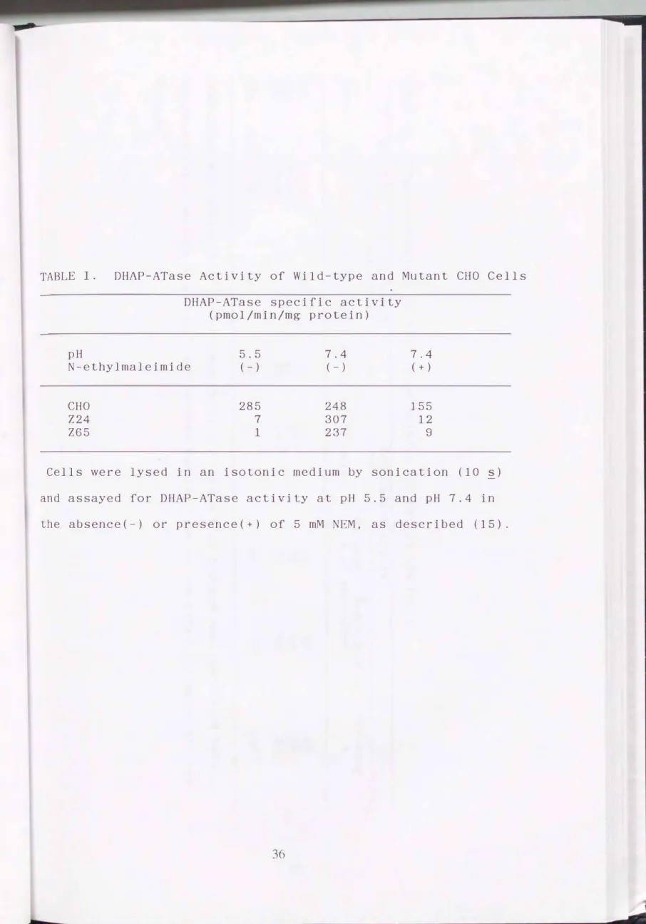
CHO-K1 cells treated with EMS were cul ti va ted on filter paper, lysed and assayed for DHAP-ATase converting [32P]DHAP with palmitoyl-CoA to acid-insoluble palmitoyl-[32P]DHAP. Assays were carried out at pH 5.5 in the presence of NEM to inh·b·t any residual activity of microsomal DIIAP-ATase (15). Mutants defective in this enzyme were identi�ied by comparing the autoradiogram with the pattern of colonies stained with Coomassie blue. Two mutants, Z24 and Z65, were isolated from -25, 000 colonies grown from five stocks of EMS-treat d CHO cells. Both mutants were severely deficient in DHAP-ATase activity, i.e. less than 2% of the activity of wild-type cells (Table I). When measured at pH 7. 4, co nsiderab l e DIIAP-ATase activity ( 95 - 125%
of wild-type activity) was detected in the mutants. With the addition of NEM, DHAP-ATase was inhibited in the wild-type cells by 40%, whereas that in the two mutants was all but abolished.
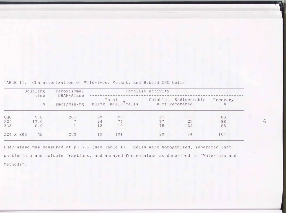
Thus, the two mutants were deficient in EM-insensitive peroxisomal DHAP-ATase, while NEM-sensitive DHAP-ATase remained active at pH 7.4. The deficiency in peroxisomal DHAP-ATase was not lethal to the mutants. Ce 1 growth, represented as doubling time, was not affected in the Z65, whereas that of Z24 was reduced to nearly half, presumably because Z24 was almost twice in size, as compared to the wild type and Z65 cells (Table II).
Morphological and Biochemical Ana ysis of Mutants
Cells were fixed and cytochemically stained with DAB specific for catalase. lectron microscopic analysis
demonstrated that the wild-type CHO cell contained numerous -0.15 11m catalase-containing particles, peroxisomes (Fig. 1 CHO, P).
No DAB-positive particle was detected in any microscopic section of Z24 and Z65 mutant cells examined ( Z24 and Z65). Other intracellular organelles in both mutants apppeared to be morphologically normal.
Catalase activity was nearly twice as high in the Z24 cells as in the wild-type; the specific activity in the homogenate of Z24 was much the same as that in the wild-type CHO cells (Table II), no doubt due to differences in cell size (see Fig. 7). In the mutant cell 265, catalase activity was about half that in the wild-type cells. Catalase was probably less in amount in the Z65, as two catalase polypeptides were observed, one of which may be inactive (see below). The subcelluler fractionation study revealed that about 80% of the catalase activity of Z24 and Z65 was not sedimentable, whereas 75% of that in the wild-type cells was particle-associated (Table II). The residual activity in the supernatant from the wild-type cells presumably reflects the catalase leaked from the broken peroxisomes during homogenization (33), while that of the particulate fraction in the mutants may represent the unbroken cells. This would suggest that catalase is present, in an active form, in the cytosol of the Z24 and 265.
In the immunoblot analysis, rabbit antibodies used against rat liver peroxisomal proteins cross-reacted with CHO cell proteins.
CHO cells, (Fig. 2A) .
Anti-catalase antibody recognized the polypeptide of which was slightly smaller than that of rat liver In both mutants, this protein was present mostly in
the cytosolic fraction, whereas it was detected in peroxisome- containing particulate fraction from the wild-type cells. Thus, the intracelluler localization of cata ase activity was confirmed, in both parent and mutant cells. n the Z65, the catalase took the form of a fuzzy band (see below).
In the rat liver, AOx is a heterodimer consisting of 75-kD A, 53-kD B and 22-kD C polypeptide components, all of which were detected by immunoblots with anti-rat liver AOx antibody (Fig.2B, lane 1). In the parental CHO cells, the. antibody reacted with
polypeptides comigrating with A and B components of rat liver AOx, respectively; C component was slightly visible (lane 2). In the mutant Z24 and Z65 cells, only A component was detected (lanes 3 and 4). The faint band in Z24, with a mobility similar to that of the B component was likely to be nonspecific (see Fig.
3A).
Biosynthesis of Peroxisomal Proteins
To examine the biogenesis of peroxisomal proteins in the wild-type and mutant cells, metabolic labeling of cells was carried out for 48 h with [35s]methionine. Immunoprecipitates of several proteins with specific antibodies against rat liver peroxisomal proteins were analyzed by SDS-PAGE and fluorography (Fig. 3). All the anti-rat liver peroxisomal protein antibodies used recognized the CHO counterparts, as seen in the immunoblot.
[35s]catalase was present to the same extent in immunoprecipi tates from mutants Z24 and Z65 and parent cells (lanes 7-9, arrowhead). With a short exposure, the [35s]catalase in Z65 was discerned as two bands, one with an authentic size and the other with a slightly smaller mass (not shown).
Three polypeptides with apparent masses of 75, 53 and 22 kD were detected by anti-AOx antibody in the wild-type cells, whereas only the 75-kD compoment was found in a small quantity, in both Z24 and Z65 mutants (Fig. 3, lanes 10-12). These three proteins found in CHO cells had a mobility on SDS-PAGE that was indistinguishable from the A, B, and C components of rat liver AOx, respectively, as observed in the immunoblot. Anti-3- ketoacyl-CoA thiolase antibody immunoprecipitated a 41-kD
[35s]polypeptide in the parent cells, whereas only the 44-kD protein was detected in Z24 and Z65 (lanes 1-3, solid and open arrowheads) . These two proteins comigrated w · th the thiolase precursor and mature protein of rat liver, respectively (not shown) .
Rabbit antiserum against 70 IMP of rat liver peroxisomes recognized a [ 35s] polypeptide of 70 kD in both wild-type and mutant cells (Fig. 3, lanes 4-6). A [35s]protein with a mass of -79 kD was equally present in all types of cells, by immunoprecipitation with anti-rat liver bifunctional protein, hydratase-dehydrogenase (lanes 13-15); it comigrated on SDS-PAGE with rat liver bifunctional protein (not shown).
The [ 35s] polypeptides of CHO cells, immunoprecipi tated by anti-peroxisomal proteins of rat liver, were all displaced dur·ng immunoprecipitation by purified rat liver proteins, thereby indicating that the CHO cell proteins were spec if· cally immunoprecipi tated (not shown). I interpreted these events to mean that the immunoprecipitated proteins of CHO cells were the counterparts corresponding to those of rat liver peroxisomes.
I next examined the kinetics of labeling of peroxisomal proteins in pulse/chase radio-labeling experiments. Cells were pulse-labeled with [35s]methionine for 1 h in a methionine-free medium, and the radioactivity was chased for 1, 3, 8, and 24 h in normal Ham's F12 medium (Fig. 4A-D). In the wild-type cells, immunoprecipitation with anti-AOx antibody revealed a
[35s]protein, identified as the 75-kD A form, as described above, then disappeared with an apparent half-life of 2-3 h (panel CHO).
The [ 35s] polypeptide, found to be the B . component of AOx, was barely seen at 0 h but increased in amount with time. The [35s]protein comigrating with the AOx-C component was little evident after only an 8-h chase, presumably because of the low content of methinione, as noted for rat liver AOx (34). On the other hand, in both mutants, only the A component polypeptide was seen at 1 h-labeling ( c hase, 0 h) of cells and disappeared with
an apparent half-life of less than one hour. either B nor C
polypeptide was apparent (panel Z24 and Z65). The faint band in Z24 and Z65 with a mass similar to that of AOx-B was apparently nonspecific, as it was not displaced by purified AOx during the immunoprecipitation (not shown). These results show that AOx is synthesized as component A and proteolytically converted to B and C subunits in the wild-type cells, whereas the AOx A component is synthesized in the mutants as in the wild-type cells but is not processed to B and C, and then rapidly degrades. This finding of conversion of A to B and C components is in good agreement with reports on the biosynthesis of rat liver AOx, in vivo and in vitro (11, 35, 36).
Immunoprecipitation with anti-3-ketoacyl-CoA thiolase
antibody yielded 44-kD and 41-kD [35s]polypeptides in the wild
type cells (Fig. 4B, panel CHO, open and solid arrowheads). The larger polypeptide disappeared within 1 h in the chase, with a concomitant increase in the 41-kD protein. n the mutants, the 44-kD [35s]polypeptide was synthesized and remained in the same form. The radioactivity of the band gradually decreased during the 24-h chase (panels Z24 and Z65). These findings are interpreted to mean that 3-ketoacyl-CoA thiolase was synthesized as a larger precursor of 44 kD, in both wild-type and mutant cells, then processed to 41-kD mature form in the wild-type but remained as the 44-kD precursor in the mutants.
[35s]catalase was immunochemically detected in all types of cells (Fig. 4C). The radioactivity of the polypeptide increased with the time of chase, although the significance of this was not clear. One possible explanation is re-utilization by the cells
of [35s]methionine which was depleted in the medium. Catalase was observed in Z65 as a fuzzy band.
The anti-70 IMP antibody 'mmnunoprecipitated [35s]protein, identified as 70 IMP (see above), from both parent and mutant cells (Fig. 4D). The 70 IMP was stable for at least up to 24 h during the chase, in the mutants and the wild-type cell.
Cell-free Synthesis of Peroxisomal Proteins
RNA from the wild-type and mutant cells was translated in vitro with [ 35s ]methionine as the label; several peroxisomal proteins were respectively immunoprecipitated from the translation products by antibodies against peroxisomal proteins of the rat liver. [35s]catalase was detected in all types of cells, which
migrated in SDS-PAGE catalase (Fig. 5A).
more rapidly than did the 60-kD rat liver Two polypeptide bands were seen in the mutant Z65; the larger band comigrated with authentic catalase and the other migrated slightly faster (solid and open arrowheads). These results are consistent with those obtained from the metabolic labeling of cells (Figs. 3 and 4) as well as the immunoblotting (Fig. 2A), albeit the two polypeptides not being clearly discerned. Accordingly, the lower protein is unlikely to be a degradation product from the authentic polypeptide. One allele of catalase gene may be mutated by treatment with EMS.
Immunoprecipitation with anti-AOx antibody yielded a single [35s]polypeptide possessing electrophoretic mobility indistinguishable from that of rat liver AOx A component (Fig.
SB). Catalase and AOx were both present to a greater extent in the mutant Z24 than in the wild-type and mutant Z65. The 3- ketoacyl-CoA thiolase translation product was detected in each type and had the same molecular mass as that of the rat liver 44- kD thiolase precursor (Fig. 5C). These three enzyme proteins, i.e. AOx, catalase, and thiolase, were much less abundant in the CHO cells than in rat liver (compare lanes 1 with 2-4).
I then examined the biosynthesis of two peroxisomal integral membrane proteins, 70 IMP and 22 IMP. Immunoprecipitation of in vitro translation products with anti-rat liver 70 IMP antiserum gave a [35s]polypeptide band in all three types of cells, Practically comigra ting with rat liver 70 IMP (Fig. 5D) . Among the cell-free products, [35s]polypeptide with the same mobility as rat liver 22 IMP was similarly immunoprecipitated by anti-rat
liver 22 IMP antiserum (Fig. 5E, lanes 1-4). [35s]labeled 22 IMP of the parent and mutant cells was displaced during immunoprecipitation by isolated cold 22 IMP of rat liver, thereby indicating that the polypeptide band was the 22 IMP translation product of CHO cells (lanes 5-8).
Complementation Analysis of Mutant Cell Lines
To analyze genetics of the two mutant cell lines, we carried out a complementation test by means of cell fusion with polyethylene glycol. After a combination of fusions among the wild-type and mutant cells, the hybridized cells were selected by growing in the HAT plus ouabain medium.
fusion was -1%.
The efficiency of cell
To observe the intracellular localization of catalase, we examined the latency of catalase by means of digitonin-titration (Fig. 6). Total catalase activity (as 100%) of each type of cell was measured in the presence of 1% Triton X-100; 10-20% activity was detected in the absence or at 10 �g/rnl of d'gitonin for both wild-type and mutant cells (Fig. 6A). At the concentration of 25
�g/ml of digitonin, ca. 25% of catalase was noted for the parent cells, while -50% was detected for the mutants. Full catalase activity was obtained at 100 � g/ml for both mutants, but only 35% catalase was observed in the case of wild-type cells.
Treatment with 200 and 300 � g/ml of digitonin released 65% and 100% activity, respectively, for the wild-type CHO cells. At the concentration over 100 � g/ml of digitonin, 100-115% of catalase was constantly detected for the mutants. These results indicate that the catalase of the mutants is completely released at 100
f.l g/ml of digitonin, whereas near] y 65% of the catalase remains latent in the wild-type CHO cells. This striking difference in the latency of catalase seems useful to distinguish cell type.
The variants of mutants Z24 and Z65 resistant to thioguanine (TGr) and ouabain ( Ouar) showed the same profile of catalase latency as seen in the parent cells (Fig. 6B). When the mutant cells Z24 and Z65 were respectively fused with the wild-type cell, both hybrids showed, upon treatment with digitonin, nearly the same profile of catalase activity as .seen in the wild-type CHO cells, thereby indicating that these mutants are recessive to the wild-type cells (Fig. 6B).
Catalase activity of the hybrid between the mutant Z24 and Z65 was detected at a rate of 30-40% at 100 J1 g/ml of digitonin and full activity was released at 300 Jlg/ml, thereby indicating the catalase latency of
Z24
x Z65 hybrid(Z24
x Z65TGrouar and Z24TGrouar x Z65) was close to that of the wild-type cells. This was confirmed in the subcellular fractionation study (Table II).The latency of catalase was restored after cell hybridization between Z24 and Z65. Thus, the mutants Z24 and Z65 apparently belong to different complementation groups. Cell fusion between homologous types of cells, i.e. Z24 x Z24TGrouar and Z65 x Z65TGrouar showed that the cata ase latency was the same as observed in the respective cell mutant, before the fusion, hence the hybridization procedure itself did not alter the property of the cells (Fig. 6C).
Morphological analyses of the wild-type, mutant, and hybrid cells were performed by
catalase antibody and
means of immunocytochemistry with anti
horseradish peroxidase-labeled second
antibody (Fig. 7). Numerous partie es immunoreactive with anti-
catalase antibody were found in the wild-type cells, but no particle was seen in either mutant, Z24 or Z65. There the cytosol was stained with diaminobenzidine. In the hybrid of Z24 with Z65TGrouar, catalase-positive particles were as numerous as in the wild-type cells (Fig. 7, Z24 x Z65). The results are
consistent with findings in the measurements of catalase latency described above.
DHAP-ATase activity of the hybrid of Z24 with Z65TGrouar was restored to nearly that seen in the wild-type (Table II).
To investigate the biosynthesis of peroxisomal proteins in the hybrid of Z24 with Z65TGrouar, radio-lab ling experiments were carried out (Fig. 8). Unlike observations of the mutants,
[35s]polypeptides corresponding to A, B and little C components of AOx as well as the 41-kD mature thiolase were detected by imrnunoprecipi tation after 48 h of continuous labeling of the fused cells (Fig. 8, continuous label). Pulse/chase radiolabeling experiments revealed AOx initially in the 75-kD A form; B and C components increased with time during the chase for 24 h, with a concomitant decrease in the A component (Fig. 8, pulse/ chase) .
1 h-labeling
Immunoprecipitation with anti-thiolase antibody at (chase, 0 h) yielded two [35s]polypeptides corresponding to a arger precursor and to a mature polypeptide, respectively. The thiolase precursor disappeared with an apparent half-life within 1 h; only the 41-kD mature form was evident after a 3-h chase. Catalase and 70 IMP were also synthesized and remained as observed in the wild-type CHO cells (not shown).
Therefore, the hybrid of two mutants apparently synthesizes and processes peroxisomal proteins, as so do the wild-type cells.
All these observations taken together show that the genetic lesions in these two mutants differ and that the mutations are recessive.
DISCUSSION
Genetic disorders in which peroxisomes are deficient and/or their functions are impaired have be n investigated (2, 3, 4, 5).
The biogenesis of peroxisomes appears to be severely impaired in Zellweger syndrome, a prototype and the most severe disorder among the peroxisomal diseases (17).
I isolated CHO cell mutants defective in peroxisomal DHAP
ATase activity, the enzyme responsible .for the synthesis of ether-glycerolipids such as plasmalogens. The two mutants I obtained both possessed only 2% or less of the wild-type DHAP
ATase activity, consistent with the data of Zoeller and Raetz ( 15) who isolated several CHO cell mutants. The frequency of
mutation by EMS-treatment described here, one mutant out of 104 cells, is consistent with the generally accepted concept (37). I characterized morphological and biochemical
mutants. Peroxisomes were never detected
properties in either
of of
the the mutants. Catalase is synthesized and is active as in the wild
type cells, but is localized in the cytosol. Several peroxisomal proteins, including /3 -oxidation enzymes, are likewise synthesized as in the wild-type cells. AOx is synthesized as a 75-kD A polypeptide component but is not converted to B and C components and is rapid y degraded, thereby implying that the conversion depends on the presence of peroxisomes. This may be explained by the finding by several groups, including ours, that the proteolytic cleavage of A form to B and C occurs inside the peroxisomes (11, 34).
Thiolase is synthesized as a larger precursor and is
processed to its mature form in the wild-type cells, as was noted for the rat liver ( 26, 36), whereas it remains as a larger precursor in both mutants. This suggests that processing of thiolase occurs with translocation to the peroxisomes, as is the case with rat liver thiolase (36). It is noteworthy that the proteolytic processing of thiolase proceeds much more rapidly than the conversion of component A of AOx to B and C (Fig. 5).
The bifunctional protein was equa ly present in all types of cells (Fig. 4). This enzyme, however, has been reported to be greatly reduced or absent in human peroxisome-deficient disorders such as 2ellweger syndrome ( 17). t may be stable in the CHO mutants.
Peroxisomal integral membrane proteins are a so synthesized, one of which, 70 IMP is as stable in the mutant cells as in the wild-type, but the subcellular localization in the mutants is unknown. 22 IMP was detected when total RNA was translated (Fig.
5), however, I did not observe 22 IMP in the radio-cell labeling experiment. Whether 22 IMP is degraded or stable in the mutant cells was not determined.
Together, the mutants appear to be defective in the assembly of peroxisomes.
Complementation analyses revealed that the mutation(s) in mutants 224 and 265 are recessive to parental cells. The mutants belong to different complementation groups, because cell fusion between two mutants resulted in a hybrid where the morphological and biochemical properties of peroxisomes are restored. As seen in the wild-type cells, catalase is particle-bound; DHAP-ATase
activity is fully active; the biosynthesis and processing of
enzymes are normal. This implies that the genetic lesion(s) in the two mutants reside most likely on the different genes needed for assembly of the peroxisomes. It was noted by Allen et al.
(38) that all of the CHO mutants isolated by Zoeller and Raetz (15) appear to belong to the same complementation group.
It is generally agreed that peroxisomal proteins are synthesized on free polyribosomes
posttranslationally transported to
in the cytosol and preexisting peroxisomes, whereupon new peroxisomes are formed by growth and division (6, 7). Several possibi ities can be considered for the impairment of peroxisome assembly in the mutants we have described. First, peroxisomal constituent proteins are synthesized but cannot be transported, because there is no membrane vesicle formed to be targeted. Second, cytosolic protein factors necessary for import are defective. Peroxisomal membrane polypeptides are synthesized and remain, but the localization is not clear. Third, membrane proteins may locate on other endomembranes, hence peroxisomal membrane vesicles do not form. Another possibility is that membrane proteins are indeed localized in peroxisomal membrane vesicles but they are defective in translocation of newly synthesized peroxisomal content proteins. Santos et al. (39) suggested that such peroxisomal ghost vesicles seemed to be present in fibroblasts from a patient with Zellweger syndrome.
These peroxisomal membrane vesicles would not be recognizable by conventional catalase-cytochemistry. Finally, it is
possible that a combination of these factors may be involved.
also
Plasmalogens may possibly be essential for the formation of
peroxisomal membrane vesicles, however, we found that supplementation of plasmalogens to the CHO eel mutants, 224 and Z65 did not lead to formation of peroxisomes (data not shown), a
finding consistent with the results of Zoeller et al. (40).
All the features of the mutants described above are similar to those noted in biopsy samples and fibroblasts from Zellweger patients (3, 17). Among human autosomal recessive peroxisome diseases such as Zellweger syndrome and neonatal adrenoleukodystrophy, five complementation groups have thus far been characterized by means of somatic cell fusion followed by the measurement of DHAP-ATase and particle-bound catalase activities (41), and the number is on the increase (42). Thus, it is of interest to determine to which complementation group the mutants Z 24 and Z65 belong. The CHO cell mutants are a pertinent model which can be used to study molecular bases and primary defects of these human peroxisomal diseases and related syndromes that are characteristic in dysfunctions of peroxisomes. These mutant ce 1 lines will also facilitate identification and characterization of gene ( s) and gene product ( s) essential for assembly of fully functional peroxisomes.
REFERENCES
1. de Duve, C., and P. Baudhuin. 1966. Peroxisomes (microbodies
and related particles). Physiol. Rev. 46:323-357.
2. Schutgens, R.B.H., H.S.A. Heymans, R.J.A. Wanders, H. van den Bosch, and J.M. Tager. 1986. Peroxisomal disorders: a newly recognized group of genetic diseases. Eur. J. Pediatr.
144:430-440.
3. Moser, H.W. 1987. New approaches in perGxisomal disorders.
Dev. Neurosci. 9:1-18.
4. Goldfischer, S., and J.K. Reddy. 1984. Perox·somes
(microbodies) in cell pathology. Int. Rev. � Pathol.
26:45-84.
5. Zellweger, H., P. Maertens, D. Superneau, and W. Wertelecki.
1988. History of the cerebrohepatorenal syndrome of Zellweger and other peroxisomal disorders. Southern Med. J. 81:357-364.
6. Lazarow, P.B., andY. Fujiki. 1985. Biogenesis of peroxisomes. Annu. Rev. Cell Biol. 1:489-530.
7. Borst, P. 1986. How proteins get into microbodies
(peroxisomes, glyoxysomes, glycosomes). Biochem. Biophys.
Acta 866:179-203.
8. Fujiki, Y., and P.B. Lazarow. 1985. Post-translational import of fatty acyl-CoA oxidase and catalase into peroxisomes of rat liver in vitro. J. Biol. Chern. 260:5603-5609.
9. Imanaka, T., G.M. Small, and P.B. Lazarow. 1987.
Translocation of acyl-CoA oxidase into peroxisome requires ATP hydrolysis but not membrane potential. �Cell Biol.
105:2915-2922.
10. Small, G.M., and P.B. Lazarow. 1987. Import of carboxy- terminal portion of acyl-CoA oxidase into peroxisomes of Candida tropicalis.
�
Cell Bio . 105:247-250.11. Miyazawa, S., H. Hayashi, M. Hi
j
ikata, N. Ishii, S. Furuta, H. Kagamiyama, T. Osumi, and T. Hashimoto. 1987. Complete nucleotide sequence of eDNA and predicted amino acid sequence of rat acyl-CoA oxidase.�
Biol. Chern. 262:8131-8137.12. Gould, S.J., G.-A. Keller, and S. Subramani. 1987.
Identification of a peroxisomal targeting signal at the carboxy terminus of firefly luciferase. J. Cell Biol.
105:2923-2931.
13. Gould, S.J., G.-A. Keller, and S. Subramani. 1988.
Identification of peroxisoma targeting signals located at the carboxy terminus of four peroxisomal proteins. J. Cell Biol. 107:897-905.
14. Small, G.M., T. Imanaka, and P.B. Lazarow. 1988. Acyl-CoA
oxidase contains two targeting sequences each of which can mediate protein import into peroxisomes. EMBO. J. 7:1167- 1173.
15. Zoeller, R.A., and C.R.H. Raetz. 1986. Isolation of animal cell mutants deficient in plasmalogen biosynthesis and peroxisome assembly. Proc. Natl. Acad. Sci. USA. 83:5170- 5174.
16. Ha
j
ra, A.K., and J.E. Bishop. 1982. Glycerolipid biosynthesis in peroxisomes via the acyl dihyroxyacetone phosphatepathways. Ann.
� �
Acad. Sci. 386:170-182.17. Wanders, R.J.A., H.S.A. Heymans, R.B.H. Schutgens, P.G.
Barth, H. van den Bosch, and J.M. Tager. 1988. Peroxisomal disorders in neurology.
�
Neurolog. Sci. 88:1-40.18. Leighton, F., B. Poole, P.B. Lazarow, and C. de Duve. 1969.
The synthesis and turnover of rat liver peroxisomes. I.
Fractionation of peroxisome proteins. J. Cell Biol. 41:521- 535.
19. Fu
j
iki, Y., S. Fowler, H. Shio, A.L. Hubbard, and P.B.Lazarow. 1982. Polypeptide and phospholipid composition of the membrane of rat liver peroxisomes: oomparison with endoplasmic reticulum and mitochondrial membranes. J. Cell Biol. 93:103-110.
20. Fu
j
iki, Y., R.A. Rachubinski, and P.B. Lazarow. 1984.Synthesis of a ma
j
or integral membrane polypeptide of rat liver peroxisomes on free polysomes. Proc. Natl. Acad. Sci.USA. 81:7127-7131.
21. Schlossman, D.M., and R.M. Bell. 1976. Triglycerol synthesis in isolated fat cells. J. Biol. Chern. 251:5738-5744.
22. Baudhuin, P., H. Beaufay, Y. Rahman-Li, O.Z. Sellinger, R.
Wattiaux, P. Jacques, and C. de Duve. 1964. Tissue
fractionation studies. 17. Intracellular distribution of monoamine oxidase, aspartate aminotransferase, alanine aminotransferase, D-amino acid oxidase and catalase in rat- liver tissue. Biochem. J. 92:179-184.
23. Wanders, R.J.A., M. Kos, B. Roest, A.J. Mei
j
er, G. Schrakamp, H.S.A. Heymans, W.H.H. Tegelaers, H. van den Bosch,R.B.H.Schutgens, and J.M. Tager. 1984. Activity of peroxisomal enzymes and intracellular distribution of catalase in Zellweger syndrome. Biochem. Biophys. Res.
Commun. 123:1054-1061.
24. Berthet, J., and C. de Duve. 1951. Tissue fractionation studies. I. The existence of a mitochondria-linked,
enzymatically inactive form of acid phosphatase in rat-liver tissue. Biochem. J. 50:174-181.
25. Ullrich, A., J. Shine, J. Chirgwin, R. Pictet, E. Tischer, W.J. Rutter, and H.M. Goodman. 1977. Rat insulin genes:
construction of plasmids containing the coding sequences.
Science 196:1313-1319.
26. Fujiki, Y., R.A. Rachubinski, R.M. Mortensen, and P.B.
Lazarow. 1985. Synthesis of 3-ketoacyl-CoA thiolase of rat liver peroxisomes on free polyribosomes as a larger
precursor. Induction of thiolase mR A activity by clofibrate.
Biochem. J. 226:697-704.
27. Pelham, H.R.B., and R.J. Jackson. 1976. An efficient mR A
dependent translation system from reticulocyte lysates. Eur.
J. Biochem. 67:247-256.
28. Kucherlapati, R.S., R.M. Baker, and F.H. Ruddle. 1975.
Ouabain as a selective agent in the isolation of somatic cell hybrids. Cytogenet. Cell Genet. 14:362-363.
29. Sato, K., M. Ikenaga, and S. Sano. 1982. Kinetic analysis of polyethylene glycol-induced ce 1 fusion in cultured human fibroblasts: its application to genetic complementation
analysis of xeroderma pigmentosum. Med. J. Osaka Univ. 33:19- 28.
30. Laemmli, U.K. 1970. Cleavage of structual proteins during the assembly of the head of bacteriophage T4. Nature 227:680-685.
31. Burnette, W. N. 1981. Western blotting: electrophoretic transfer of proteins from sodium dodecyl sulfate
polyacrylamide gels to unmodified nitrocellulose and
radiographic detection with antibody and radiolabeled protein A. Anal. Biochem. 112:195-203.
32. Lowry, H. , N. J. Rosebrough, A. L. Farr, and R. J. Randall.
1951. Protein measurement with the Folin phenol reagents. J.
Biol. Chern. 193:265-275.
33. Lazarow, P. B. , and C. de Duve. 1973. The· synthesis and
turnover of rat liver peroxisomes. V. Intracellular pathways of catalase synthesis.
�
Cell Biol. 59:507-524.34. Miyazawa, S. , T. Osumi, T. Hashimoto, K. Ohno, S. Miura, and Y. Fujiki. 1989. Peroxisome targeting signal of rat liver acyl-Coenzyme A oxidase resides at the carboxy terminus. Mol.
Cell. Biol. 9:83-91.
35. Furuta, S. , S. Miyazawa, and T. Hashimoto. 1982. Biosynthesis of enzymes of peroxisomal �-oxidation. J. Biochem. 92:319- 326.
36. Miura, S. , M. Mori, M. Takiguchi, M. Tatibana, S. Furuta, S.
Miyazawa, and T. Hashimoto. 1984. Biosynthesis and intracellular transport of enzymes of peroxisomal �
oxidation. J. Biol. Chern. 259:6397-6402.
37. Thompson, L. H. , and R. M. Baker. 1973. Isolation of mutants of cultured mammalian cells. Methods Cell Biol. 6:209-281.
38. Allen, L. H. , R. A. Zoeller, and C. R. H. Raetz. 1987. Chinese hamster ovary cell mutants lacking peroxisomes. J. Cell Biol.
105:157a.
39. Santos, M. J. , T. Imanaka, H. Shio, G. M. Small, and P. B.
Lazarow. 1988. Peroxisomal membrane ghosts in Zellweger syndrome-- aberrant organelle assembly. Science 239:1536- 1538.
40. Zoeller, R. A. , O. H. Morand, and C. R. H. Raetz. 1988. A possible role for plasmalogens in protecting animal cells against photosensitized killing. J. Biol. Chern. 263:11590- 11596.
41. Brul, S. , A. Westerveld, A. Strijland, R. J. A. Wanders, A. W.
Schram, H. S. A. Heymans, R. B. H. Schutgens, H. van den Bosch, and J. M. Tager. 1988. Genetic heterogeniety in the
cerebrohepatorenal (Zellwegcr) syndrome and other inherited disorders with a generalized impairment of peroxisomal
functions. J. Clin. Invest. 81:1710-1715.
42. Shimozawa, N., T. Tsukamoto, Y. Suzuki, T. Ori·,
Y. Shirayoshi, T. Mori andY. Fujiki. 1992. A human gene responsible for Zellweger syndrome that affects peroxisome assembly. Science 255, 1132-1134.
TABLE I. DHAP-ATase Activity of Wild-type and Mutant CHO Cells
pH
DHAP-ATase specific activity (pmol/min/mg protein)
5.5 7.4
N-ethylmaleimide ( -) ( -)
CHO 285 248
Z24 7 307
Z65 1 237
7.4 ( + )
155 12 9
Cells were lysed in an isotonic medium by sonication (10 �) and assayed for DHAP-ATase activity at pii 5.5 and pH 7.4 in the absence(-) or presence(+) of 5 mM NEM, as described (15).
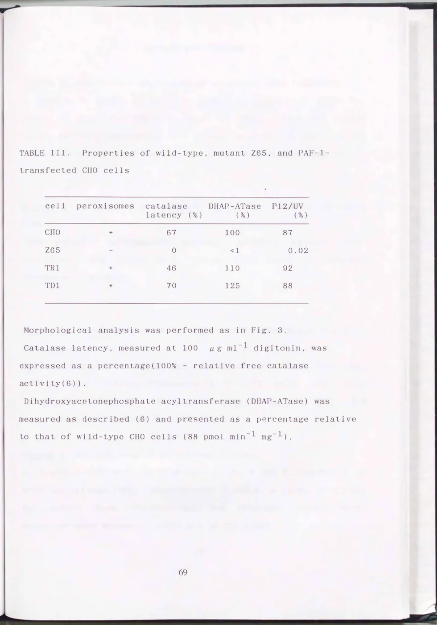
TABLE II. Characterization of Wild-type, Mutant, and Hybrid CHO Cells Peroxisomal Catalase activity
Doubling
time DHAP-ATase ---
CHO Z24 Z65
Z24 x Z65
9.6 17.0 9.9 ND
h pmol/min/mg
285 1 1 233
Total
mU/mg mU/107cells
20 35
24 11
12 19
19 101
Soluble Sedimentable Recovery
% of recovered %
25 75 86
11 23 88
18 22 96
26 7 4 107
DHAP-ATase was measured at pH 5.5 (see Table I). Cells were homogenized, separated into particulate and soluble fractions, and assayed for catalase as described in "Materials and Methods".
r-�
LEGENDS FOR FIGURES
Figure
l·
Electron microscopy of wild-type and mutant CHO cells, with catalase cytochemistry. Cytochemical reaction for catalase was carried out with DAB. Peroxisomes (P) were seen only in the wild-type CHO cell. CHO, wild-type CHO cell; 224 and 265, CHO cell mutants 224 and 265, respectively. Bar , 0 . 3 J1 m.Figure 2. Immunoblot analysis of wild-type and mutant CHO cells.
Cells were homogenized and fractionated as described in
"Materials and methods". Immunoblot was carried out with rabbit
antisera against rat liver cata ase (A) and rat liver acy -CoA
oxidase (B), respectively. Antibodies were detected by [125I]protein A. A and B are composites of two autoradiographic
exposures of a single gel, respectively: lane 1 was exposed for 5 h, the remainder for 18 h. A, lanes: 1, liver homogenate (15 /1 g) of a rat treated with a hypolipidemic drug, clofibrate; 2, postnuclear supernatant fraction (P S, 100 Jlg) of wild-type CHO cells; 3 and 4, particulate and supernatant fractions, respectively, from P S ( 100 J1 g) of parent cells 5, PNS ( 100 /1 g) of mutant 224 cells; 6 and 7, particulate and supernatant fractions, respectively, from P S of 224 cells; 8-10, cell fractions as in lanes 5-7, but prepared from the mutant 265. B, lanes: 1, rat liver homogenate (15 JLg); 2-4, PNS (100 JLg) of wild-type, 224, and 265 cells, respectively. Open arrowheads indicate the components A (75 kD), B (53 kD), and C (22 kD) of rat liver AOx. STD, standard markers in kD; H, homogenate; P S, postnuclear supernatant fraction; P, particulate fraction; S,
supernatant fraction.
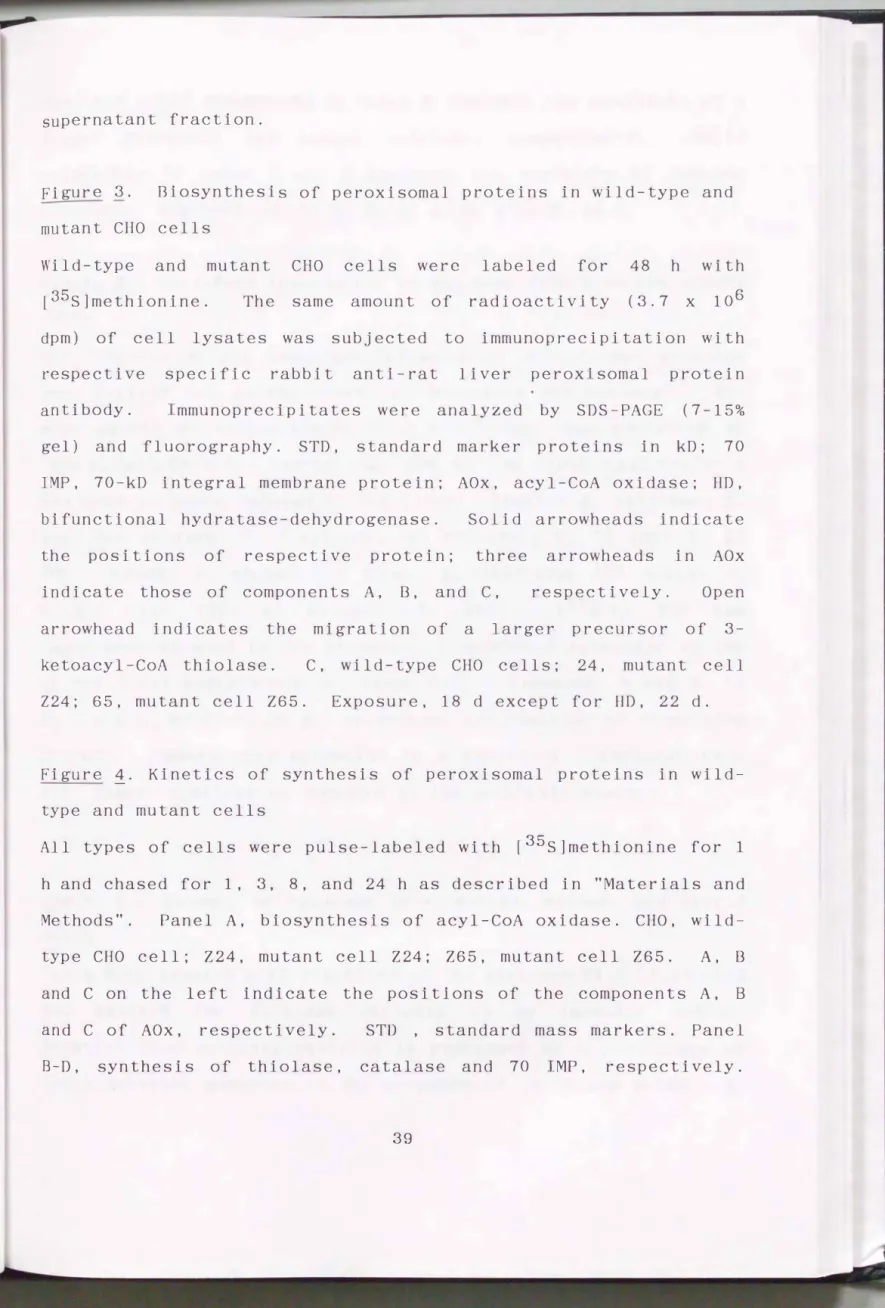
Figure 3. Biosynthesis of peroxisomal proteins in wild-type and mutant CHO cells
Wild-type and mutant CHO cells were labeled for 48 h with [35s]methionine. The same amount of radioactivity (3.7 x 10⁶ dpm) of cell lysates was subjected to immunoprecipi tat ion with respective specific rabbit anti-rat liver perox'somal protein antibody. mmunoprecipi tates were analyzed by SDS-PAGE ( 7-15%
gel) and fluorography. STD, standard marker proteins in kD; 70 IMP, 70-kD integral membrane protein; AOx, acy -CoA oxidase; HD, bifunctional hydratase-dehydrogenase. Solid arrowheads indicate the positions of respective protein; three arrowheads in AOx indicate those of components A, B, and C, respectively. Open arrowhead indicates the migration of a arger precursor of 3- ketoacyl-CoA thiolase. C, wild-type CHO cells; 24, mutant cell 224; 65, mutant cell Z65. Exposure, 18 d except for HD, 22 d.
Figure
i·
Kinetics of synthesis of peroxisomal proteins in wildtype and mutant cells
All types of cells were pulse-labeled with [35s]methionine for 1 h and chased for 1, 3, 8, and 24 h as described in "Materials and Methods". Panel A, biosynthesis of acyl-CoA oxidase. CHO, wild
type CHO cell; Z24, mutant cell Z24; Z65, mutant cell Z65. A, B and C on the left indicate the positions of the components A, B and C of AOx, respectively. STD , standard mass markers. Panel B-D, synthesis of thiolase, catalase and 70 IMP, respectively.
open and solid arrowheads in Panel B indicate the positions of a larger precursor and mature thiol se, respectively. Solid arrowheads in panel C and D indicate the positions of labeled proteins. Exposure: A, 11 d; B, 41 d; C, 4 d; D, 11 d.
Figure 5. Cell-free translation of RNA from wild-type and mutant cells.
RNA translation and immunoprecipitation of peroxisomal proteins were carried out as described in "Materials ·and Methods". The same amount of radioactivity ( 2. 6 x 10⁶ dprn) was subjected to immunoprecipi tat ion, except that 20% of the input radioactivity was used in lanes related to rat liver. anels: A, catalase; B, acyl-CoA oxidase; C, 3-ketoacyl-CoA thiolase; D, 70 IMP; E, 22 IMP. Lanes:
mutant cell
1, normal 224; 4'
rat liver; 2, wild-type CHO mutant cell 265. [35s]22
cells; 3, IMP was immunoprecipitated in the presence of unlabeled authentic 22 IMP of rat liver peroxisomes (E, lanes 5-7). Exposure; A and B, 11 d; C and D, 54 d; E, 20 d. Arrowhead, the position of respective
protein. Upward open arrowhead in A indicates a catalase band with higher mobility as compared to the authentic monomer.
Figure 6.
cells.
Latency of catalase in wild-type, mutant, and hybrid
Cells were treated with digitonin at the concentration indicated and assayed for catalase activity in an isotonic medium.
Relative free catalase activity is expressed as a percentage of total activity measured in the presence of 1% Triton X-100. A,
wild-type (CHO,
0
and two mutants (224,�
; 265,0 )
. B,variants of the mutants, 224TGrouar
( �
) and 265TGrouar (0 )
; hybrid cells, wild-type x 224TGrouar•
and wild-type x Z65TGrouar( •
) . C, heterologous hybrids, 224 x 265TGrouar( y)
and 265 x 224TGrouare
) ; homologous hybrids, 224 x Z24TGrouar( �
) and 265 x 265TGrouar( 0 )
.Figure 7. Immunocytochemical analysis of wild-type, mutant , and hybrid cells.
Wild-type (CHO), mutants (224 and 265), and hybrid (224 x Z65TGrouar) cells were respectively stained for catalase by the immunocytochemical procedure described in "Materials and Methods", and examined under an Olympus BH-2 microscope. Bar, 50
11m.
Figure 8. Biosynthesis of peroxisomal proteins in hybrid cells.
Hybrid cells ( 224 x 265TGrouar) selected in HAT plus ouabain medium after fusion wer labeled with [35s]methionin and analyzed as described in Figs. 3 and 4. STD, mol cular mass standards; TH, thiolase. A, B, and C on the left indicate the components A, B, and C of AOx, respectively.
arrowheads are as in Fig. 4. Exposure, 7 d.
Open and solid
Fig. 1
F g. 2
A
CatalaseRAT CHO Z24 Z65
STD -H- PNS P S ;;;:;-;-s ;;-;s
2QO-
2 3 4 5 6 7 8 9 10
8
Acyt-CoA Oxkiase" 0 "' "
STD �� v� 'VrJ; 'Vro
200-
97- 68- 45-
26-
18- 14-
1 2 3 4
F g. 3
ttiolase 701M=> catalase A 0 x H D
STO C 24 65 C 24 65 C 24 65 C 24 65 C 24 65
2 3 4 5 6 7 8 9 10 11 12 13 14 15
Fig. 4
A Ac�A Oxidase CHO Z24 Z65
0\ase(h) 0 1 3 8 24 0 1 3 8 24 0 1 3 8 24
c-
STD -97
-28 -18 -14
CHO ��
Chue(h) 0 I 3 8 24 0 I 3 � 24 0 I 3 8 24
8 c Catalase D
701MP
F g. 5
A catalase 8 A Ox c thlolase D 701MP E 221MP
STO 2QO-
97-
•s-
26-
18- 1·-
2 3 4 1 2 3 4 1 2 3 4 1 2 3 4 1 2 3 4 5 6 7 8 '
,..._
0 0
';:100 ..-
>
<! v
� 50
OJ� ru u OJ �
lL
Fig. 6
A
100 200
B c
300 0 100 200 300 0 100 200 300
0 igi ton in ( }19 /ml)
Fig. 7
1" ••
. �.
' .
]{.:: '
;• ,I
· . .,
F g. 8
Pulse/ Cha se Contiruous Label A 0 x thiolase(TH) AOx TH
0\ase(h) 0 1 3 8 24 0 1 3 8 24
A-
B- -45--
-26- c-
-18- -14-
Chapter II
eDNA cloning and characterization of peroxisome assembly factor-1
Abbreviations
AOx, acyl-coenzyme A oxidase; CHO, Chinese hamster ovary;
DHAP-ATase, dihydroxyacetonephosphate acyltransferase;
FCS, fetal calf serum; IAA, iodoacetamide; 22 IM , 22-kD integral
membrane protein; MEM, minimum essential medium; OTC, ornithine transcarbamylase; PAF-1, peroxisome assembly factor-1;
Pl2/UV, 12-(1'-pyrene)dodecanoic acid/ultraviolet;
TG, 6-thioguanine.
SUMMARY
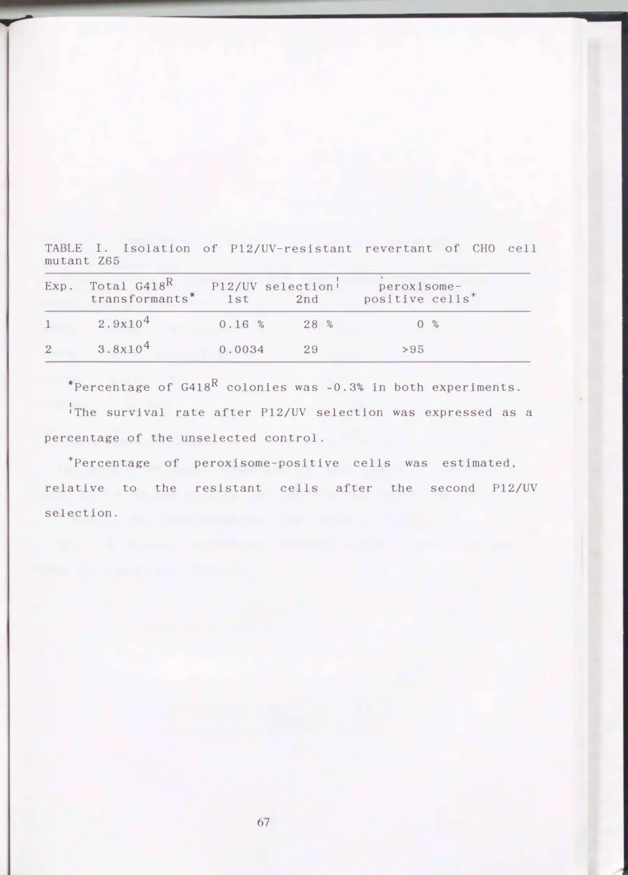
To reveal the molecular mechanism of peroxisome biogenesis, eDNA which could restore peroxisome assembly in a peroxisome
deficient CHO cell mutant, 265 was isolated. A eDNA library of rat liver was constructed in the mammalian expression vector, pcD2. 265 cells were transfected with the cD A library, and peroxisome-restored revertant cells were selected by the P12/UV method which kills peroxisome-deficient mutant· cells selectively.
Five eDNA clones were recovered from the revertant cells. Three of them contained a eDNA fragment in common, and were able to complement the 265 cell in another round of transfection.
Sequencing revealed that this eDNA fragment encoded a novel 35 kDa protein (termed peroxisome assembly factor-1 or PAF-1) consisting of
suggested that Transfection of
305 amino acid residues. Hydropathy analysis two hydrophobic regions were present in PAF-1.
PAF-1 eDNA could restore not only peroxisomes morphologically, but
such as the defects ketoacyl-CoA thiolase.
also complement biochemical abnormalities in processing of acyl-CoA oxidase and 3 Using anti-PAF-1 antibody, I revealed that PAF-1 was localized in the peroxisome fraction and behaved as an integral membrane protein. These results indicate that PAF-1 is an
biogenesis.
essential membrane component for peroxisome