九州大学学術情報リポジトリ
Kyushu University Institutional Repository
DEVELOPMENT OF CELL CULTURE PLATFORMS BY
ENZYMATIC FUNCTIONALIZATION OF REDOX-RESPONSIVE HYDROGELS
ワヒユ, ラマダン
http://hdl.handle.net/2324/4110491
出版情報:九州大学, 2020, 博士(工学), 課程博士 バージョン:
権利関係:
i
DEVELOPMENT OF CELL CULTURE PLATFORMS BY ENZYMATIC FUNCTIONALIZATION OF
REDOX-RESPONSIVE HYDROGELS
Wahyu Ramadhan
Department of Chemical Systems & Engineering Faculty of Engineering
Kyushu University
2020
ii
TABLE OF CONTENTS
CHAPTER 1 GENERAL INTRODUCTION ... 1
1.1 Hydrogel scaffold and the polymer choice ... 1
1.2 Methods of synthesis of hydrogels ... 6
1.3 Designation of horseradish peroxidase (HRP)-catalyzed hydrogels ... 8
1.3.1 Horseradish peroxidase (HRP) ... 8
1.3.2 Horseradish peroxidase (HRP)-mediated hydrogel formation through cross-linking between phenol groups ... 10
1.3.3 Timeline and research evolution of the HRP-mediated hydrogelation as a cell culture scaffold ... 10
1.4 Aim and outline of the thesis ... 17
1.5 References ... 18
CHAPTER 2 ENZYMATICALLY PREPARED DUAL FUNCTIONALIZED HYDROGELS WITH GELATIN AND HEPARIN TO FACILITATE CELLULAR ATTACHMENT AND PROLIFERATION ... 27
2.1 Introduction ... 27
2.2 Experimental ... 30
2.2.1 Materials. ... 30
2.2.2 Synthesis of Hepa-SH and Gela-SH. ... 31
2.2.3 Arginine density of gelatin. ... 32
2.2.4 Heparin loading amount in hydrogel system. ... 33
2.2.5 Fabrication of hydrogels. ... 34
2.2.6 Measurement of gelation time and rheological properties. ... 35
2.2.7 Swelling behavior of PEG/Gela/Hepa_hydrogels. ... 35
2.2.8 ζ-potential measurements. ... 36
2.2.9 Adhesion and proliferation of cells on hydrogels. ... 36
2.2.10 Immobilization strategies of growth factors. ... 37
2.2.11 Antiproliferative effects of native heparin and Hepa-SH. ... 39
2.2.12 Effect of various Hepa-SH concentrations on the numbers of NIH3T3 cells treated with exogenous bFGF. ... 39
2.2.13 Measurement of the loading capacity of bFGF on PEG/Gela/Hepa_ hydrogels and the release profile of bFGF from the loaded hydrogels. ... 40
2.2.14 Statistical analysis. ... 40
2.3 Results and discussion ... 40
2.3.1 Synthesis and characterization of dual functionalized hydrogels ... 40
iii
2.3.2 Cell proliferation assay on dual functionalized hydrogels ... 46
2.3.3 In situ immobilization of bFGF in dual functionalized hydrogels ... 50
2.3.4 Administration of GFs in different hydrogel systems for adherent cell culture ... 54
2.4 Conclusion ... 57
2.5 References ... 57
CHAPTER 3 CONSTRUCTION OF HIGHER-ORDER CELLULAR MICRO- STRUCTURES BY A SELF-WRAPPING CO-CULTURE STRATEGY USING A REDOX-RESPONSIVE HYDROGEL ... 62
3.1 Introduction ... 62
3.2 Experimental ... 65
3.2.1 Materials. ... 65
3.2.2 Fabrication of the redox responsive hydrogel. ... 66
3.2.3 Cell lines and cell-culture conditions. ... 66
3.2.4 Preparation of the NIH3T3 cell sheet, HepG2 spheroids, HUVECs and collagen beads. ... 67
3.2.5 Observation of the wrapping process and cell viability. ... 67
3.2.6 Immunofluorescence staining of HUVECs ... 69
3.2.7 Statistical tests. ... 70
3.3 Results and discussion ... 70
3.3.1 Kinetic analysis of the detachment of a cell sheet from the redox- responsive hydrogel. ... 71
3.3.2 Self-wrapping behaviour of the cell sheet upon detachment from the redox-responsive hydrogel. ... 74
3.3.3 Viability of co-cultured cells in the wrapped cellular structure. ... 78
3.3.4 Metabolism of co-cultured cells inside the wrapped cellular structure. . 83
3.4 Conclusions ... 87
3.5 References ... 87
CHAPTER 4 REDOX-RESPONSIVE FUNCTIONALIZED HYDROGEL MARBLE FOR THE GENERATION OF CELLULAR SPHEROIDS ... 94
4.1 Introduction ... 94
4.2 Experimental ... 96
4.2.1 Materials. ... 96
4.2.2 Fabrication and Hydrogelation of HM. ... 97
4.2.3 Equilibrium swelling ratio (QM) and gel content. ... 97
4.2.4 Gelation time. ... 98
4.2.5 Rheological evaluation. ... 98
iv
4.2.6 Cell lines and cell culture conditions. ... 98
4.2.7 Evaluation of cultured cells and spheroids. ... 99
4.2.8 Morphology analysis of hydrogel marbles. ... 100
4.2.9 Statistical test. ... 101
4.3 Results and Discussion ... 101
4.3.1 Physicochemical properties of hydrogel marbles prepared by HRP catalysis under various Gela-SH concentrations ... 102
4.3.2 Formation and distribution of HepG2 cellular aggregates in hydrogel marbles ... 106
4.3.3 Evaluation of cellular functions in HMs ... 111
4.4 Conclusion ... 115
4.5 References ... 116
CHAPTER 5. CONCLUSIONS ... 120
5.1 Summary ... 120
5.2 Future prospects ... 120
ACKNOWLEDGEMENTS ... 125
1 CHAPTER 1 GENERAL INTRODUCTION 1.1 Hydrogel scaffold and the polymer choice
Untangling complex biological systems is still a major challenge for scientists despite over a hundred years of modern researches1. Mimicking a specific convoluted system is one of the best approaches to understand and valorize it for research purposes and applications benefitting a variety forms of life2,3. In recent years, bottom- up approaches including cellular scaffolding have been studied in tissue engineering, which could be the best path forward to initially mimicking the complexity of cellular environment. A rise in demand of this technology to treat chronic diseases is expected to drive a research, impelling the market growth through to 2027. Grand View Research Inc. has reported a perceived tendency of cell culture market in 2027. The world cell culture market value is forecasted to attain USD 3.2 billion, broadens at a compound annual growth rate of 11.3%4.
Fig. 1.1 Summary of different technological cell culture technology approaches. Reproduce by permission from ref5 Copyright 2018 Elsevier.
Over the last decade, a variety of cell culture platforms have been developed to achieve versatile cell culture system as a research model, especially for the preclinical drug discovery and the development of tissue engineering itself. The cell culture technology was initiated by two-dimensional (2D) monolayer cell culture under
2
adherent conditions. At the same time, three-dimensional (3D) cell culture systems have been designed to sufficiently mimicking the physiological conditions of natural structures of the tissue. Current approaches in 3D cell culture platform are mainly represented by two strategies, scaffold-free and scaffold-based platform, where each having their advantages and posing intrinsic limitations (Fig 1.1). By integrating the lack of each system, it is desirable to constructively generate a smart scaffold while covering their drawbacks.
Today, one of promising smart scaffolds for cell culture is hydrogel, known as potential bio-based polymeric materials with dynamic chemical, physical, and structural properties6,7. Hydrogels have extensively studied by many researchers and recognized as the most popular scaffold in the past 30 years. The search for the words
“hydrogel” and “hydrogel-cell culture” in the PubMed and Scopus database shows a significantly increase in the number of the issued articles (Fig 1.2).
Fig. 1.2 The growing trend in published articles identified during the past 30 years with the keyword “hydrogel” and “hydrogel-cell culture” for the past 10 years.
Due to their unique three-dimensional (3D) hydrophilic network that is cross-linked and swollen, hydrogels serve as a wet environment or provide fluid to recapitulate and mimic the natural extracellular matrix (ECM) or many tissues in the body8–10. Therefore,
3
either hard or soft networks of hydrogels have shown an important class to have good compatibility. Hydrogels have been designed to create specific properties of biomaterial scaffolds for embedding bioactive molecules and other biological entities, particularly as a cell culture platform (Fig. 1.3).
Fig. 1.3 Representation of the general strategies of hydrogel as a cell-laden scaffold. A.
Synthetic polymer create and develop the environments that mimic tissues or natural structure.
B. Hydrogel matrix able to alter with protein (such as cell adhesion peptide or growth factors) which is mimic properties of the ECM. C. Cells are live in communities, so the chemotaxis can be modified by attaching biochemical entities to a hydrogel that can mimic the natural interaction. D. The mechanical properties of hydrogel can be modified by controlling the cross- linking density and the ligand. E. Synthetic hydrogel (left; orange matrix) provide a 3D environment for cell culture scaffold but with a lack of activated integrins and receptors of cell (brown). Besides, hydrogel composed natural polymer (right) serve the growth factors (orange) and integrin sites (green) that can bind with the cell surface receptors. F. Engineered synthetic hydrogel that integrated and modified well-deveined natural compounds and chemical moieties. Fig. A-D are reproduced by permission from ref6. Copyright 2016 Springer Nature. Fig. E-F are reproduced by permission from ref11. Copyright 2009 John Wiley & Sons, Inc.
In the initial report of hydrogels, the scaffold was employed to encapsulate cells by natural polymers, such as fibrin, elastin, collagen, hyaluronic acid, gelatin, and other proteinaceous based polymer12–15, namely natural hydrogel. Generally, natural hydrogel (biopolymer) can be categorized by constituents such as polysaccharide, protein, protein/polysaccharide hybrid polymers, DNA and decellularized matrices16–
18. Several key class of natural hydrogel and their properties are described in Table 1.
4
Table 1. Key classes of natural compounds used as the main polymer of hydrogel fabrication, and their main advantages and drawback properties in the cell culturing.
Polymer Class Advantages Disadvantages
Alginate Polysaccharides - Reactive handles for functionalization
- Rapid gelation with divalent cations
- Ease of use for 3D printing - Abundant
- Poorly adhesive
- Cation leaching leads to dissolution
- Non-biodegradable
Chitosan Polysaccharides - Adhesive and antimicrobial - Low immunogenicity - Abundant
- Poor solubility at neutral pH
HA Polysaccharides - Bioactive and biocompatible - Binds growth factors and
cytokines
- Reactive handles for functionalization
- Rapidly degraded in vivo - Low stability without cross-
linking
Chondroitin Sulfate
Polysaccharides - Bioactive and biocompatible - Binds growth factors and
cytokines
- Reactive handles for functionalization
- Low stability without cross- linking
- Rapidly degraded in vivo
Collagen Proteinaceous - Adhesive and bioactive - Mimics native ECM - Abundant and
biodegradable
- Contamination can lead to immunogenicity
- Mechanical stability lost during processing - Assembly sensitive to
modification Gelatin Proteinaceous - Adhesive and bioactive
- Tolerant of functionalization - Abundant and
biodegradable
- Mechanically weak - Requires cross-linking - Contamination can lead to
immunogenicity Silk Proteinaceous - High mechanical strength
and elasticity - Low immunogenicity - Adhesive
- Slow gelation
ELPs Proteinaceous - Tunable structure and sequence
- Thermoresponsive (LCST) - Recombinant expression
- Low stability without cross- linking
Reproduce by permission from ref19. Copyright 2020 Royal Society of Chemistry (RSC).
As shown in Table 1, although the studies of natural hydrogels have been established early success in cell and tissue culture, the mechanical properties, gelation time, and degradation rate were difficult to control. Moreover, natural compounds have diverse contents, and batch-to-batch variability in compositions and biochemical properties lead to significant uncertainty in cellular experiments20. For these reasons, in recent years, synthetic materials possessing more tunable and well-
5
defined structures have been explored to create hydrogels for cell and tissue culture7,21. Synthetic polymers also show high gel strength, high capacity of water absorption, long shelf life and generally simpler well-defined structures than that of natural polymers, the introduction of cross-linking substrates is thus controllable.
Eventually, synthetic polymers possessing in hydrogels fabrication can be exploited to generate the adjustable design especially in their degradation and functionalization properties for the advanced cell/tissue culture applications19.
Fig. 1.4 Commonly used synthetic polymer as the hydrogel precursor for cell culture application.
In the class of synthetic material examined thus far22–25, many polymers have been used as hydrogel precursors that can be classified by chemical entities such as polyvinyl, polyester, poly(ethylene oxide) and other synthetic polymer (Fig 1.4). One promising hydrogel’s backbone is polyethylene glycol (PEG)26,27. PEG is the most commonly investigated polymer used to make synthetic hydrogels as an FDA- approved material. Its chemical and biological inertness, highly hydrophilic nature, controllable and homogenous microstructure as well as a wide range of polymer
6
architectures are attainable by synthetic chemistry28,29. Moreover, along with the ease of derivatization in which end-functionalization of PEG enable the inclusion of various chemical cues that can be applied for efficiently crosslinking reaction19.
1.2 Methods of synthesis of hydrogels
Hydrogels are mostly constructed by the crosslinking reaction of polymeric matrix.
Currently, the most attractive way to manipulate the shape, activity, and biocompatibility of a synthetic hydrogel is by altering the cross-linking site, either by physical or chemical means30 (Fig .1.5). Physically cross-linked hydrogels are formed by weak and reversible intermolecular interactions, such as ionic cross-linking, hydrogen bonds, hydrophobic interactions of thermal inclusion mediated on upper Critical Solution Temperature (UCST)/Lower Critical Solution Temperature (LCST), and ultrasonication assisted formation of sol-to-gel phase transition31,32. The most important advantage of this system is its low cytotoxicity because of the absence of a chemical reaction. However, physical hydrogels have limited mechanical properties owing to the weak interaction involved in their cross-linking points.
On the contrary, chemical cross-linking is attained by covalent bond formation such as click chemistry33, free-radical photopolymerization34 and enzyme-mediated cross-linking35. The chemical crosslinking strategy serves higher stability and mechanical properties than that of the physical crosslinking methods; consequently, chemical hydrogels are more advisable for long-term cell culture and tissue engineering applications. However, from the viewpoint of cytocompatibility, there are a couple of shortcomings, including photoinitiators and irradiation used in photopolymerization, which are potentially detrimental to cell survival, causing tissue damage36 and deactivating the incorporated proteins37.
7
Fig. 1.5 The most widely used crosslinking strategies for hydrogel construction. Including crystallization crosslinked hydrogel, ionic crosslinking, UV crosslinking, LCST/UCST hydrogel, dual crosslinking and enzymatic catalyzed crosslinking. Reproduced with permission from ref32. Copyright 2019 The Royal Society of Chemistry.
Among these existing strategies, an emerging attractive approach for the rational and feasible design of hydrogels is enzyme-mediated hydrogelation. The enzyme- catalyzed hydrogelation realizes a mild cross-linking reaction, making it suitable with the incorporation of therapeutic proteins, drugs and typically for living cells38–40. The kinetic manipulation of hydrogel formation could be realized by controlling the catalytic behavior of enzymes in crosslinking reaction by adjusting reaction prameters. Despite hydrogel formation by an enzymatic reaction is a relatively recent concept, the interest
8
of an enzyme-based cross-linking is not only providing the strong and dynamic covalent bond but also exhibit the fast gelation under physiologically relevant and mild oxidative conditions.
So far, several enzymes have been studied for their abilities to control and provide advanced hydrogelation systems, such as lysyl oxidase38, transglutaminase41, sortase42, laccase43,44, phosphatases45, ß-lactamase46, plasma amine oxidase47, thrombin48, thermolysin49, kinase/phosphatase50,51, phosphopantetheinyl transferase52, tyrosinase53, ɑ-chymotrypsin54, and peroxidases55. Among these group of enzymes, peroxidases that catalyze a variety of oxidative transformations using hydrogen peroxide or other peroxides as oxidants56, and horseradish peroxidase (HRP) extracted and isolated from horseradish roots is one of the most studied, favored and used enzymes in hydrogel fabrication57–59. HRP is widely applied as a biocatalyst due to the fast reaction kinetics, moderate substrate specificity, and ability to control the cross-linking density, that can be tailored by simply altering the precursor reactants56,57,60–62. In fact, another enzyme demonstrates weak mechanical properties, lower biorthogonality, less specific binding site and lack of immunogenicity.
Conversely, because HRP is plant-based derived peroxidase that can offer relatively low immunogenicity risk, thus HRP has been authorized by the U.S. Food and Drug Administration (FDA) for biomedical applications63 and commercially accessible. It was predicted that HRP could become the greatest enzyme for the next decade64.
1.3 Designation of horseradish peroxidase (HRP)-catalyzed hydrogels
Hydrogels have transitioned from being a static and passive material to a dynamic, bio-based, and stimuli-responsive biomaterial for use in cell-seeding technology65,66. Recent advances towards such biologically active hydrogels have been directed to design biomaterials with superior feature for higher-order cell culture
9
than conventional 2D culture. As previously described, in the term of cellular scaffold development, HRP is promising biocatalyst because of its hydrogelation abilities. Thus, many HRP-mediated hydrogel systems have been proposed for 2D or 3D cellular scaffolds, which were deeply discussed in a recent review by Sakai and Nakahata57, such as cell-laden microcapsules, solid- and hollow-core hydrogel fibers, approaching in single-cell hydrogel coating and biofabrication in 3D bioprinting.
1.3.1 Horseradish peroxidase (HRP)
HRP is an oxidoreductase comprising of 308 amino acid residues, 4 disulfide bonds between cysteine residues, a single heme group [iron(III) protoporphyrin IX]
and two calcium ions (Fig 1.6-A)67. Basically, HRP-catalyzed reaction is described by the following equation, in which AH and AH2 imply a radical product and its reducing substrate, respectively67,68.
H2O2 + 2AH2
!"#
$⎯& 2H2O + 2AH
The reaction cycle initiates from the binding of H2O2 to the vacant octahedral position on the iron atom of HRP (Fig. 1.6-A), and subsequently the oxidized HRP (compound I/II) oxidizes a reducing substrate and returns to its original form.
Consequently, the created phenol radicals allow covalent bond formation within aromatic rings structure (Fig. 1.6-B)68. HRP recognizes indoles, sulfonates, phenylamines, and phenol as reducing substrates, which are converted to radicals to react with each other via a radical coupling reaction. These reducing substrates are introduced to polymers for the formation of hydrogels via cross-linking. Details of the HRP structure and its complex catalytic mechanism have been elegantly described in the recent literature56,67,69–73.
10
Fig.1.6 A. The heme as an active center of peroxidases structure. B. The catalytic cycle of horseradish peroxidase (HRP). A Reproduce from Refs.73 with permission from Elsevier 2019.
B. Reproduce from ref68 with permission from John Wiley & Sons, Ltd. 2014
1.3.2 Horseradish peroxidase (HRP)-mediated hydrogel formation through cross-linking between phenol groups
The crosslinkable substrate is one of the most important considerations to increase the stability of polymer crosslinking through HRP-catalyzed hydrogelation. In fact, despite many functional groups, for instance phenolic acids, aromatic phenols, and amines can be applied as the reducing substrate67. Currently, the introduction of phenolic groups is the most commonly used method to improve HRP-mediated hydrogelation in many polymers63 because it is a common form of industrial waste68. Various phenol derivatives are introduced to the backbone of hydrogels such as tyrosine74, tyramine75, phenylalanine, 4-hydroxyhneyl acetic acid76 and hydroxyphenyl propionic acid77,78 to form cross-linking in the hydrogels.
11
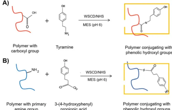
Precursor that works in phenol-mediated cross-linking has wide range of polymer type, either natural or synthetic moieties could be conjugated and modified through its sites63. Conjugation of phenol moieties in hydrogel network can be selected according to the chemical cues of each polymer. Generally, water soluble carbodiimide hydrochloride (WSDC) is utilized in the reaction between carboxyl group and a primary amine (e.g. tyramine)79 (Fig. 1.7-A). An alternative method is also using WSDC within the reaction of polymer consisting amine group and propionic acid (Fig. 1.7-B).
Fig. 1.7. Schematics illustration of synthesis of phenol polymer using water soluble carbodiimide hydrochloride (WSDC/NHS). Reproduce from ref79 with permission from Royal
Chemistry Society 2018.
From the viewpoint of the compatibility of the main polymer, the bound phenol moieties in HRP system work in many types of polymers because of the reasonable design and substrate specificity of HRP. Natural compounds for instance collagen80, hyaluronic acid81, gelatin82, chitosan83, silk84, chondroitin sulfate85, dextran86, alginate87, and various combinations among them have been used as compatible substrates polymer in HRP-mediated hydrogelation. However, as described in the
12
previous section due to the low mechanical properties of natural-based polymer, it will predict limits of the application on the biomedical field where the mechanical aspect is crucial. To overcome the limitation, the synthetic polymer has been obtained to construct hydrogel backbone, and among all the synthetic material, functionalized polyethylene glycol (PEG) is widely involved as a base polymer for HRP-mediated hydrogelation due to highly controllable and homogenous microstructure28,29.
1.3.3 Timeline and research evolution of the HRP-mediated hydrogelation as a cell culture scaffold
Historically, the first report of the valorization of an HRP-catalyzed oxidation in a hydrogel system was reported by Kaplan et al. in 2002, where they demonstrated the fabrication of poly(aspartic acid) modified with phenol as a gel precursor using an HRP catalysis system55. Since then, many researchers have followed the same concept to construct hydrogels based on HRP catalysis either using natural polymer or synthetic polymer22,23,87–96,24,55,80–85.
A significant improvement was reported by Kurisawa and coworkers in 200576 in the application of a hyaluronic acid-tyramine conjugate to produce an injectable hydrogel, where HRP induced oxidative coupling, resulting in hydrogelation. In their report, the classical HRP cycle could interact with hydrogen peroxide (H2O2) to form highly intermediate product as oxidants. The results show HRP mediated hydrogelation can support the injection of hydrogel as a minimally invasive technique.
However, in general, these development of HRP-mediated hydrogelation systems involves H2O2 in the equal ratio as HRP amount to the fabrication of cell-laden hydrogel scaffold (Fig. 1.8).
13
Fig.1.8. HRP-catalyzed crosslinking reaction with phenol-rich polymer and H2O2 as a common cell encapsulation strategy in the fabrication of hydrogel in the tissue engineering field.
Reproduce from ref68 with permission from John Wiley & Sons, Ltd. 2014.
It should be pointed out that HRP-catalyzed cross-linking of several reaction type has been reported and discussed in thousands of research papers in various points of view56,57,70,73,79. However, from the viewpoint of cytotoxicity, removal of residual H2O2 in the HRP-mediated hydrogelation system for mild conditions to cells and increase in the cytocompatibility of hydrogel system are required.
Despite the development of HRP-mediated preparation hydrogel has been conducted since more than twenty years ago, the trend of published paper during 2000 to 2010 certainly reveals that the study and progress of HRP-hydrogels are still at an early development phase, against with the considerably steps taken to totally remove the use of H2O2 in its system (Fig 1.9). The continuous work has been settled to adapt
14
and integrate the facile HRP preparation system with the another chemical/biological entity as well as the significantly effort to advance the different compartment in cell culture application.
Fig 1.9. Timeline chart of the evolution of HRP-mediated preparation of hydrogels based on natural and synthetic polymers and the current goal of this study. Reproduced and redrawn from ref64 with permission from the Springer Nature 2020.
In the previous section, it was discussed that H2O2 is necessary in the HRP- mediated hydrogelation. However, the amount of H2O2 is crucial concern for harsh impact in cytocompatibility, thus the consumption of H2O2 amount should be decreased. However, the low H2O2 dosage and the short cell exposure time to H2O2
may not provide milder condition especially when the system uses as the cellular scaffold. The further approach to solve the problem is using glucose oxidase (GOx), where oxidizing glucose to glucono-δ-lactone by consuming oxygen could suppress the high concentration of H2O292. Nevertheless, the remaining GOx in the system might still produce H2O2 and react with glucose, thus predict gives negative impact to the cell. Indicating in the in vivo application this technique may face potential problem, as the glucose is a common molecule in ECM, GOx in the hydrogel will subsequently oxidize the excess of glucose molecules and create undesired H2O2.
15
Another important report came from Singh and colleagues, where they found that the thiol groups incorporated into the polymer promoted hydrogelation induced by HRP, without recruitment of H2O293
. Under aerobic condition, mixture of HRP and thiol can generate hydrogelation reaction of phenol compound via autooxidation in the redox sensitive hydrogel. This basic gelation concept is in the presence of thiol, thinly radical was dimerized to form disulfides or interact with the disulfide radicals after reacting with oxygen. However, in this system the hydrogelation condition occurs in the pH 8.5, which may not suitable with the physiological condition in ECM. Moreover, the gelation time of the polymer solution was also slow (41-110 min), even at high HRP concentration and high concentration of polymeric substrates.
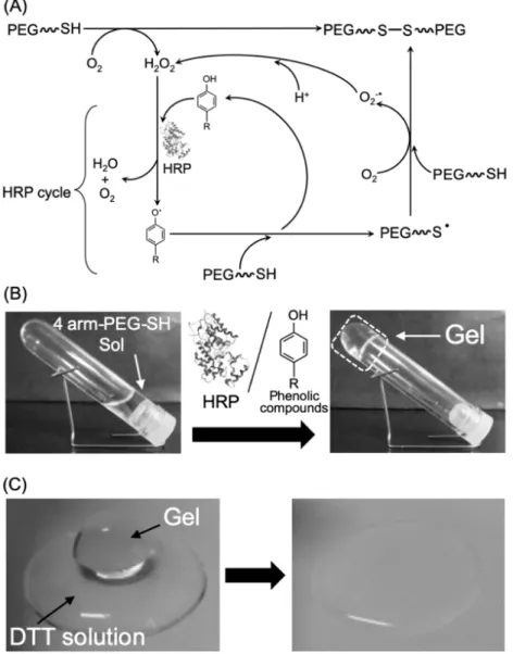
To date, the next impactful research developed from our group, where the dramatic improvement of the HRP-catalyzed hydrogelation of thiolated polymers is realized94. The main concept of the gelation system was the improvement of phenolic compounds as the substrates for HRP catalysis. The rate of second-order constant of HRP compound II with the general thiol substrate cysteine is < 50 M-1 s-1 97, while that with phenolic compounds is usually in the range of 103–107 M-1 s-1 98, indicating that the inclusion of thiolated polymers as an HRP substrate would lead to much slower gelation kinetics than phenolated ones. Nevertheless, in the incorporation of phenolic substrates, radical exchange between the phenol radicals produced by HRP catalysis and thiols occurs with a rate constant at pH 7.15 is reportedly 106 M-1 s-1. Therefore, in the HRP-cycle, the presence of single electron oxidation from thiol occurs when generated the phenol radicals and that thiols radical then transformed to disulfides after interacting with the oxygen. Eventually, the resultant phenolic compounds should accelerate the HRP-mediated fabrication of disulfide-crosslinked hydrogels without exogenous H2O264 (Fig. 1.10).
16
Fig. 1.10. HRP-mediated hydrogel system without exogenous H2O2 using thiolated polymers.
(A) Proposed scheme of hydrogelation in the presence of a phenolic compound as a direct HRP substrate. (B) Hydrogel formation using 4-arm-PEG-SH, HRP, and tyramine at pH 7.4.
(C) Degradation of the redox-responsive hydrogel by soaking in DTT solution for 15 min.
Reproduced from ref64 with permission from the Springer Nature 2020.
This strategy was the first report of disulfide-cross-linked hydrogels prepared by HRP catalysis at neutral pH without the addition of exogenous H2O2. In this HRP hydrogelation system, the inclusion of phenolic compound is aid and amplify the autooxidation of the thiol groups. Indicating this phenolic compound plays a significant and crucial role as the direct substrate in the HRP catalytic cycle and in the preparation of the redox-responsive disulfide-cross-linked hydrogels. Obviously, the design of formation and degradation of the disulfide cross-linking in this system can effectively
17
encapsulate and release living cells, which might be highly beneficial for the development of cell culture platform64.
1.4 Aim and outline of the thesis
The aim of this research is to integrate and develop the facile HRP preparation system in redox responsive hydrogel with the inclusion of chemical/biological entities and with different shapes of compartment to advance the cell-cell and cell-matrix interactions in cell culturing.
In Chapter 1, a general introduction was described on the development of scaffold to better mimic the natural life form and the increase of hydrogel research as a cell culture platform in history which constructed based on the variety of polymer. A brief review of the polymer choice and the crosslinking strategy for precious hydrogelation in hydrogel was included as well. The designation of HRP as the selected biocatalyst in hydrogelation and their improvement year by year as the cellular friendly scaffold was also discussed.
In Chapter 2, the feasibility of using HRP hydrogelation system in the PEG-based hydrogel as the main polymer was discussed with functionalized gelatin and heparin.
By varying the gelatin type and heparin concentration, the capture of growth factors in hydrogel system and cell adherence as well as the rapid fabrication of cell sheet on the redox responsive hydrogel were studied.
In Chapter 3, the performance of obtained cell sheet on the redox responsive hydrogel is utilized to wrap and encapsulate other cells and biological entities to form heterogeneous of multicellular structure. Cell sheet detachment from hydrogel and their wrapping behavior induced by cysteine were studied. By changing the density of cell or the number of carcinoma spheroids, the cell number of endothelial cell and the collagen beads number, the properties and the resultant of heterogenous 3D cellular
18 structure were investigated.
In Chapter 4, functionalized gelatin in facile HRP-mediated preparation was adapted to transform the liquid marble system to hydrogel marble as a scaffold for 3D cell culture. With this new hydrogel marble system, the carcinoma spheroid fabrication process was described.
Finally, in Chapter 5 the findings of this research are outlined and discussed further research direction related to the next development of HRP hydrogels as a scaffold and their prospect in medical application and tissue engineering fields.
1.5 References
1. Ma’ayan, A. Complex systems biology. J. R. Soc. Interface 14, (2017).
2. Raman, R. & Bashir, R. Biomimicry, Biofabrication, and Biohybrid Systems: The Emergence and Evolution of Biological Design. Adv. Healthc. Mater. 6, 1–20 (2017).
3. Patterson, J., Martino, M. M. & Hubbell, J. A. Biomimetic materials in tissue engineering. Mater. Today 13, 14–22 (2010).
4. Grand View Research. Three-dimensional Cell Culture Market Size, Share &
Trends Analysis Report by Technology (Scaffold Based, Scaffold Free, Bioreactors), by Application (Cancer, Drug Development), by End Use, by Region, and Segment Forecasts, 2020 – 2027. (2020).
5. Ovsianikov, A., Khademhosseini, A. & Mironov, V. The Synergy of Scaffold-Based and Scaffold-Free Tissue Engineering Strategies. Trends Biotechnol. 36, 348–357 (2018).
6. Green, J. J. & Elisseeff, J. H. Mimicking biological functionality with polymers for biomedical applications. Nature 540, 386–394 (2016).
7. Lee, S. C., Kwon, I. K. & Park, K. Hydrogels for delivery of bioactive agents: A historical perspective. Adv. Drug Deliv. Rev. 65, 17–20 (2013).
8. Kopeček, J. Swell gels. Nature 417, 389–391 (2002).
9. Hoffman, A. S. Hydrogels for biomedical applications. Adv. Drug Deliv. Rev. 54, 3–12 (2002).
19
10. Kang, D. H., Kim, D., Wang, S., Song, D. & Yoon, M. H. Water-insoluble, nanocrystalline, and hydrogel fibrillar scaffolds for biomedical applications. Polym.
J. 50, 637–647 (2018).
11. Tibbitt, M. W. & Anseth, K. S. Hydrogels as extracellular matrix mimics for 3D cell culture. Biotechnol. Bioeng. 103, 655–663 (2009).
12. Schneider-Barthold, C., Baganz, S., Wilhelmi, M., Scheper, T. & Pepelanova, I.
Hydrogels based on collagen and fibrin - Frontiers and applications.
BioNanoMaterials 17, 3–12 (2016).
13. Janmey, P. A., Winer, J. P. & Weisel, J. W. Fibrin gels and their clinical and bioengineering applications. J. R. Soc. Interface 6, 1–10 (2009).
14. Chen, Y. Front-matter. Hydrogels Based Nat. Polym. i–iii (2020).
doi:10.1016/b978-0-12-816421-1.00018-5
15. Hughes, C. S., Postovit, L. M. & Lajoie, G. A. Matrigel: a complex protein mixture required for optimal growth of cell culture. Proteomics 10, 1886–1890 (2010).
16. Catoira, M. C., Fusaro, L., Di Francesco, D., Ramella, M. & Boccafoschi, F.
Overview of natural hydrogels for regenerative medicine applications. J. Mater.
Sci. Mater. Med. 30, (2019).
17. Kirchmajer, D. M., Gorkin, R. & In Het Panhuis, M. An overview of the suitability of hydrogel-forming polymers for extrusion-based 3D-printing. J. Mater. Chem. B 3, 4105–4117 (2015).
18. Varghese, S. A., Rangappa, S. M., Siengchin, S. & Parameswaranpillai, J. Natural polymers and the hydrogels prepared from them. Hydrogels Based on Natural Polymers (Elsevier Inc., 2019). doi:10.1016/B978-0-12-816421-1.00002-1
19. Spicer, C. D. Hydrogel scaffolds for tissue engineering: The importance of polymer choice. Polym. Chem. 11, 184–219 (2020).
20. Caliari, S. R. & Burdick, J. A. A practical guide to hydrogels for cell culture. Nat.
Methods 13, 405–414 (2016).
21. McKinnon, D. D., Kloxin, A. M. & Anseth, K. S. Synthetic hydrogel platform for three-dimensional culture of embryonic stem cell-derived motor neurons. Biomater.
Sci. 1, 460–469 (2013).
22. Park, K. M., Shin, Y. M., Joung, Y. K., Shin, H. & Park, K. D. In situ forming hydrogels based on tyramine conjugated 4-Arm-PPO-PEO via enzymatic oxidative reaction. Biomacromolecules 11, 706–712 (2010).
23. Lee, S. H. et al. Enzyme-mediated cross-linking of Pluronic copolymer micelles for
20
injectable and in situ forming hydrogels. Acta Biomater. 7, 1468–1476 (2011).
24. Sakai, S. et al. Polyvinyl alcohol-based hydrogel dressing gellable on-wound via a co-enzymatic reaction triggered by glucose in the wound exudate. J. Mater.
Chem. B 1, 5067–5075 (2013).
25. Sun, Y., Deng, Z., Tian, Y. & Lin, C. Horseradish peroxidase-mediated in situ forming hydrogels from degradable tyramine-based poly(amido amine)s. J. Appl.
Polym. Sci. 127, 40–48 (2013).
26. Tan, H. & Marra, K. G. Injectable, biodegradable hydrogels for tissue engineering applications. Materials (Basel). 3, 1746–1767 (2010).
27. Patel, G. & Dalwadi, C. Recent Patents on Stimuli Responsive Hydrogel Drug Delivery System. Recent Pat. Drug Deliv. Formul. 7, 206–215 (2013).
28. Macdougall, L. J., Pérez-Madrigal, M. M., Arno, M. C. & Dove, A. P. Nonswelling Thiol-Yne Cross-Linked Hydrogel Materials as Cytocompatible Soft Tissue Scaffolds. Biomacromolecules 19, 1378–1388 (2018).
29. Ulery, B. D., Nair, L. S. & Laurencin, C. T. Biomedical applications of biodegradable polymers. J. Polym. Sci. Part B Polym. Phys. 49, 832–864 (2011).
30. Buwalda, S. J. et al. Hydrogels in a historical perspective: From simple networks to smart materials. J. Control. Release 190, 254–273 (2014).
31. Kuo, C. K. & Ma, P. X. Ionically crosslinked alginate hydrogels as scaffolds for tissue engineering: Part 1. Structure, gelation rate and mechanical properties.
Biomaterials 22, 511–521 (2001).
32. Hu, W., Wang, Z., Xiao, Y., Zhang, S. & Wang, J. Advances in crosslinking strategies of biomedical hydrogels. Biomater. Sci. 7, 843–855 (2019).
33. Crescenzi, V., Cornelio, L., Di Meo, C., Nardecchia, S. & Lamanna, R. Novel hydrogels via click chemistry: Synthesis and potential biomedical applications.
Biomacromolecules 8, 1844–1850 (2007).
34. Choi, J. R., Yong, K. W., Choi, J. Y. & Cowie, A. C. Recent advances in photo- crosslinkable hydrogels for biomedical applications. Biotechniques 66, 40–53 (2019).
35. Hu, B. H. & Messersmith, P. B. Rational Design of Transglutaminase Substrate Peptides for Rapid Enzymatic Formation of Hydrogels. J. Am. Chem. Soc. 125, 14298–14299 (2003).
36. Mironi-Harpaz, I., Wang, D. Y., Venkatraman, S. & Seliktar, D.
Photopolymerization of cell-encapsulating hydrogels: Crosslinking efficiency
21
versus cytotoxicity. Acta Biomater. 8, 1838–1848 (2012).
37. Steinhilber, D. & Haag, R. Multifunctional Dendritic Polyglycerol Nano- and Microgels for Encapsulation and Release of Functional Biomacromolecules. 20, 7545 (2011).
38. Moreira Teixeira, L. S., Feijen, J., van Blitterswijk, C. A., Dijkstra, P. J. & Karperien, M. Enzyme-catalyzed crosslinkable hydrogels: Emerging strategies for tissue engineering. Biomaterials 33, 1281–1290 (2012).
39. Buwalda, S. J., Vermonden, T. & Hennink, W. E. Hydrogels for Therapeutic Delivery: Current Developments and Future Directions. Biomacromolecules 18, 316–330 (2017).
40. Kurisawa, M., Chung, J. E., Yang, Y. Y., Gao, S. J. & Uyama, H. Injectable biodegradable hydrogels composed of hyaluronic acid-tyramine conjugates for drug delivery and tissue engineering. Chem. Commun. 4312–4314 (2005).
doi:10.1039/b506989k
41. Yung, C. W., Bentley, W. E. & Barbari, T. A. Diffusion of interleukin-2 from cells overlaid with cytocompatible enzyme-crosslinked gelatin hydrogels. J. Biomed.
Mater. Res. - Part A 95, 25–32 (2010).
42. Cambria, E. et al. Covalent Modification of Synthetic Hydrogels with Bioactive Proteins via Sortase-Mediated Ligation. Biomacromolecules 16, 2316–2326 (2015).
43. Huber, D. et al. Anti-inflammatory and anti-oxidant properties of laccase- synthesized phenolic-O-carboxymethyl chitosan hydrogels. N. Biotechnol. 40, 236–244 (2018).
44. Huber, D. et al. Chitosan hydrogel formation using laccase activated phenolics as cross-linkers. Carbohydr. Polym. 157, 814–822 (2017).
45. Yang, Z. et al. Enzymatic formation of supramolecular hydrogels. Adv. Mater. 16, (2004).
46. Yang, Z. et al. Using β-lactamase to trigger supramolecular hydrogelation. J. Am.
Chem. Soc. 129, 266–267 (2007).
47. Bakota, E. L., Aulisa, L., Galler, K. M. & Hartgerink, J. D. Enzymatic cross-linking of a nanofibrous peptide hydrogel. Biomacromolecules 12, 82–87 (2011).
48. Rowe, S. L., Lee, S. Y. & Stegemann, J. P. Influence of thrombin concentration on the mechanical and morphological properties of cell-seeded fibrin hydrogels.
Acta Biomater. 3, 59–67 (2007).
22
49. Toledano, S., Williams, R. J., Jayawarna, V. & Ulijn, R. V. Enzyme-triggered self- assembly of peptide hydrogels via reversed hydrolysis. J. Am. Chem. Soc. 128, 1070–1071 (2006).
50. Hai, Z., Li, J., Wu, J., Xu, J. & Liang, G. Alkaline Phosphatase-Triggered Simultaneous Hydrogelation and Chemiluminescence. J. Am. Chem. Soc. 139, 1041–1044 (2017).
51. Yang, Z., Liang, G., Wang, L. & Xu, B. Using a kinase/phosphatase switch to regulate a supramolecular hydrogel and forming the supramolecular hydrogel in vivo. J. Am. Chem. Soc. 128, 3038–3043 (2006).
52. Mosiewicz, K. A., Johnsson, K. & Lutolf, M. P. Phosphopantetheinyl transferase- catalyzed formation of bioactive hydrogels for tissue engineering. J. Am. Chem.
Soc. 132, 5972–5974 (2010).
53. Kim, E. H., Lim, S., Kim, T. E., Jeon, I. O. & Choi, Y. S. Preparation of in situ Injectable Chitosan/Gelatin Hydrogel Using an Acid-tolerant Tyrosinase.
Biotechnol. Bioprocess Eng. 23, 500–506 (2018).
54. Xie, Y. et al. Enzyme-substrate interactions promote the self-assembly of amino acid derivatives into supramolecular hydrogels. J. Mater. Chem. B 4, 844–851 (2016).
55. Sofia, S. J., Singh, A. & Kaplan, D. L. Peroxidase-catalyzed crosslinking of functionalized polyaspartic acid polymers. J. Macromol. Sci. - Pure Appl. Chem.
39 A, 1151–1181 (2002).
56. Lopes, G. R., Pinto, D. C. G. A. & Silva, A. M. S. Horseradish peroxidase (HRP) as a tool in green chemistry. RSC Adv. 4, 37244–37265 (2014).
57. Sakai, S. & Nakahata, M. Horseradish Peroxidase Catalyzed Hydrogelation for Biomedical, Biopharmaceutical, and Biofabrication Applications. Chem. - An Asian J. 12, 3098–3109 (2017).
58. Bae, J. W., Choi, J. H., Lee, Y. & Park, K. D. Horseradish peroxidase-catalysed in situ -forming hydrogels for tissue-engineering applications. J. Tissue Eng. Regen.
Med. 9, 1225–1232 (2015).
59. Guebitz, G. M. & Nyanhongo, G. S. Enzymes as Green Catalysts and Interactive Biomolecules in Wound Dressing Hydrogels. Trends Biotechnol. 36, 1040–1053 (2018).
60. Lee, F., Bae, K. H. & Kurisawa, M. Injectable hydrogel systems crosslinked by horseradish peroxidase. Biomed. Mater. 11, 14101 (2015).
23
61. Shakya, A. K. & Nandakumar, K. S. An update on smart biocatalysts for industrial and biomedical applications. J. R. Soc. Interface 15, (2018).
62. Krainer, F. W. & Glieder, A. An updated view on horseradish peroxidases:
recombinant production and biotechnological applications. Appl. Microbiol.
Biotechnol. 99, 1611–1625 (2015).
63. Guebitz, G. M. & Nyanhongo, G. S. Enzymes as Green Catalysts and Interactive Biomolecules in Wound Dressing Hydrogels. Trends Biotechnol. 36, 1040–1053 (2018).
64. Wakabayashi, R., Ramadhan, W., Moriyama, K., Goto, M. & Kamiya, N.
Poly(ethylene glycol)-based biofunctional hydrogels mediated by peroxidase- catalyzed cross-linking reactions. Polym. J. (2020). doi:10.1038/s41428-020- 0344-7
65. Ohya, Y. Temperature-responsive biodegradable injectable polymer systems with conveniently controllable properties. Polym. J. 51, 997–1005 (2019).
66. Kambe, Y., Tokushige, T., Mahara, A., Iwasaki, Y. & Yamaoka, T. Cardiac differentiation of induced pluripotent stem cells on elastin-like protein-based hydrogels presenting a single-cell adhesion sequence. Polym. J. 51, 97–105 (2019).
67. Veitch, N. C. Horseradish peroxidase: A modern view of a classic enzyme.
Phytochemistry 65, 249–259 (2004).
68. Bae, J. W., Choi, J. H., Lee, Y. & Park, K. D. Horseradish peroxidase-catalysed in situ -forming hydrogels for tissue-engineering applications. J. Tissue Eng. Regen.
Med. 9, 1225–1232 (2014).
69. Rodríguez-López, J. N. et al. Mechanism of reaction of hydrogen peroxide with horseradish peroxidase: Identification of intermediates in the catalytic cycle. J. Am.
Chem. Soc. 123, 11838–11847 (2001).
70. Zakharova, G. S., Uporov, I. V. & Tishkov, V. I. Horseradish peroxidase:
Modulation of properties by chemical modification of protein and heme. Biochem.
76, 1391–1401 (2011).
71. Kobayashi, S., Uyama, H. & Kimura, S. Enzymatic Polymerization. Chem. Rev.
101, 3793–3818 (2001).
72. Wang, L. S., Chung, J. E. & Lee, F. (12) Patent Application Publication (10) Pub.
No.: US 2010/0074956 A1. 1, (2010).
73. Hoang Thi, T. T., Lee, Y., Le Thi, P. & Park, K. D. Engineered horseradish
24
peroxidase-catalyzed hydrogels with high tissue adhesiveness for biomedical applications. J. Ind. Eng. Chem. 78, 34–52 (2019).
74. Ulery, B. D., Nair, L. S. & Laurencin, C. T. Biomedical applications of biodegradable polymers. J. Polym. Sci. Part B Polym. Phys. 49, 832–864 (2011).
75. Lim, K. S. et al. Promoting Cell Survival and Proliferation in Degradable Poly(vinyl alcohol)-Tyramine Hydrogels. Macromol. Biosci. 15, 1423–1432 (2015).
76. Kurisawa, M., Chung, J. E., Yang, Y. Y., Gao, S. J. & Uyama, H. Injectable biodegradable hydrogels composed of hyaluronic acid-tyramine conjugates for drug delivery and tissue engineering. Chem. Commun. 4312–4314 (2005).
doi:10.1039/b506989k
77. Zaviskova, K. et al. Injectable hydroxyphenyl derivative of hyaluronic acid hydrogel modified with RGD as scaffold for spinal cord injury repair. J. Biomed. Mater. Res.
- Part A 106, 1129–1140 (2018).
78. Sgambato, A., Cipolla, L. & Russo, L. Bioresponsive Hydrogels: Chemical Strategies and Perspectives in Tissue Engineering. Gels 2, 28 (2016).
79. Khanmohammadi, M. et al. Horseradish peroxidase-catalyzed hydrogelation for biomedical applications. Biomater. Sci. 6, 1286–1298 (2018).
80. Kuo, K. C. et al. Bioengineering vascularized tissue constructs using an injectable cell-laden enzymatically crosslinked collagen hydrogel derived from dermal extracellular matrix. Acta Biomater. 27, 151–166 (2015).
81. Raia, N. R. et al. Enzymatically crosslinked silk-hyaluronic acid hydrogels.
Biomaterials 131, 58–67 (2017).
82. Sakai, S., Hirose, K., Taguchi, K., Ogushi, Y. & Kawakami, K. An injectable, in situ enzymatically gellable, gelatin derivative for drug delivery and tissue engineering.
Biomaterials 30, 3371–3377 (2009).
83. Sakai, S., Yamada, Y., Zenke, T. & Kawakami, K. Novel chitosan derivative soluble at neutral pH and in-situ gellable via peroxidase-catalyzed enzymatic reaction. J. Mater. Chem. 19, 230–235 (2009).
84. Zhou, B. et al. Self-Crosslinking of Silk Fibroin Using H2O2-Horseradish Peroxidase System and the Characteristics of the Resulting Fibroin Membranes.
Appl. Biochem. Biotechnol. 182, 1548–1563 (2017).
85. Chen, F. et al. An Injectable Enzymatically Crosslinked Carboxymethylated Pullulan/Chondroitin Sulfate Hydrogel for Cartilage Tissue Engineering. Sci. Rep.
6, 1–12 (2016).
25
86. Jin, R., Hiemstra, C., Zhong, Z. & Feijen, J. Enzyme-mediated fast in situ formation of hydrogels from dextran-tyramine conjugates. Biomaterials 28, 2791–2800 (2007).
87. Sakai, S. & Kawakami, K. Synthesis and characterization of both ionically and enzymatically cross-linkable alginate. Acta Biomater. 3, 495–501 (2007).
88. Kim, Y. J. & Uyama, H. Biocompatible hydrogel formation of gelatin from cold water fish via enzymatic networking. Polym. J. 39, 1040–1046 (2007).
89. Sun, Y., Deng, Z., Tian, Y. & Lin, C. Horseradish peroxidase-mediated in situ forming hydrogels from degradable tyramine-based poly(amido amine)s. J. Appl.
Polym. Sci. 127, 40–48 (2013).
90. Moriyama, K., Minamihata, K., Wakabayashi, R., Goto, M. & Kamiya, N.
Enzymatic preparation of streptavidin-immobilized hydrogel using a phenolated linear poly(ethylene glycol). Biochem. Eng. J. 76, 37–42 (2013).
91. Moriyama, K., Wakabayashi, R., Goto, M. & Kamiya, N. Characterization of enzymatically gellable, phenolated linear poly(ethylene glycol) with different molecular weights for encapsulating living cells. Biochem. Eng. J. 93, 25–30 (2014).
92. Sakai, S., Komatani, K. & Taya, M. Glucose-triggered co-enzymatic hydrogelation of aqueous polymer solutions. RSC Adv. 2, 1502–1507 (2012).
93. Singh, S., Topuz, F., Hahn, K., Albrecht, K. & Groll, J. Embedding of active proteins and living cells in redox-sensitive hydrogels and nanogels through enzymatic cross-linking. Angew. Chemie - Int. Ed. 52, 3000–3003 (2013).
94. Moriyama, K., Minamihata, K., Wakabayashi, R., Goto, M. & Kamiya, N.
Enzymatic preparation of a redox-responsive hydrogel for encapsulating and releasing living cells. Chem. Commun. 50, 5895–5898 (2014).
95. Moriyama, K., Wakabayashi, R., Goto, M. & Kamiya, N. Enzyme-mediated preparation of hydrogels composed of poly(ethylene glycol) and gelatin as cell culture platforms. RSC Adv. 5, 3070–3073 (2015).
96. Moriyama, K., Naito, S., Wakabayashi, R., Goto, M. & Kamiya, N. Enzymatically prepared redox-responsive hydrogels as potent matrices for hepatocellular carcinoma cell spheroid formation. Biotechnol. J. 11, 1452–1460 (2016).
97. Obinger, C., Burner, U. & Ebermann, R. Generation of Hydrogen Peroxide by Plant Peroxidases Mediated Thiol Oxidation. Phyt. - Ann. Rei Bot. 37, 219–226 (1997).
26
98. Dunford, H. B. & Adeniran, A. J. Hammett ϱσ correlation for reactions of horseradish peroxidase compound II with phenols. Arch. Biochem. Biophys. 251, 536–542 (1986).
27
CHAPTER 2 ENZYMATICALLY PREPARED DUAL FUNCTIONALIZED HYDROGELS WITH GELATIN AND HEPARIN TO FACILITATE CELLULAR ATTACHMENT AND PROLIFERATION
2.1 Introduction
Mimicking the structure and function of natural extracellular matrices by synthetic materials is of great interest to promote the field of biomaterial scaffolds for tissue engineering and numerous studies have shown the potential of hydrogels as cell culture platforms1. The most attractive feature of synthetic hydrogels is that their physicochemical properties can be manipulated by altering the chemical components of the water-swollen three-dimensional (3D) polymeric network2. Many different approaches have been proposed to prepare biologically active hydrogels3. A major obstacle in the fabrication of engineered hydrogels is development of methods for in situ crosslinking of gel precursors without impairing bioactive agents. Enzyme- mediated hydrogelation is a recent popular approach for this purpose because of the mild crosslinking reaction conditions and compatibility with drugs, therapeutic proteins, and living cells4,5. As a relatively recent concept, several enzymes have been found to perform advanced hydrogelation in scaffold design6. Because horseradish peroxidase (HRP)-mediated crosslinking provides mild gelation conditions (e.g., physiological conditions), it is one of the best studied enzymes to trigger hydrogelation in a variety of biomedical applications including tissue engineering7,8 and a range of natural and synthetic polymers have been designed for HRP-mediated crosslinking9.
Properly functionalized 4-arm polyethylene glycol (PEG) is widely employed as a base polymer for hydrogelation10. The incorporation of bioactive factors into scaffolds should facilitate cells to adhere and grow11. Recently, our group developed an HRP- catalyzed gelation system to prepare a redox-responsive PEG-based hydrogel
28
consisting of a thiolated synthetic polymer. Formation of the disulfide bonds proceeds by simply mixing thiolated 4-arm PEG (PEG-SH), HRP, and small phenolic compounds without exogenous addition of H2O212. This hydrogelation system is cytocompatible and could prepare 3D spheroids of human liver cancer (HepG2) cell line13 or fabricate a 2D cell monolayer (i.e., cell sheet14) of fibroblast (L929) cells using the bioactive hydrogel co-crosslinked with PEG-SH and thiolated gelatin (Gela-SH)15. Gelatin-based materials are an excellent bioactive compound to support the proliferation of cells with correct biological signals for cellular activity16. Gelatin is also suitable to sustain cellular adhesion and proliferation, because it contains the cellular binding motif Arg-Gly-Asp (RGD)17. Gelatin itself, has been extensively studied and widely used in a range of hydrogel scaffolds because of their biocompatibility and biodegradability18. In addition, chemically modified gelatin was employed to tune mechanical properties of hydrogel19. Nevertheless, the incorporation of bioactive signaling compounds into hydrogel networks to mimic an ECM should expand the properties of this simple HRP-mediated hydrogelation system to the formation of engineered tissues20.
In addition to the increase in cellular adhesion that is closely related to cell-cell and cell-matrix interactions, growth factors (GFs) influence cell behaviors markedly. In well-established cell culture techniques, because of the fast degradation of signaling compounds, which reduces their stimulation, periodic addition of GFs to culture medium is mandatory21. However, GFs solely in medium can reduce their bioactivities because of the difficulty in conserving their native state and controlling the orientation for adequate interaction with target receptors. However, immobilization of GFs on a substrate for direct contact to the cell surface is presumed to decrease downregulation of cell receptors and support intracellular signal transduction22. A simple method to
29
provide biomimetic functionality in a PEG-SH-based hydrogel is to introduce another polymeric component that accommodates GFs in the hydrogel network. Heparin is a highly sulfated, anionic polysaccharide of repeating disaccharide (1,4)-linked glucosamine and uronic acid residues and know to bind GFs23. Importantly, it protects GFs from denaturation and enzymatic degradation in vivo. I thus selected heparin as a bioactive component to capture basic fibroblast growth factor (bFGF), because it accelerates the regeneration of several tissues such as skin, bone, cartilage, and nerves. bFGF is also a potent mitogen and chemotactic factor for human fibroblasts24. The first report on the heparin immobilization into hydrogel by Tae and coworkers25 was followed by a broad-spectrum of studies26,27. The incorporation of heparin was accomplished either via covalent conjugation or via non-covalent interaction to the polymeric network in hydrogel25,28.
As shown above, both gelatin and heparin have been actively incorporated in hydrogels to acquire the superior intrinsic feature such as cellular adhesiveness and immobilization of bioactive molecules. However, only a specific combination of gelatin and heparin has been used so far (for example, type-A gelatin and heparin29 or type- B gelatin and heparin30). To the best of our knowledge scientific and systematic comparison has not been available on the combined use of gelatin and heparin in HRP-mediated preparation of PEG-based hydrogels.
In this manuscript, our goal was to develop a simple but effective method to manufacture PEG-SH-based hydrogels functionalized with various biochemical properties, cellular attachment (Gela-SH), and a GF-capturing ability (Hepa-SH) to enhance cellular adhesiveness and proliferation. The gelatin type that can maintain physical and biological properties of hydrogel is explored. I also hypothesize that thiolation chemistry via HRP-mediated hydrogelation allows the stable incorporation
30
of gelatin and heparin into hydrogels, leading to accelerate formation of higher order cellular aggregates.
Here, the effect of combined incorporation of different type of Gela-SH and Hepa- SH into PEG-based hydrogels on adherent cell culture is systematically evaluated.
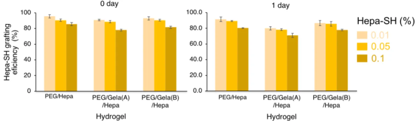
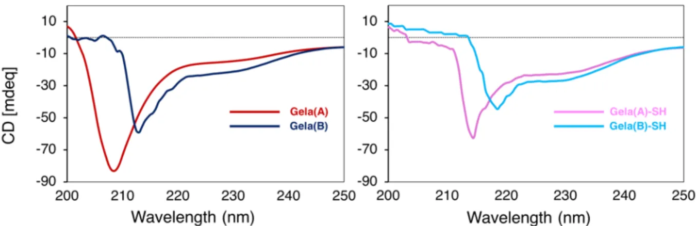
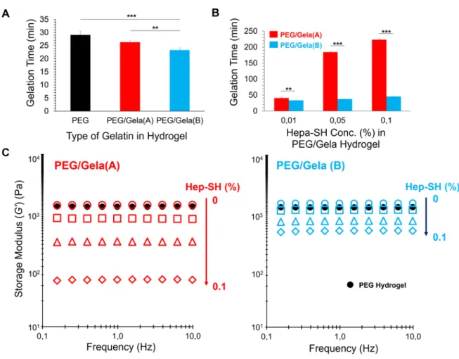
Evidently, the type of gelatin strongly affected the biological activity of hydrogels.
Specifically, thiolated type-B gelatin had much higher compatibility with Hepa-SH compared with thiolated type-A gelatin, resulting in a shorter gelation time and higher storage modulus of hydrogels observed in physicochemical characterization. Finally, incorporation of GFs into dual functionalized hydrogels under optimized conditions was validated by the proliferation and morphological change of NIH-3T3 cells and HUVECs seeded on the hydrogels and accelerated formation of 2D cellular sheets for cell sheet-based tissue engineering.
2.2 Experimental 2.2.1 Materials.
PTE-200 SH (Sunbright®) {4arm PEG, -[(CH2)2-SH]4, MW 20 kDa} was purchased from NOF Corporation (Tokyo, Japan). Glycyl-L tyrosine hydrate and 1-ethyl-3-(3 dimethyl aminopropyl) carbodiimide (EDC) were purchased from Tokyo Chemical Industry (Tokyo, Japan). HRP (100 U/mg), 1,4-dithiothreitol (DTT), and heparin sodium salt (200 U/mg, MW 15 kDa) were purchased from Wako Pure Chemical Industries (Osaka, Japan). 1-Hydroxybenzotriazole (HOBt) was purchased from Watanabe Chemical Industry (Hiroshima, Japan). Gelatin from porcine skin (type A, acid-treated gelatin) and gelatin from bovine skin (type B, alkaline-treated gelatin) were purchased from Sigma Aldrich. 5,5′-Dithiobis (2-nitrobenzoic acid) (DTNB) and a Cellstain-double staining kit were purchased from Dojindo (Kumamoto, Japan).
31
NIH3T3 mouse fibroblasts was obtained from the Riken Cell Bank (Tsukuba, Japan), and human umbilical vein endothelial cells (HUVECs) was purchased from KURABO (Osaka, Tokyo). Recombinant human basic fibroblast growth factor (bFGF, RSD) was acquired from Funakoshi (Tokyo, Japan). Trypan blue (0.4%), minimum essential medium (MEM), GlutaMAX™-I, and 10% fetal bovine serum (FBS) were purchased from Thermo Fisher Scientific (Waltham, USA). Cysteamine (2-mercaptoethylamine hydrochloride), cystamine, 1% antibiotic-antimycotic, trypsin 0.25%/1 mM EDTA, and Dulbecco’s phosphate buffered saline [D-PBS (-)] were purchased from Nacalai Tesque (Kyoto, Japan). EGM-2 supplemented with FBS, hydrocortisone, growth factors, such as bFGF, VEGF, R3-IGF-1, and hEGF, ascorbic acid, and GA-1000 was supplied by Lonza (Walkersville, USA). Human FGF DuoSet ELISA kit was acquired from R&D systems (Minneapolis, USA). Milli-QÒ water was used in experiments.
2.2.2 Synthesis of Hepa-SH and Gela-SH.
Briefly, heparin was dissolved in Milli-Q water at 10 mg/mL, and then EDC and HOBt were added. The molar ratio of reactants was 1:1:1:2 (heparin:HOBt:EDC:
cysteamine)25,31. The pH of the reaction mixture was adjusted to 6.8 with 0.1 M NaOH and/or HCl solutions, and the reaction was allowed to continue for 5 h with stirring at room temperature. Then, a 10-fold molar excess of DTT (moles per COOH of heparin) was added to reduce the disulfide groups and generate free thiol groups. The reaction was performed for 3 h at pH 7.5 that was adjusted to pH 3.5 by addition of 1 N HCl.
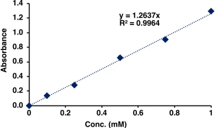
The solution was dialyzed against HCl (pH 3.5) containing 100 mM NaCl, followed by lyophilization. Gela-SH was prepared by following the protocol in our previous study15. Free thiol groups in gelatin samples were measured by Ellman’s reagent assay. DTNB reacted with a free sulfhydryl group to yield a mixed disulfide and 2-nitro-5-thiobenzoic acid (TNB). The absorbance of Gela-SH samples was measured with a microplate
32
reader (Power Wave X, Bio-Tec Instruments Inc., USA) at 412 nm. A cysteine solution was used to estimate free thiols. A calibration curve to quantify free thiol groups was obtained by measuring the absorbance of known concentrations of cysteine solutions (Fig. 2.1). Hereafter, Gela-SH samples obtained from type A and B gelatin are abbreviated as Gela(A)-SH and Gela(B)-SH, respectively.
Fig. 2.1. Calibration curve obtained by measuring the absorbance of the cysteine solution.
2.2.3 Arginine density of gelatin.
The amount of free arginine side chains is measured using a fluorometric technique adapted from previous studies33,34. The arginine density in Gel-SH (from bovine or porcine gelatin) was quantified by reacting arginine groups with 9,10- phenanthrenequinone to produce a fluorescent compound. Several diketo compounds, such as 2-amino-1H-phenanthrol[9,10-d] imidazole and 9,10- phenanthrenequinone, form a stable fluorescent compound upon reaction with arginine. 9,10-phenanthrenequinone has been shown to react with arginine and related compounds containing quanidinium groups. Briefly, 1 mg/mL gelatin or gel-SH was mixed with 300 μL of an ethanol solution of 9,10-phenanthrenequinone (150 μM) and 50 μL of an NaOH aqueous solution (2 N). The mixture was incubated at 60 °C in the dark for 3 h. Then, 200 μL of the gelatin solution was mixed with 200 μL HCl (1.2
y = 1.2637x R² = 0.9964
0.0 0.2 0.4 0.6 0.8 1.0 1.2 1.4
0 0.2 0.4 0.6 0.8 1
Absorbance
Conc. (mM)