INTRODUCTION
P
eriostin, also known as osteoblast-specific factor 2, is a 93.3-kDa extracellular matrix (ECM) protein, cloned from a murine osteoblast-like cell line in 1993 [1]. Periostin has been found to play an important role in tumor growth and to up-regulate various cancers [2]. Periostin is a ligand for integrin such as αVβ1, αVβ3, αVβ5, and α6β4, and activates the Akt/PKB and FAK-mediated signaling pathways. Periostin hasbeen reported to increase the adhesion, angiogenesis, invasion and metastasis of cancer cells [3-5]. It also induces neovascularization and supports tumor growth by inducing vascular endothelial growth factor recep- tor expression in vascular endothelial cells, while also promoting the survival of vascular endothelial cells through the Akt pathway [6-8]. Periostin supports on- cogenesis not only by activating intracellular pathways but also through its effect on ECM desmoplasia. The desmoplastic stroma of a malignant neoplasm consti- Summary: Periostin is an extracellular matrix N-glycoprotein that is a major constituent of the desmoplastic stroma around solid tumors. Periostin promotes tumor invasion and metastasis via epithelial-mesenchymal tran- sition. The aims of this study were to evaluate periostin expression immunohistochemically and quantitatively in patients with non-small cell lung cancer (NSCLC) and to assess any associations with clinical features and prog- nosis. A total of 184 specimens of NSCLC tissue were investigated, including 134 adenocarcinomas, 39 squamous cell carcinomas, and 11 other histologic subtypes. The intra-tumoral periostin expression area in each captured field was calculated using the image processing integration software WinROOF. The mean periostin expression score was classified as high or low by the median value of its expression area. Univariate analysis demonstrated that gender, tumor size, T status, N status, stage, histologic type, smoking habits, percent vital capacity, 1% forced expiratory volume, and pleural invasion were each significantly associated with periostin scores. Multivariate analysis revealed that high periostin expression score was an independent prognostic factor significantly associ- ated with decreased cancer-specific survival (HR, 3.65; 95% CI, 1.04–12.84; P=0.0439). We concluded that intra- tumoral periostin expression was an independent prognostic factor for NSCLC.
Key words periostin, non-small cell lung cancer, survival, prognostic factor, multivariate analysis
Periostin Expression in Non-Small Cell Lung Cancer:
Clinical Significance
DAIGO MURAKAMI, SHINZO TAKAMORI, AKIHIKO KAWAHARA*, MASAHIRO MITSUOKA, MASAKI KASHIHARA, KOICHI YOSHIYAMA, RYOICHI MATSUMOTO,
SHINTARO YOKOYAMA, KIMINORI FUJIMOTO**, ATSUSHI KAWAGUCHI†, KENJI IZUHARA‡ AND YOSHITO AKAGI
Department of Surgery, Kurume University School of Medicine, *Department of Diagnostic Pathology, Kurume University Hospital,
**Department of Radiology, Kurume University School of Medicine, Kurume, 830-0011 Japan
†Center for Comprehensive Community Medicine, ‡Division of Medical Biochemistry, Department of Biomolecular Sciences, Faculty of Medicine, Saga University, Saga, 849-8501 Japan.
Received 20 April 2017, accepted 11 September 2017 J-STAGE advance publication 27 November 2017
Edited by TOMOAKI HOSHINO
Corresponding author: Shinzo Takamori M.D., Ph.D., Department of Surgery, Kurume University School of Medicine, 67 Asahi-machi, Kurume-city, Fukuoka, 830-0011, Japan. Tel: +81-942-31-7566, Fax: +81-942-34-0709, E-mail: [email protected]
Abbreviations: CSS, cancer-specific survival; ECM, extracellular matrix; FEV, forced expiratory volume; HE, hematoxylin-eosin; NSCLC, non-small cell lung cancer; WHO, World Health Organization.
Original Contribution
tutes a tumor microenvironment that supports tumor growth and invasion. Periostin forms a plexus of ECM by binding to tenascin-C and fibronectin [9, 10]. The excessive production of ECM components such as periostin may be involved in generating a microenvi- ronment that supports the tumor [11]. Importantly, periostin is involved in the epithelium-mesenchymal transition of carcinoma cells [12-14]. Periostin was found to be over-expressed in various types of human cancers, including neuroblastoma, head and neck can- cers, nasopharyngeal carcinoma, thyroid carcinoma, ovarian cancer, breast cancer, pancreatic ductal adeno- carcinoma, and non-small cell lung carcinoma (NSCLC) [2,15,16]. NSCLC is the most common type of lung cancer, and its development and progression is a com- plex process in which the tumor microenvironment plays an important role. In this study, we immunohis- tochemically and quantitatively investigated periostin expression in NSCLC, and evaluated its clinical sig- nificance.
MATERIALS AND METHODS Patients and Samples
The study was approved by the Ethics Committee of Kurume University (Research No. 11018). Speci- mens from a total of 184 consecutive patients with stage I–III NSCLC who underwent complete resection without preoperative therapy at Kurume University Hospital between 2007 and 2010 were reviewed. The patients consisted of 101 men and 83 women, with a median age of 68 years (age range: from 15 to 87 years).
Tumor samples were classified according to the 2004 World Health Organization (WHO) histological clas- sification and the 2010 7th TNM edition of the UICC.
There were 134 adenocarcinomas, 39 squamous cell carcinomas, and 11 other histologic types (including 4 pleomorphic carcinomas, 2 typical carcinoids, 1 carci- nosarcoma, 1 mucoepidermoid carcinoma, and 3 un- classified tumors). There were 79 patients at stage IA, 44 at stage IB, 21 at stage IIA, 13 at stage IIB, 20 at stage IIIA, and 7 patients at stage IIIB disease (Table 1).
Inpatient, outpatient and surgical medical records were reviewed for each patient. The gender, age, tu- mor size, TNM staging, WHO histological classifica- tion, pleural invasion, smoking status, pulmonary func- tion, including percent vital capacity (%VC) and forced expiratory volume (FEV) 1%, and survival period were each investigated for all patients. Preoperative therapy was not performed in any patient. Postopera- tive adjuvant therapy including chemotherapy, radia- tion, or other therapy was performed in conformity
with established criteria. Review of the postoperative clinical course was performed using the outpatient medical records and by inquiries by telephone or let- ter. A follow-up survey was completed in full by pa- tients with a return rate of 100%. Primary tumor spec- imens were immediately fixed in 10% formalin and embedded in paraffin. Serial 5-μm sections were pre- pared from each specimen, and used for routine hema- toxylin-eosin (HE) staining and immunohistochemis- try.
Immunohistochemistry for periostin
The monoclonal antibody used and the method of TABLE 1.
Patients Characteristics
Characteristic Number (n = 184) %
SexMale 101 54.9%
Female 83 45.1%
Age (years)
Median [range] 68 [25-87]
< 65 77 41.8%
>_ 65 107 58.2%
Tumor status
T1a 59 32.1%
T1b 29 15.8%
T2a 62 33.7%
T2b 12 6.5%
T3 20 10.9%
T4 2 1.0%
Lymph node status
N0 144 78.3%
N1/N2/N3 17/18/5 21.7%
Stage
IA/ IB 79/44 66.8%
IIA/ IIB 21/13 18.5%
IIIA/ IIIB 20/7 14.7%
Histology
Adenocarcinoma 134 72.8%
Squamous cell carcinoma 39 21.2%
Other histologic type 11 6.0%
(Pleomorphic carcinoma 4, Typical carcinoid 2, Carcino- sarcoma 1, Mucoepidermoid carcinoma 1, Unclassified 3) Pleural invasion
pl0 125 67.9%
pl1/ pl2/ pl3 35/14/10 32.1%
immunohistochemistry have been reported previously [17,18]. Paraffin-embedded tissue samples were cut into 5 μm sections that were captured on a coated glass slide and labeled with anti-periostin antibody using the ChemMate ENVISION method (Dako Cytomation, Glostrup, Denmark). Intrinsic peroxidase activity was blocked by treating the sections with peroxidase-block- ing reagent for 5 min. After washing with Tris-buff- ered saline for 10 min, the rat anti-human periostin monoclonal antibody of immunoglobulin G (clone no.
SS19B or SS5D, produced in the laboratory of Biomo- lecular Sciences, Saga Medical School, Saga, Japan) was diluted 1:100 and applied to the sections. The his- tological specimens were incubated at room tempera- ture for 30 min, washed with phosphate-buffered sa- line (PBS) for 15 min, and then incubated with labeled polymer-HRP secondary antibody for 30 min at room temperature. After washing with PBS for 10 min, the slides were visualized using 3, 3’-diaminobenzidine.
Immunohistochemical results were evaluated by the authors without knowledge of the patients’ clinical data.
Computer-assisted measurements of periostin expres- sion areas
The greatest dimensions of the extracted specimens were evaluated. We photographed five fields of vision, and the periostin expression area in each field was ex- amined microscopically at ×100 magnification. The image processing integration software WinROOF Ver- sion 5.7 (Mitani Corp., Tokyo, Japan) was used to ob- jectively quantify the areas of periostin expression in each field. This analysis of the periostin expression us- ing WINROOF used a semi-quantitative method. The results were digitized and calculated as units μm2/
field. The average of the five areas of periostin expres- sion was analyzed for periostin expression score in each individual case, and the mean value of periostin expression in the five fields was calculated as the over- all periostin expression value.
Statistical analysis
All data regarding the clinical and histopathologi- cal variables were grouped using standard thresholds for laboratory parameters. Continuous data were pre- sented as median and range, and category data were presented as counts and percentages. These data in- cluded gender, age, tumor size, tumor status, lymph node status, stage, histology, smoking status, %VC, FEV1%, and pleural invasion. The correlation be- tween groups of patients and their periostin expression scores were analyzed using the Wilcoxon test for trends.
Univariate and multivariate analyses with calculated
hazard ratios (HRs) and 95% confidence intervals (CIs) were performed using Cox’s proportional hazards re- gression model to determine the association between each prognostic factor and clinical parameter. In the multivariate analyses, the survival analysis, tumor size, tumor status, lymph node status and pleural inva- sion were excluded because they were highly corre- lated with stage. Kaplan–Meier curves were used to plot cancer-specific survival (CSS), and these were compared using the log-rank test. To define the cut-off of periostin expression for the Harrell’s concordance index, receiver operating characteristic analysis for survival data was computed. The CSS was defined as time duration until death due to lung cancer from the date of surgery, or until the most recent follow-up date for surviving patients. All analyses were performed using SAS 9.3 software (SAS Institute, Cary, NC, USA). P values below 0.05 were considered statisti- cally significant.
RESULTS
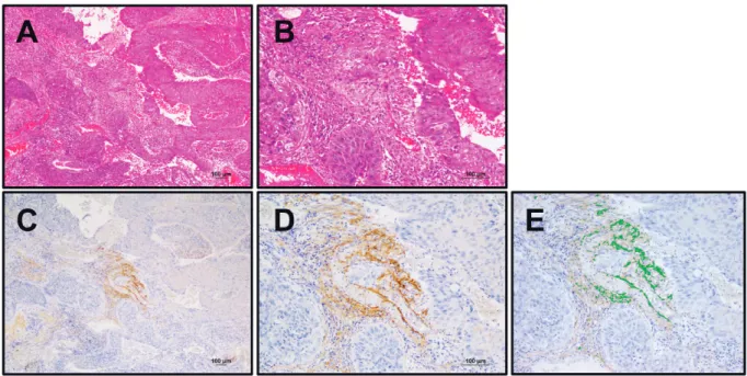
In normal lung tissue, periostin was weakly ex- pressed in parts of the bronchiolar basement membrane but not in pulmonary cells or alveolar macrophages. In lung cancer tissue, periostin was expressed in the mes- enchymal areas, but not in the cancer cells. Lung ade- nocarcinoma sections were HE stained (Fig. 1A, 1B) and immunohistochemically stained for periostin (Fig 1C, 1D). Periostin expression was found in the intra- tumoral stroma, while weak or negative staining was found in the peri-tumoral stroma and in the tumor epi- thelium. Similar results were observed in the squa- mous cell carcinoma sections (HE: Fig. 2A, 2B; peri- ostin: Fig 2C, 2D). The image processing integration software WinROOF showed similar areas of periostin expression in the two tumor types (Fig. 1E, Fig. 2E).
The periostin expression score ranged from 2.1 to 311,350.7, and the median value was 43,265.5 (Fig.
3A, 3B). Periostin expression scores were classified as high or low using this median value as a cut-off.
Univariate analysis showed significant associa- tions between periostin expression levels and male gen- der, tumor size, high T status, positive N status, high stage, squamous cell carcinoma, smoking, low %VC, low FEV1%, or positive pleural invasion (Table 2).
Cumulative CSS for the 184 patients according to periostin expression scores are shown in Figure 3. The 5-year CSS rate in patients with low periostin expres- sion was 94.4%, but fell significantly to 77.6% in pa- tients with high periostin scores (Fig. 4).
Univariate analysis of CSS demonstrated that age,
tumor size, T status, N status, stage, histology, %VC, positive pleural invasion or periostin expression score were each significantly associated with CSS. Multi- variate analysis revealed that high periostin expres-
sion score, age, and stage were independent prognos- tic factors associated with CSS (HR, 3.65; 95% CI, 1.04–12.84; p=0.0439) (Table 3).
Fig. 2. Representative images of lung squamous cell carcinomas. A. Hematoxylin and eosin stained sec- tion (×40). B. Hematoxylin and eosin stained section (×100). C. Periostin immunohistochemistry (×40).
D. Periostin immunohistochemistry (×100). E. Periostin expression labeled by WinROOF. The WinROOF labeled areas show similar periostin expression.
Fig. 1. Representative images of lung adenocarcinomas. A. Hematoxylin and eosin stained section (×40).
B. Hematoxylin and eosin stained section (×100). C. Periostin immunohistochemistry (×40). D. Periostin immunohistochemistry (×100). E. Periostin expression labeled by WinROOF. The WinROOF labeled areas show similar periostin expression.
DISCUSSION
In this study, we used the image processing inte- gration software WinROOF to objectively and quanti- tatively investigate periostin expression. The immu- nostaining results were captured as digital images, and then analyzed using WinROOF with macroinstruc-
tions for analyzing each captured area either immuno- labeled with chromogen or counterstained with hema- toxylin. The immunostained carcinoma cells were evaluated based on staining intensity and revealed periostin labeling index. The labeling index correlated highly to the immunohistochemical results for NSCLC.
Periostin expression was observed in intra-tumoral stro- TABLE 2.
Univariate Analysis between Clinicopathological Features and Periostin Expression
Variables Number Median Periostin
Expression Score P value
SexMale 101 68176.24 0.0001*
Female 83 22910.08
Age (years)
< 65 77 37496.60 0.4023
>_ 65 107 51841.20
Tumor size
< 2.5 cm 90 25622.11 0.0003*
>_ 2.5 cm 94 71171.85
Tumor status
T1/T2 162 37596.74 0.0035*
T3/T4 22 96187.39
Lymph node status
N0 144 33486.56 0.0049*
N1/N2/N3 40 81785.24
Stage
I 123 29306.78 0.0006*
II/III 61 76481.50
Histology
Adenocarcinoma 134 36660.67 0.0251*
Squamous cell carcinoma 39 72357.32
Smoking Status
Never-smoker 84 25622.11 0.0005*
Ever-smoker 100 65296.62
%VC < 80 4 138187.40 0.0290*
>_ 80 180 39482.10
FEV1.0%
<70 61 69986.38 0.0448*
>_70 123 37507.74
Pleural invasion
pl0 125 29306.78 0.0045*
pl1/ pl2/ pl3 59 72357.32
Abbreviations: VC, vital capacity; FEV1.0%, forced expiratory volume
*P <0.05
ma, while weak or negative staining was found in the peri-tumoral stroma and in the tumor epithelium. The
periostin expression areas were similar in both adeno- carcinomas and squamous cell carcinomas, showing that cytometric image analysis using WinROOF was applicable for immunohistochemical determination of periostin status in NSCLC.
Univariate analysis in the present study showed that male gender, tumor size, high T status, positive N sta- tus, high stage, squamous cell carcinoma, smoking status, low %VC, low FEV1%, and positive pleural in- vasion were each significantly associated with peri- ostin expression scores. These findings are consistent with a previous report by Takanami et al., who showed that periostin expression was significantly correlated with tumor size, lymph node status, stage, lymphatic invasion, microvessel density, and with lymphatic mi- crovessel density [5], and with a second study which found that periostin expression was significantly cor- related with male gender, high stage, high pleural in- vasion, tumor size, and with tumor relapse [14]. How- ever, in the present study, we found for the first time that histological type and pulmonary function, includ- ing %VC and FEV1%, were also correlated with peri- ostin expression scores. This suggests that periostin expression could potentially be used to predict disease progression in respiratory diseases. Our univariate analysis revealed that gender was associated with per- iostin expression. Other studies have found no signifi- cant correlation between periostin expression score and Fig. 3. Calculated periostin expression in resected lung tumors. A. Histogram of periostin expression
scores. B. Five-number summary in box plot indicates minimum, lower quartile, median, upper quartile, and maximum. Periostin expression scores were divided into high or low by the median value.
Fig. 4. Kaplan–Meier survival curves according to high or low periostin expression. For cancer-specific survival, patients with high periostin scores showed significantly lower survival rates than patients with low periostin scores.
gender [14,15]. In molecular analysis by Morra et al., periostin mRNA was up-regulated in NSCLC tissue compared with normal lung tissue, with significantly higher scores in the adenocarcinoma compared with the squamous cell carcinoma, while protein scores were correlated with squamous cell carcinoma and with larger tumor size [16]. Our results similarly showed that periostin expression scores were higher in squamous cell carcinoma than in adenocarcinoma, and higher in large tumors than small tumors. These findings suggest that intra-tumor stroma in squamous cell carcinoma and in larger tumors could lead to high periostin ex- pression scores.
With regard to survival analysis, Sasaki et al. have reported that NSCLC patients with periostin expres- sion had significantly poorer survival than patients without periostin expression [15]. Takanami et al. has found that the five-year survival rates were worse in patients with periostin expression than in patients with- out periostin expression [5]. Soltermann et al. showed that high periostin expression was a prognostic factor for decreased progression-free survival, in univariate analysis [14]. Our multivariate analysis revealed that high periostin expression score, age, and stage were independent prognostic factors associated with CSS.
Taken together, these findings indicate that periostin
expression was significantly associated with survival in NSCLC patients.
Sasaki et al. reported that there was no correlation between serum periostin levels and gender, stage, bone metastasis, N status or T status, but NSCLC patients with high periostin levels had significantly poorer sur- vival than patients with normal periostin levels. They concluded that serum periostin levels were an inde- pendent prognostic factor in NSCLC [19]. An in vitro study by Hong et al. showed that the mean value of serum periostin in NSCLC patients was significantly higher than that in normal healthy volunteers, but se- rum periostin levels in NSCLC patients had no associa- tion with gender, age, pathological type, TNM stage, lymph node status, tumor size, or invasiveness. They suggested that periostin played an important role dur- ing NSCLC progression and development [20]. Kudo et al. has described the roles of periostin in tumor de- velopment, and suggested the usefulness of periostin as a therapeutic and diagnostic target [21].
In the present study we investigated periostin ex- pression in NSCLC immunohistochemically and quan- titatively, and demonstrated that intra-tumoral peri- ostin expression was an independent prognostic factor in NSCLC. We also found for the first time that histo- logical type and pulmonary function, including %VC TABLE 3.
Univariate and Multivariate Analysis on Cancer-Specific Survival for Periostin Expression
Variables Univariate Analysis P value Multivariate Analysis P value
HR 95% CI HR 95% CI
Sex (female vs. male) 2.04 0.77-5.37 0.1489
Age (< 65vs. >_65 years) 4.14 1.21-14.21 0.0240 3.67 1.06-12.64 0.0394*
Tumor size (< 2.5vs. >_ 2.5 cm) 8.38 1.93-36.26 0.0045 Tumor status (T1/T2 vs. T3/T4) 7.22 2.89-18.04 <0.0001
Lymph node status 2.73 1.10-6.79 0.0308
(N0 vs. N1/N2/N3)
Stage (I vs. II/III) 6.51 2.34-18.07 0.0003 4.92 1.74-13.92 0.0027*
Histology 2.77 1.03-7.45 0.0435
(adenoca. vs. squamous cell ca.)
Smoking status 1.33 0.54-3.32 0.5361
(never-smoker vs. ever-smoker)
%VC ( >_80 vs. <80) 8.62 1.92-38.73 0.0050 FEV1.0% ( >_70 vs. <70) 1.91 0.78-4.70 0.1588 Pleural invasion (pl0 vs. pl1/pl2/pl3) 2.95 1.19-7.33 0.0200
Periostin expression (low vs. high) 5.96 1.73-20.47 0.0046 3.65 1.04-12.84 0.0439*
Abbreviations: HR, hazard ratio; 95% CI, 95% confidence interval, VC, vital capacity; FEV1.0%, forced expira- tory volume *P <0.05
and FEV1%, were also correlated with periostin ex- pression scores. We believe that these findings may open the way to new types of therapeutic interventions as future studies clarify the mechanisms by which per- iostin mediates cancer cell growth and survival.
DISCLOSURE STATEMENT: The authors have no conflict of interest to declare.
REFERENCES
1. Takeshita S, Kikuno R, Tezuka K, Amann E. Osteoblast- specific factor 2: cloning of a putative bone adhesion protein with homology with the insect protein fasciclin I. Biochem J 1993; 294:271-278.
2. Ruan K, Bao S, Ouyang G. The multifaceted role of peri- ostin in tumorigenesis. Cell Mol Life Sci 2009; 66:2219- 2230.
3. Gillan L, Matei D, Fishman DA, Gerbin CS, Karlan BY, et al. Periostin secreted by epithelial ovarian carcinoma is a ligand for alpha(V)beta(3) and alpha(V)beta(5) integrins and promotes cell motility. Cancer Res 2002; 62:5358-5364.
4. Baril P, Gangeswaran R, Mahon PC, Caulee K, Kocher HM, et al. Periostin promotes invasiveness and resistance of pancreatic cancer cells to hypoxia-induced cell death: role of the beta4 integrin and the PI3k pathway. Oncogene 2007; 26:2082-2094.
5. Takanami I, Abiko T, Koizumi S. Expression of periostin in patients with non-small cell lung cancer: correlation with angiogenesis and lymphangiogenesis. Int J Biol Markers 2008; 23:182-186.
6. Kühn B, del Monte F, Hajjar RJ, Chang YS, Lebeche D, et al. Periostin induces proliferation of differentiated cardio- myocytes and promotes cardiac repair. Nat Med 2007;
13:962-969.
7. Bao S, Ouyang G, Bai X, Huang Z, Ma C, et al. Periostin potently promotes metastatic growth of colon cancer by augmenting cell survival via the Akt/PKB pathway. Cancer Cell 2004; 5:329-339.
8. Shao R, Bao S, Bai X, Blanchette C, Anderson RM, et al.
Acquired expression of periostin by human breast cancers promotes tumor angiogenesis through up-regulation of vas- cular endothelial growth factor receptor 2 expression. Mol Cell Biol 2004; 24:3992-4003.
9. Kii I, Nishiyama T, Li M, Matsumoto K, Saito M, et al.
Incorporation of tenascin-C into the extracellular matrix by periostin underlies an extracellular meshwork architecture.
J Biol Chem 2010; 285:2028-2039.
10. Takayama G, Arima K, Kanji T, Toda S, Tanaka H, et al.
Periostin: a novel component of subepithelial fibrosis of bronchial asthma downstream of IL-4 and IL-13 signals. J Allergy Clin Immunol 2006; 118:98-104.
11. Erkan M, Kleeff J, Gorbachevski A, Reiser C, Mitkus T, et al. Periostin creates a tumor-supportive microenvironment in pancreas by sustaining fibrogenic stellate cell activity.
Gastroenterology 2007; 132:1447-1464.
12. Yan W, Shao R. Transduction of a mesenchyme-specific gene periostin into 293T cells induces cell invasive activity through epithelial-mesenchymal transformation. J Biol Chem 2006; 281:19700-19708.
13. Morra L and Moch H. Periostin expression and epithelial- mesenchymal transition in cancer: a review and an update.
Virchows Arch 2011; 459:465-475.
14. Soltermann A, Tischler V, Arbogast S, Braun J, Probst- Hensch N, et al. Prognostic significance of epithelial-mes- enchymal and mesenchymal-epithelial transition protein expression in non-small cell lung cancer. Clin Cancer Res 2008; 14:7430-7437.
15. Sasaki H, Lo KM, Chen LB, Auclair D, Nakashima Y, et al. Expression of periostin, homologous with an insect cell adhesion molecule, as a prognostic marker in non-small cell lung cancers. Jpn J Cancer Res 2001; 92:869-873.
16. Morra L, Rechsteiner M, Casagrande S, von Teichman A, Schraml P, et al. Characterization of periostin isoform pat- tern in non-small cell lung cancer. Lung Cancer 2012;
76:183-190.
17. Takayama G, Arima K, Kanaji T, Toda S, Tanaka H, et al.
Periostin: a novel component of subepithelial fibrosis of bronchial asthma downstream of IL-4 and IL-13 signals. J Allergy Clin Immunol 2006; 118:98-104.
18. Fujimoto K, Kawaguchi T, Nakashima O, Ono J, Ohta S, et al. Periostin, a matrix protein, has potential as a novel sero- diagnostic marker for cholangiocarcinoma. Oncol Rep 2011; 25:1211-6.
19. Sasaki H, Dai M, Auclair D, Fukai I, Kiriyama M, et al.
Serum level of the periostin, a homologue of an insect cell adhesion molecule, as a prognostic marker in nonsmall cell lung carcinomas. Cancer 2001; 92:843-848.
20. Hong L, Sun H, Lv X, Yang D, Zhang J, et al. Expression of periostin in the serum of NSCLC and its function on proliferation and migration of human lung adenocarcinoma cell line (A549) in vitro. Mol Biol Rep. 2010; 37:2285-2293.
21. Kudo Y, Siriwardena BS, Hatano H, Hatano H, Ogawa I, Tanaka T. Periostin: novel diagnostic and therapeutic target for cancer. Histol Histopathol 2007; 22:1167-1174.