INTRODUCTION
Bone mineral density (BMD) accounted for ap-proximately 75-80% of the variance in bone strength (1). It is certain that BMD is one of the important de-terminants of bone strength. However, in our clinical experience, we have encountered some patients with high or normal BMD who had suffered an atraumatic
bone fracture. On the other hand, some patients with low BMD have never had a bone fracture. Previous studies have shown that one of the determinants of bone strength, apart from BMD or bone mineral content, is the three-dimensional trabecular microstructure of bones (2-4). In order to predict bone strength and bone quality, we need new trustworthy parameters of bone quality. At present, a definition of bone quality has been difficult and controversial.
This review focused on the biophysic analysis of bone quality as a predictor of bone strength. We present two biophysic techniques, Fourier transformed infrared (FTIR) spectroscopy and phosphorus-31 solid-state
REVIEW
Biophysic evaluation of bone quality -application of Fourier
transform infrared spectroscopy and phosphorus-31 solid-state
nuclear magnetic resonance
spectroscopy-Shinjiro Takata
1),
Akira Shibata
2),
Hiroshi Yonezu
3),
Toshihide Yamada
4),
Mitsuhiko Takahashi
1), Aziz Abbaspour
1), and Natsuo Yasui
1) 1)Department of Orthopedics, and2)
Faculty of Pharmaceutical Sciences, Institute of Health Biosciences, The University of Tokushima Graduate School, Tokushima, Japan ;3)
Department of Orthopedic Surgery, Shikoku Chuo Hospital, Ehine Japan ; and 4)
Formulation Research Institute, Otsuka Pharmaceutical Co., Ltd., Tokushima, Japan
Abstract : In this review, we focus on findings obtained with biophysic techniques, Fourier transformed infrared (FTIR) spectroscopy and phosphorus-31 solid-state nuclear magnetic resonance (31P solid-state NMR) spectroscopy, which may allow us to evaluate bone quality and to predict bone strength. FTIR measures the absorption energy that produces an increase in the vibrational or rotational energy of atoms or groups of atoms within the molecule. FTIR spectroscopy allows us to examine the relative amount of minerals and matrix content and the arrangement of apatite and organic matrix. FTIR spectroscopy should become an important tool, because the relative amount of minerals and the arrangement of apatite and organic matrix could be a measure for evaluating bone quality.31P solid-state NMR spectroscopy is useful for evaluating the quality of bone and predicting bone strength by calculating the spine-lattice relaxation time (T1) of bone.31P solid-state NMR imaging can be used to measure quantitatively the mass of hydroxyapatite. The T1relaxation time of both bone and deficient hydroxyapatite was much longer than that of pure hydroxyapatite. T1relaxation time is one of the promising indices of bone quality. J. Med. Invest. 51 : 133-138, August, 2004
Keywords : Fourier transform infrared spectroscopy, phosphorus-31 solid-state nuclear magnetic resonance spec-troscopy, aging, bone quality, peripheral quantitative computed tomography
Received for publication April 26, 2004 ; accepted July 23, 2004. Address correspondence and reprint requests to Shinjiro Takata, M.D.,Ph.D., Department of Orthopedics, Institute of Health Bi-osciences, The University of Tokushima Graduate School, Kuramoto-cho, Tokushima 770-8503, Japan and Fax : +81-88-633-0178.
The Journal of Medical Investigation Vol. 51 2004 133
nuclear magnetic resonance (31
P solid-state NMR) spec-troscopy, for evaluating bone quality. These techniques should become important in evaluating bone quality in the near future.
FOURIER TRANSFORM INFRARED (FTIR)
SPECTROSCOPY AND FTIR IMAGING (FTIRI)
FTIR spectroscopy allows us to examine the molecu-lar structure and conformation of biological macromole-cules because it measures the absorption energy, which produces an increase in the vibrational or rotational energy of atoms or groups of atoms within the molecule (3, 5, 6). The information obtained from FTIR spec-troscopy includes the relative amount of minerals and matrix content, the arrangement of apatite and organic matrix (7, 8), and carbonate to phosphate ratio (9). Paschalis et al. (7) used FTIR microspectroscopy to analyze single osteons in the iliac crest of normal hu-mans, and showed that the mineral/matrix ratio in-creased from the center to the periphery of the osteon. If the relative amount of minerals and the arrangement of apatite and organic matrix are useful in evaluating bone quality, FTIR spectroscopy should become an important tool.
The spectra bands of interest in FTIR are both min-eral bands and organic bands (Fig.1). The spectral resolution was 4 cm-1
. The abscissa range was 900-1800 cm-1
, covering the phosphate band and the amide
1 band. According to previous studies (5,10), peak positions near 1020 cm-1
and near 1650 cm-1
were as-signed to the PO43-stretching vibrations of apatite and
the C=O (amide 1) stretching vibration of the bone organic matrix, respectively. Data were Fourier trans-formed and averaged after 50 scans. The mineral / matrix ratio was calculated from the ratio of absorbance of the phosphate band at 1020cm-1
to that of the amide 1 band at 1650 cm-1
.
Recently, we reported the mineral/matrix ratio of the rat femur, to clarify effects of disuse on bone (11). The infrared spectra were recorded by the FTIR spec-trometer (FTIR-1720, Perkin-Elmer, Norwalk, USA) (Fig.2). The samples were examined by the KBr tech-nique (6, 12). In our previous study (11), rat femurs were removed and soft tissue, periosteum and bone marrow were immediately cleaned off. The midshaft of the
Fig.1 Typical spectra of the femoral shaft of rats obtained by FTIR. Peak positions near 1020 cm-1
and near 1650 cm-1were assigned to the PO4 3-stretching vibrations of apatite and the C=O (amide
1)stretching vibrations of the bone organic matrix, respectively. The mineral / matrix ratio was calculated from the ratio of the absorbance of the phosphate band at 1020 cm-1to that of the amide
1 band at 1650 cm-1.
Fig.2 FTIR spectrometer (FTIR-1720, Perkin-Elmer, Norwalk, USA).
S. Takata et al. Biophysic evaluation of bone quality
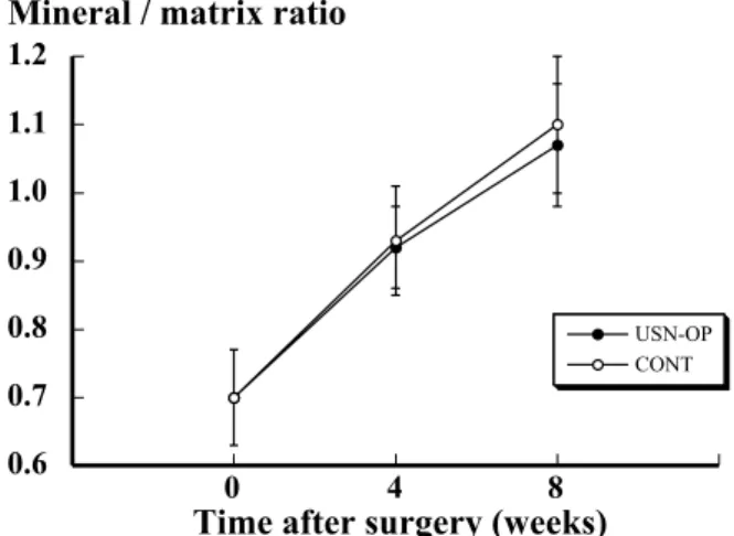
diaphysis was frozen in liquid nitrogen and lyophilized for 24 hours to remove all water, and then ground into liquid nitrogen. The mineral / matrix ratio increased 4 weeks and 8 weeks after the surgery, and we could not find significant changes in the mineral / matrix ratio of the rat femur between the disuse bone and control bone, indicating that bone quality as reflected by the mineral / matrix ratio in disused bone does not worsen (Fig.3).
Figure 4 shows the mineral / matrix ratio by FTIR and the bone mineral density by peripheral quantitative computed tomography of the femurs of male Wistar-derived albino rats aged 6 weeks and 36 weeks. The results show that the mineral / matrix ratio and bone
mineral density of the femurs of rats ageds 36 weeks were significantly greater than those of rats aged 6 weeks (p<0.0001). This suggested that the mineral / matrix ratio assessed by FTIR spectroscopy becomes an im-portant indicator of the maturation or aging of bone. FTIR imaging (FTIRI) was used to investigate the therapeutic effects of estrogen on bone quality in early postmenopausal women, and showed definite changes in bone properties at the molecular level following hormone replacement therapy (13). Moreover, FTIRI analyses showed that recombinant human parathyroid hormone treatment decreased the mineral crystal ma-turity and collagen cross-link ratio (pyridinoline/de-hydrodihydroxylysinonorleucine) on both periosteal and endosteal surfaces (14). Boskey et al. (15) showed an increased collagen maturity in both the cortical and trabecular bone of the osteonectin-null mice using FTIRI.
31
P SOLID-STATENMRSPECTROSCOPY AND
IMAGING
31P solid-state NMR spectroscopy using magic angle spinning has been used to study the chemical struc-ture of bone mineral (6, 16 -18).31
P solid-state NMR spectra were obtained on a custom-built pulse spec-trometer operated at 119.1 and 128.6 MHz for phos-phorus (19). Powdered samples of bone were tightly packed into a ceramic double-bearing rotor. The sample spinning rates were 2.0KHz and 2.16KHz for facili-tating comparisons of line intensities among samples
Fig.3 Time course of changes in mineral/matrix ratio of the
femur in unilateral sciatic neurectomy-operated (USN-OP) and control (CONT) groups (11). The mineral/matrix ratio did not differ significantly between these two groups at 4 and 8 weeks after the surgery. The values are means±SD (n=5).
Fig.4. Bone mineral density (A) and mineral/matrix ratio (B) of rat femurs aged 6
weeks and 36 weeks. The bone mineral density of femurs aged 36 weeks was significantly higher than that of femurs aged 6 weeks (p<0.0001). The mineral/matrix ratio of femurs aged 36 weeks was significantly greater than that of femurs aged 6 weeks (p<0.0001). The values are means±SD (n=5). Student’s unpaired t test was used to evaluate differences between these two groups.
and between spectrometers. 31
P solid-state NMR imaging can be used to measure quantitatively the mass of hydroxyapatite, a synthetic calcium phosphate used as an orthopedic implant material. This technique can be used to follow non-invasively the resorption and remodeling of calcium phosphate implants in vivo (20). Marchandise et al. (21) used magic angle sample spinning31
P solid-state NMR spectroscopy to study the NMR parameters of hy-droxyapatite, calcium-deficient hydroxyapatite and beta-tricalcium phosphate. The results showed that the spec-trum of rabbit bone is similar to that of deficient hy-droxyapatite, and that the T1relaxation times of both bone and deficient hydroxyapatite were much longer than that of pure hydroxyapatite.
Dawson et al. (22) applied31
P solid-state NMR
spec-troscopy to characterize and quantitate bone mineral and a synthetic apatite, to establish a model for bioab-sorption studies, and suggested that31
P solid-state NMR is a suitable technique for the in vitro analysis of bone specimens.31
P solid-state NMR spectroscopy allows us to examine a number of synthetic crystalline and noncrystalline CaPO4. Aue et al.(23) showed that iso-tropic and anisoiso-tropic chemical shifts together with proton-suppression techniques can be used to differ-entiate synthetic CaPO4compounds from one another. This technique permitted us to exclude certain CaPO4 solid phases as major or minor phases in bone, but it has also made it possible to define much more clearly the nature of the mineral phases in bone.
In our recent study, an 8.5 -Tesla superconducting spectrometer (CMX-960, CHEMAGNETICS, INC., Colorado) was used to obtain31
P solid-state NMR spec-tra (Fig.5). Casella et al. showed that the chemical shift of phosphorus of normal human bone is 3.3±0.1 ppm (6). Figure 6 shows a typical spectrum of the femoral shaft of male Wistar-derived albino rats obtained by31
P solid-state NMR spectroscopy in our recent study. The chemical shift of phosphorus of the rat femur is ap-proximately 3.3ppm. We measured the T1relaxation time of male rat femurs aged 6 weeks and 36 weeks to study the effects of aging and development on T1 relaxation time and bone strength (Figs.7 A, B). The T1relaxation time and bone strength of the male rat femurs aged 36 weeks were significantly greater than those aged 6 weeks (p<0.0001). These results suggest
that T1relaxation time is one of the promising indices
Fig.5 An 8.5 -Tesla superconducting spectrometer (CMX-960,
CHEMAGNETICS, s INC., Colorado).
Fig.6 A typical spectrum of the rat femoral shaft of obtained by31P solid-state NMR. An
8.5-Tesla superconducting spectrometer was used to obtain this spectrum. The chemical shift of phosphorus is 3.3 ppm.
S. Takata et al. Biophysic evaluation of bone quality
of bone strength and bone quality.31
P solid-state NMR spectroscopy is a promising technique for evaluating the quality of bone and predicting bone strength by calculating the T1relaxation time of bone.
REFERENCES
1. Smith CB, Smith DA : Relationship between age, mineral density and mechanical properties of human femoral compacta. Acta Orthop Scand 47(5) : 496 -502, 1976
2. Ito M, Nishida A, Nakamura T, Uetani M, Hayashi K : Differences of three-dimensional trabecular microstructure in osteopenic rat models caused by ovariectomy and neurectomy. Bone 30(4) : 594-598, 2002
3. Guo XE, Kim CH : Mechanical consequence of trabecular bone loss and its treatment : A three-dimensional model simulation. Bone 30(2) : 404-411, 2002
4. Muller R, Van Campenhout H, Van Damme B, Van Der Perre G, Dequeker J, Hildebrand T, Ruegsegger P : Morphometric analysis of human bone biopsies : A quantitative structural comparison of histological sections and micro-computed to-mography. Bone 23(1) : 59-66, 1998
5. Very JM, Gibert R, Guilhot B, Debout M, Alexandre C : Effect of aging on the amide group of bone matrix, measured by FTIR spectrophotometry, in adult subjects deceased as a result of violent death. Calcif Tissue Int 1997 : 60 : 271-275
6. Cassella JP, Barrie PJ, Garrington N, Ali SY : A Fourier transform infrared spectroscopy and solid-state NMR study of bone mineral in osteogenesis imperfecta. J Bone Miner Metab 18 : 291-296, 2000 7. Paschalis EP, Dicarlo E, Betts F, Shermann P, Mendelsohn R, Boskey AL : FTIR microspec-troscopic analysis of human osteonal bone. Calcif Tissue Int 59 : 480-487, 1996
8. Yonezu H, Ikata T, Takata S, Shibata A : Effects of sciatic neurectomy on the femur in growing rats : application of peripheral quantitative com-puted tomography and Fourier transform infrared spectroscopy. J Bone Miner Metab 17 : 259-265, 1999
9. Gadeleta SJ, Boskey AL, Paschalis E, Carlson C, Menschik F, Baldini T, Peterson M, Rimnac CM : A physical, chemical, and mechanical study of lumbar vertebra from normal, ovariectomized, and nandrolone decanoate-treated cynomolgus monkeys (Macaca fascicularis). Bone 27(4) : 541-550, 2000
10. Rey C, Renugopalakrishnan V, Collins B, Glimcher MJ : Fourier transform infrared spectroscopic study of the carbonate ions in bone mineral during aging. Calcif Tissue Int 49 : 251-258, 1991 11. Yonezu H, Takata S, Shibata A : Effects of
unilat-eral sciatic neurectomy on growing rat femur as assessed by peripheral quantitative computed tomography, Fourier transform infrared spectros-copy and bending test. J Med Invest 51:96 -102, 2004
12. Weiss P, Bohic S, Lapkowske M, Daculsi G :
Fig.7 Bone strength (A) and T1relaxation time (B) of male rat femurs. The bone strength
of femurs aged36weeks was significantly greater than that of rat aged6weeks (p<0.0001). T1relaxation time of femurs aged 36 weeks was significantly greater than that of femurs
aged 6 weeks (p<0.0001). The values are means±SD (n=5). Student’s unpaired t test was used to evaluate differences between these two groups.
Application of FT-IR microspectroscopy to the study of an infectable composite for bone and dental surgery. J Biomed Mater Res 41(1) : 167-70, 1998
13. Paschalis EP, Boskey AL, Kassem M, Eriksen EF : Effect of hormone replacement therapy on bone quality in early postmenopausal women.
J Bone Miner Res 18(6) : 955-959, 2003 14. Paschalis EP, Burr DB, Mendelsohn R, Hock
JM, Boskey AL : Bone mineral and collagen quality in humeri of ovariectomized cynomolgus monkeys given in rhPTH (1-34) for 18months. J Bone Miner Res 18(4) : 769-775, 2003 15. Boskey AL, Moore DJ, Amling M, Canalis E,
Delany AM : Infrared analysis of the mineral and matrix in bones of osteonectin-null mice and their wildtype control. J Bone Miner Res18(6):1005 -1011, 2003
16. Legrand AP, Sfihi H, Bouler JM:Nuclear mag-netic resonance spectroscopy of bone substitutes. Bone 25(2) : 103S -105S, 1999
17. Kaflak-Hachulska A, Samoson A, Kolodziejski W :1
H MAS and1 H→31
P CP/MAS NMR study of human bone mineral. Calcif Tissue Int 73 : 476-486, 2003
18. Cho G, Wu Y, Ackerman JL : Detection of hydroxyl ions in bone mineral by solid-state NMR spec-troscopy. Science 300 : 1123 -1127, 2003 19. Roberts JE, Bonar LC, Griffin RG, Glimcher MJ :
Characteristics of very young mineral phases of bone by solid state 31Phosphorus magic angle sample spinning nuclear magnetic resonance and X-ray diffraction. Calcif Tissue Int 50 : 42-48, 1992
20. Ramanathan C, Ackerman JL : Quantitative solid-state NMR imaging of synthetic calcium phosphate implants. Magn Reson Med 41:1214-1220, 1999 21. Marchandise X, Belgrand P, Legrand AP :
Solid-state31
P NMR spectroscopy of bone and bone substitute. Magnet Reson Med 28 : 1-8, 1992 22. Dawson KL, Farnan IE, Constantz BR, Young
SW : Solid-state phosphorus-31 nuclear magnetic resonance differentiation of bone mineral and synthetic apatite used to fill bone defect. Invest Radiol 26 : 946-950, 1991
23. Aue WP, Roufosse AH, Glimcher MJ, Griffin RG : Solid-state phosphorus-31 nuclear magnetic reso-nance studies of synthetic solid phases of calcium phosphate : Potential models of bone mineral. Biochemistry 23 ; 6110-6114, 1984
S. Takata et al. Biophysic evaluation of bone quality