Screening of New Bioactive Compounds from Marine
Bacteria Associated with Stony Corals
Toyama Prefectural University
Amit Raj Sharma
Contents
CHAPTER 1 Introduction 1
1-1 Marine Natural Products as a Source of Pharmaceuticals 5
1-2 Natural Products from Corals 10
1-2-1 Natural Products from Soft Corals 10
1-2-2 Natural products from Stony Corals 12
1-3 Natural Products from Coral-Associated Microorganisms 14
1-3-1 Microbial Community in Corals 14
1-3-2 Bioactive Compounds from Soft Coral-Associated Microorganisms 16
1-3-3 Bioactive Compounds from Stony Coral-Associated Microorganisms 24
CHAPTER 2 General Methodology 33
CHAPTER 3 Labrenzbactin from a Coral-Associated Bacterium Labrenzia sp. C1-1 43
3-1 Background 44
3-2 Results and Discussion 46
3-2-1 Fermentation and Isolation 46
3-2-2 Structure Determination 47 3-2-3 Absolute Configuration 49 3-2-4 Bioactivity 53 3-3 Conclusion 53 3-4 Experimental Section 56 3-5 Spectral Data 62
CHAPTER 4 Isolation and Biosynthesis of an Unsaturated Fatty Acid with Unusual
Methylation Pattern from a Coral-Associated Bacterium Microbulbifer sp. C4-6 79
4-1 Background 80
4-2 Results and Discussion 82
4-2-1 Fermentation and Isolation 82
4-2-2 Structure Determination 83
4-2-3 Biosynthesis 84
4-2-4 Bioactivity 92
4-4 Experimental Section 94
4-5 Spectral Data 100
CHAPTER 5 Two Antibacterial and PPARα/γ-Agonistic Unsaturated Keto Fatty Acids from
a Coral-Associated Bacterium Micrococcus sp. C5-9 111
5-1 Background 112
5-2 Results and Discussion 115
5-2-1 Fermentation and Isolation 116
5-2-2 Structure Determination 117
5-2-3 Determination of Configuration for the C8-C9 Double Bond 119
5-2-4 Bioactivity 122 5-3 Conclusion 128 5-4 Experimental Section 131 5-5 Spectral Data 139 CHAPTER 6 Conclusion 155 Acknowledgements 161 Publication List 162
List of Abbreviations
1-BuOH 1-butanol
CFU Colony forming unit
DMSO Dimethyl sulfoxide
EtOAc Ethyl acetate
HCl Hydrochloric acid
HDL High-density lipoprotein
HEPES 4-(2-hydroxyethyl)-1-piperazineethanesulfonic acid
HPLC High performance liquid chromatography
HPLC-UV High performance liquid chromatography-ultraviolet
HR-ESI-TOFMS
High resolution-electrospray ionization-time of flight- mass spectrometry
LC-MS Liquid chromatography-mass spectrometry
MeCN Acetonitrile
MeOH Methanol
MIC Minimum inhibitory concentration
MTT 3-(4,5-dimethylthiazol-2-yl)-2,5-diphenyl tetrazolium bromide
NMR Nuclear magnetic resonance
ODS Octadecyl-silica
TFA Trifluoroacetic acid
XTT Sodium
CHAPTER 1
Introduction
Natural products are small organic molecules produced by living organisms. They are usually called secondary metabolites because these molecules are not essential for the survival of organisms. Secondary metabolites are produced from key intermediates of the primary metabolism and relatively limited in occurrence and are often unique to particular groups of organisms or even species [1]. Secondary metabolites normally represent a chemically extremely diverse group of organic molecules and include products as a consequence of nutrient limitation, compounds for defense, regulatory molecules, or signaling molecules [1]. Secondary metabolites can be classified on the basis of the primary metabolic pathway from which they are derived or in terms of their structural similarity [1].In contrast to secondary metabolites, primary metabolites are essential for life, and include proteins, carbohydrates, lipids, nucleic acids, and their precursors and these molecules are produced by metabolic pathways common to most of organisms and include all of the modes of synthesis, interconversion, or degradation involved with the production of primary metabolites [1,2].
Historically, higher plants are considered as the most important source of drugs and medicinal plants are well documented throughout human history. In the history of natural product drug discovery, synthesis of acetylsalicylic acid (aspirin), an anti-inflammatory agent derived from the salicin, the natural product originally isolated from the bark of willow trees is one of the most noted examples [3] (Figure 1-1). Investigation of Papver somniferous (opium poppy) resulted in the isolation of morphine including several alkaloids, commercially available as analgesic agent, first reported in 1800s [4]. The bark of Cinchona succirubra had been used for the centuries for the treatment of fever and malaria [5], and the active component of bark of Cinchona succirubra was identified as quinine, and commonly used as anti-malaria drug and approved by the Food and Drug Administration (FDA) in 2004 [2]. Pilocarpine, L -histidine-derived alkaloid isolated from Pilocarpus jaborandi, which has been used as a drug for the treatment of glaucoma for over centuries and an oral formulation of pilocarpine was approved by the FDA in 1998 for the treatment of dry mouth (xerostomia) [2].
Figure 1-1. Examples of plant-derived drugs.
Traditionally, microbial natural product drug discovery program includes the isolation of targeted microorganisms, fermentation of individual isolated strains, and extraction of fermentation broth with desired organic solvent and if the extract showed any activity in bioassays, then each compound from extract was isolated and the structure was elucidated, and biological activity of pure compound was evaluated [6]. The modern era of antibiotic chemotherapy was started with introduction of penicillin in 1940s in clinical trials for the treatment of bacterial infections. Penicillin was first discovered on September 3, 1928 with the somewhat serendipitous event by Sir Alexander Fleming as a secondary metabolite derived from fungus Penicillum notatum [7].After the first clinical use of penicillin in 1940s, there was a worldwide search for new antibiotics from microorganisms and bioactive natural products began [2]. Another important milestone in the history of antibiotics was the discovery of streptomycin from Streptomyces griseus, the first aminoglycoside antibiotic, in 1943 by Selman Waksman [8]. Chloramphenicol is a broad-spectrum antibiotic originally isolated from Streptomyces venezuelae in 1947. It was the first antibiotic synthesized by chemical means soon afterwards its structure was determined [9]. The first tetracycline antibiotic discovered, aureomycin, was isolated in 1948 from soil derived Streptomyces aureofaciens [10]. Erythromycin is the first macrolide antibiotic introduced into clinincal practice since 1952 was isolated from Streptomyces erythraeus [11].Vancomycin is a glycopeptide antibiotic used for the treatment of methicillin-resistant Staphylococcus aureus. It was originally discovered from
Streptomyces orientalis isolated from soil sample in 1956 [12]. Doxorubicin is one of anthracycline family of antibiotic originally isolated from Streptomyces peucetius from soil of sourthern Italy in 1957, is a potent chemotherapeutic agent to treat various types of liquid and solid tumors [13,14]. Avermectins are series of antihelmentic drugs originally discovered from fermentation broth of Streptomyces avermitilis isolated from a soil sample in Japan [15]. Most of antibiotic classes in use today were identified in the 1940-1960s, a period referred to as the antibiotic golden age [16](Figure 1-2).
Recently, the rate of discovery of novel bioactive compounds from well-established traditional terrestrial sources have fallen dramatically [17], which in some extent preclude researchers to use common source for the quest of new bioactive compounds and lead researchers to explore new, unexplored, unusual and extreme habitat such as the marine environment, for potentially new biosynthetic diversity [18].Principally, unexplored organisms represent the best way of discovery new metabolites because of divergence of biosynthetic pathways [19]. Two points should be considered when researchers seeking for new bioactive compounds such as (i) geographically untapped sources, and (ii) overlooked group of organisms [20].
1-1 Marine Natural Products as a Source of Pharmaceuticals
Natural products are organic molecules derived from living organisms such as plant, animals and microbes. They have been exploited by people for various purposes such as fragrances, food, medicine, pigments, and insecticides [21].They often traditionally called secondary metabolites and it is restricted to small molecules less than 3,000 Daltons [22]. These molecules are not essential to the normal growth, development or reproduction of an organism [23],but they are important for survival, defending (e.g. against predators or prey organisms) or attracting (e.g. odorant fragrances of plants attracting insectsattract animals for pollination and seed dispersal) other organisms [22]. These compounds are mostly genetically encoded and produced by secondary metabolic pathways [24,25].About 50-70% of all therapeutical agents in clinical use today are natural products insipired by such small molecules [26].
Oceans cover more than 70% of the Earth’s surface and host a vast species diversity, and there are still immense untapped resources available for the drug discovery [27].Some marine ecosystems such as coral reefs are thought to surpass even tropical rain forest in terms of species diversity [28]. This species diversity provided by the marine environment can be anticipated to translate into structural diversity at the level of secondary metabolite production [29,30].Marine organisms represnt the most recent source of novel bioactive natural products as compared terresterial plants and non-marine microorganisms [31].Marine natural products show a great varitey of biological activities, which play a pivotal role in the discovery of drugs for the treatment of human diseases [32,33].
Extreme enviroments are unsual, yet promising sources of new bioactive molecules [34]. Organisms residing unexplored harsh environment such as marine enviroments featured by high salinity, low oxygen concentration, extreme temperature (low and high), high pressure
and limited light availabity favored the production of therapeutically invaluable molecules. To live under such harsh conditions, organisms must physioloigally and biologically adapted for survival. Such adaptations usually involve modification of metabolic pathways of organisms resulting in production of a great variety of molecules bearing unique structures in terms of structural diversity and functional features [31].
Marine invertebrates, in particular, have proven to be major sources of marine natural products (MNPs) in clinical trials. Sponges, corals, tunicates, and molluscs are the major source of marine natural produts retrieved from invertebrates. It was not evident that the organism itself produced those natural products or symbiotic or assoictead microbes did. About 28,500 MNPs had been identified by the end of 2016 with cytotoxic and anticancer properties [31]. The great majority of antibiotics currently used have been obtained from terrestrial sources, accounting for more than 75% of all currently used antibiotics, however antibiotics from marine sources have not yet been developed into any clinical trial phases. So most of research is devoted to the devolpment in anti-cancer drugs.
When we trace the history of marine natural products with a clear impact on the development of drugs for human use, we should not forget two arabino-nucleosides spongothymidine and spongouridine. At first they were isolated in the early 1950s from the extract of sponge Tethya crypta (currently Tectitethya crypta) [35] (Figure 1-3). Two nucleosides, the anticancer cytarabine (ara-C) and antiviral vidarabine (ara-A), are derivatives of sponge dervived arabino-nucleosides were the first FDA approved marine drugs in 1969 and 1976, respectively [31]. Untill now, nine marine-derived compounds are the FDA pharmaceutical drugs, and six compounds are undergoing phase III clinical trials. Fourteen other compounds are in phase II clinical trials, and 10 compounds are in phase I and several drugs in preclinical trails. Targets of these drugs include a wide range of diseases such as cancer, viral, Alzheimer's, chronic pain, and hypertriglyceridemia [36] (Table 1-1).
Figure 1-3. Structures of arabino-nucleosides.
Marizomib (NPI-0052; salinosporamide A), a proteasome inhibitor discovered from a marine bacteria Salinospora tropica and Salinispora arenicola. It is undergoing phase I clinical trail for the treatment of solid tumors and refractory lymphoma [37]. Bryostatin, macrocyclic polyketide, was first isolated from the bryozoan Bugula neritina in 1968 and characterized chemically in 1982 and the main pharmacological mechanism of action is modulation of protein kinase C (PKC) activity. Bryostatin is currently in phase I clinical trial for the Alzheimer’s disease [38]. Plinabulin is a synthetic analogue of halimide, originally isolated from Aspergillus sp. Plinabulin is now undergoing phase III clinical trail against solid tumors and lymphomas [39]. Plocabulin, new polyketide isolated from the Madagascan sponge Lithoplocamia lithistoides [40]. Plocabulin currently in phase II trial in patients with advanced malignancies.Omega-3 acids are essential, because human do not synthesize them and three main types of omega-3 fatty acids are alpha-linolenic acid (ALA), eicosapentaenoic acid (EPA), and docosahexaenoic (DHA). EPA and DHA are found in all fish and are effective strategy for reducing hypertriglyceride levels in patients with hypertriglyceridemia [41]. Monomethyl auristatin E (MMAE) is a synthetic analog of dolastatin 10, a cytotoxic compound originally isolated from the the sea hare Dolabella auricularia. MMAE, as a highly potent microtubule inhibitor, and demonstrated wide spectrum of anticancer activity against a multitude of lymphomas, leukemia, and solid tumors [42] (Figure 1-4).
Table 1-1. Pipeline of marine pharmaceuticals until 2019 (according to https://www.midwestern.edu/departments/marinepharmacology/clinical-pipeline.xml,
accessed on 27 October 2019) [36].
٭Antibody Drug Conjugate (Monomethylauristatin E)
Compounds Chemical class organism Source Therapeutical use Status in 2019
Cytarabine Nucleoside Sponge Cancer FDA-approved
Vidarabine Nucleoside Sponge Antiviral FDA-approved
Ziconotide Peptide Cone snail Chronic pain FDA-approved
Omega-3-acid ethyl esters Omega-3-fatty acid Fish Hypertriglyceridemia FDA-approved
Eribulin mesylate Macrolide Sponge Cancer FDA-approved
Brentuximab vedotin ADC (MMAE)٭ Mollusc Cancer FDA-approved
Trabectedin Alkaloid Tunicate Cancer FDA-approved
Plitidepsin Depsipetide Tunicate Cancer FDA-approved
Polatuzuman ventodin ADC (MMAE) Mollusc Cancer FDA-approved
Plinabulin Diketopiperazine Fungus Cancer Phase III
Tetrodotoxin Guanidinium alkaloid Puffer fish Chronic pain Phase III
Lurbinectedin Alkaloide Tunicate Cancer Phase III
Marizomib Beta-lactone-γ-lactam
Bacterium Cancer Phase III
Enfortumab ADC (MMAE) Mollusc Cancer Phase III
Plocabulin Polyketide Sponge Cancer Phase II
Tisotumab Vedotin ADC (MMAE) Mollusc Cancer Phase II
Chemoinformatics insight into the difference between MNPs and terrestrial natural products (TNPs) revealed that MNPs have lower solubility and are often larger than TNPs. MNPs possess longer chains and larger rings, usually the 8- to 10- membered rings containing ester bonds, which facilitate marine organisms to adapt to the water habitat, while the scaffolds of TNPs often have more stable ring systems and bond types. MNPs contain more halogens, because halogens are prevalent in seawater, and nitrogen and less oxygen atom compare to TNPs, which is indicative of MNPs may be synthesized by more diverse biosynthetic pathways than TNPs [43].
The main drawback of MNPs for drug discovery is the supply problem because active MNPs obtained from natural sources are in very minute amounts [31]. Total chemical synthesis or semi synthesis is the most useful technique to solve this problem [31]. Biotechnological techniques such as large scale fermentation of producing microorganisms, aquaculture of invertebrates, change in nutrient regimes of media, change in physical parameter of cultivation media, cultivation such as prokaryote-prokaryote cultivation, prokaryote-eukaryote co-cultivaton, addition of chemical elicitors, addition of adsorptive polymeric resin in fermentation media and high throughput methods to streamline cultivation could be helpful for mass production of natural products from marine organisms [31,44].
1-2 Natural Products from Corals
Corals are large, diverse group of sessile marine invertebrates belonging to the phylum Cnidaria include over 7,500 species [45]. Corals are the one of the most prolific sources of marine natural products ranking next to sponges in marine invertebrates [46]. Corals are basically classified into stony and soft corals. Stony corals are reef-building scleractinian corals and contain skeleton made of calcium carbonate and soft corals lacking calcium carbonate skeleton and includes a range of species, like gorgonians, sea fans, sea whips, sea pens, sea feathers, and blue corals. Soft corals are not responsible for reef building but may live in them [47-50]. To date, there have been more than 5,800 compounds reported from corals all over the world, attributing almost 20% of the total marine natural products. These natural products are mostly representative as terpenoids and steroids [45].
1-2-1 Natural Products from Soft Corals
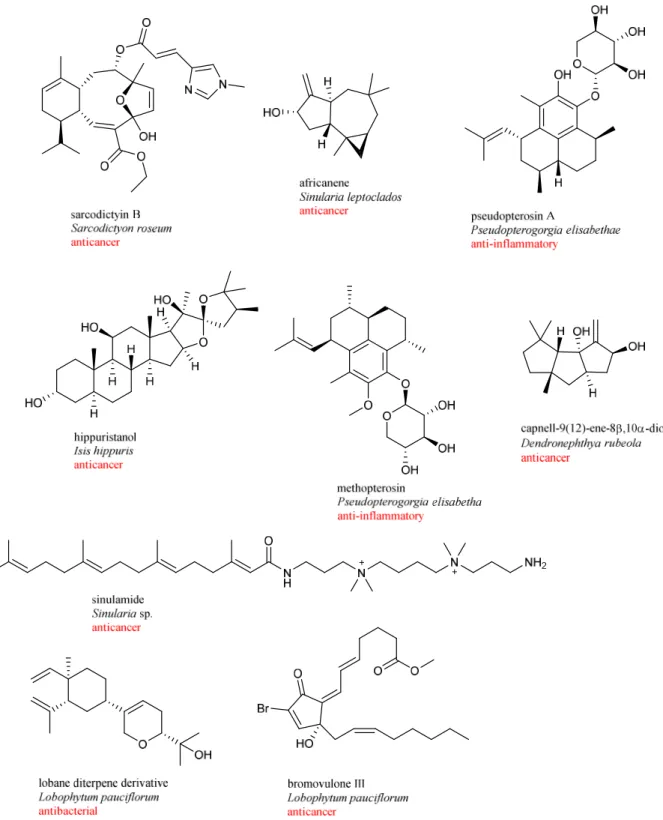
Soft corals are promising potential source of marine natural products among marine organisms and received significant attention. Many bioactive compounds with diverse
isolated from soft corals. The natural products retrieved from soft corals showed wealth of biological activities such as antimicrobial, antiviral, anticancer, antifouling and anti-inflammatory (Figure 1-5). Some of those compounds isolated from soft corals have provided a great opportunity for the development of new pharmaceuticals and antifoulants [51-59].
1-2-2 Natural Products from Stony Corals
Stony corals (or Scleractinian, hard coral, hexacoral, reef-building coral, hermatypic coral) has been rarely investigated for their ability to produce bioactive natural products, different from soft corals. The reason behind it is that researchers consider stony corals would hardly produce secondary metabolites as they consist mostly calcareous skeleton which is responsible for defensive role and thus the organic material is relatively few to the body weight. However, in soft corals secondary metabolites are produced to protect soft corals from predators because of lacking physical means of defense. New natural products obtained from stony corals usually belong to chemical classes such as alkaloids, sesterterpenes, anthraquinoids, phenols, macrolides, and acetylenic compounds. Many of them showed intriguing biological activities, such as antiviral, antibacterial, and cytotoxic activity (Figure 1-6).
Tubastrine, a new guanidinostyrene derivative was isolated from the acetone extract of stony cooral Tubastrea aurea. Tubastrine exhibited antiviral activity [60]. Brine shrimp lethality assay guided fractionation of stony coral Montipora sp. led to the discovery of N-alkyl pyridinium alkaloid, montipyridine [61]. This compound showed cytotoxic activity against panel of human cancer cell lines, A549 (human lung cancer), SK-OV-3 (human ovarian cancer), SK-MEL-2 (human skin cancer), XF498 (human CNS cancer), and HCT15 (human colon cancer) with ED50 values higer than 30 µg/mL. Aplysinopsin derivative was isolated from ethanol extract of stony coral Tubastrea aurea by solvent/solvent fractionation and silica gel column chromatography. This inhibits the first cleavage of fertilized sea urchin eggs at 2.5 µg/ml [62]. Ethanol extract of air dried sample of stony coral Tubastraea micrantha followed by defatting with hexane and extracted with CHCl3 and LH-20 purification and finally purified by RP-8 column led to the discovery of alkaloid designated tubastraine [63]. Two new macrolide derivatives, mycalolides D and E, were isolated from organic extract of stony coral Tubastraea faulkneri [64]. Mycalodie D showed modest cytotoxicity against NCI-60 cancer cell line panel with LC50 value of 0.6 µM.Two new polyacetylene carboxylic acids, montiporic acids A and B, were isolated from the stony coral Montipora digitate [65]. Montiporic acid A showed antimicrobial activity against Escherichia coli but, also cytotoxicity against P-388 murine leukemia cells with IC50 value of 5 µg/mL. Six new acetylenic compounds including montiporyne A with cytotoxic activities against human solid tumor cell lines (SKOV-3, SK-MEL-2, XF498, and HCT15) were isolated from the stony coral Montipora sp [66].
Montiporyne A exhibited cytotoxicity against human cell lines SK-MEL-2 with ED50 value of 1.4 µg/mL.
Figure 1-6. Chemical structures of natural products from stony corals.
Over the last decade, number of publications and number of new compounds from corals has been slightly decreased, whereas number of publications of new compounds from coral-associated microorganisms is slightly increasing [67]. Although corals and other marine invertebrates are rich source of bioactive natural products with a wide range of bioactivities, their pharmaceutical potential to drug discovery is often hindered by a supply problem. These marine invertebrates produce bioactive natural products in very minute amounts and it is need to collect a large amount of animal samples for extraction and ultimately drug discovery. Thus, the exploitation of bioactive natural products from corals for drug developments is still restricted by a supply problem and the need of sustainable conservation of coral reefs. So it is rational to use host-associated microorganisms for the bioactive natural products because associated microorganisms also produce a wide range of bioactive compounds.
1-3 Natural Products from Coral-Associated Microorganisms 1-3-1 Microbial Community in Corals
Corals are colonized by bacteria and other microorganisms, and the relation between corals and their colonized microbes vary from commensal to mutualistic or pathogenic or having no effect [68,69]. Corals harbor diverse and abundant microbial community, mainly consisting of eukaryota, bacteria, and archaea, as well as numerous viruses [70]. Corals live in symbiosis with unicellular dinoflagellate algae, commonly called zooxanthellae. In this association, dinoflagellate algae of genus Symbiodinium (zooxanthellae) lives within the gastrodermal cells of the coral host [70]. In this relationship, both corals and Symbiodinium are benefited. Corals provide protection of environment and compounds needed for the photosynthesis, whereas Symbiodinium furnish corals with glucose, glycerol and amino acids, which are products of photosynthesis [72]. Symbiodinium cells also provide pigmentation to corals [73] (Figure 1-7). Microorganisms are usually found in the coral surface mucus, coral tissue, and calcium carbonate skeletonand some particularly associated with Symbiodinium and each of which harbor distinct bacterial population [74]. It is assumed that coral-associated microorganisms are playing critical role in the chemical defense of their hosts by production of secondary metabolites [75]. In addition, microorganisms-associated with corals contribute to carbon cycling, sulfur cycling, nitrogen fixation, and phosphorous fixation [76]. However, some bacteria associated with corals also have negative effect on corals, including pathogenic bacteria such as Vibrio species, Aspergillus sydowii, Serratia marcescens, Thalassomonas loyan and Aurantimonas coralicida. These are the major group of bacteria responsible for the diseases of corals [77]. Study on bacteria associated with corals has been mainly focused on stony corals. Studies on the genetic diversity of bacteria associated with corals have disclosed extreme species richeness and abundance [78,79]. Diverse groups of bacteria are found from the different parts of corals [80,81]. Proteobacteria are the most abundant class of bacteria associated with corals [82].
Figure 1-7. Symbiodinium cells in the host tissues [73]. a) Stony coral with brown coloration due to the presence of Symbiodinium, b) Symbiodinium cells in the gastrovascular cavity of larva (under a microscope) of the stony coral Mussismilia hispida.
Some metabolites isolated from invertebrate-associated microbes are closely identical to compounds isolated from their hosts and sometimes associated-microbes are considered as the true producers of some marine bioactive compounds. For a long time, natural products of microbes associated with corals are largely neglected. Recently, secondary metabolites of associated microbes are attracting attention of researchers, mainly focused on associated fungi. Marine invertebrates (e.g., corals, sponges, tunicates) contain secondary metabolites in minute quantities, so it is inevitable to consume large amount of such organisms to get adequate amount of compounds for detail chemical and biological study and ultimately drug development. The concentrations of many highly active compounds in marine invertebrates are very low accounting for less than 10-6 % of their wet weight. To get approximately 1 g of ecteinascidin-743 (ET-743), an anticancer agent isolated from tunicate, about one metric ton of wet weight of tunicate Ecteinascidia turbinate must be used for extraction [83] (Figure 1-8). Therefore, exploration of new bioactive compounds for drug developments has been gradually shifted from hosts to their associated microbes. Here, bioactive new compounds from coral-associated microorganisms are categorized into two points.
Figure 1-8. Chemical structure of ecteinascidin-743 (ET-743). 1-3-2 Bioactive Compounds from Soft Coral-Associated Microorganisms
A handful of bioactive compounds were discovered from soft coral-associated bacteria, actinomycetes and fungi with a wide range of bioactivity like antimicrobial, antifungal, anticancer, antibiofilm or anti-inflammatory activity. Comparing to bacteria and actinomycetes, substantial amount of new natural products were isolated from soft coral-associated fungi, mostly belonging to genus Aspergillus followed by Penicillium.
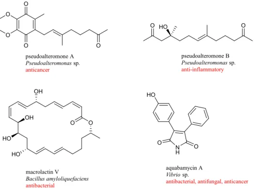
Pseudoalteromones A and B were isolated from Pseudoalteromonas collected from the cultured octocoral Lobophytum crassum [84,85]. Pseudoalteromone A, a novel ubiquinone derivative displayed cytotoxicity toward the human acute lymphoblastic leukemia cells
(MOLT-4) with IC50 value of 3.7 µg/mL. Pseudoalteromone B showed moderate inhibitory
effect on the release of elastase by human neutrophils. A new 24-membered ring lactone, macrolactin V, was isolated from culture broth of Bacillus amyloliquefaciens collected from the gorgonian coral Junceella juncea [86]. Macrolactin V exerted strong antibacterial activity against E. coli, Bacillus subtilis, and S. aureus, with MIC value of 0.1 µg/mL. Seven novel maleimide derivatives, aqabamycins A-G, were isolated from culture broth of Vibrio obtained from soft coral Sinularia polydactyla [87]. Aqabamycins A-G showed antimicrobial activity against Micrococcus luteus, Bacillus subtilis, Proteus vulgaris, and Escherichia coli and the fungus Nematospora coryli (Figure 1-9).
O O pseudoalteromone A Pseudoalteromonas sp. anticancer N H O O HO aquabamycin A Vibrio sp.
antibacterial, antifungal, anticancer
O HO O pseudoalteromone B Pseudoalteromonas sp. anti-inflammatory O O HO OH HO macrolactin V Bacillus amyloliquefaciens antibacterial O O O OH
Figure 1-9. Chemical structures of bioactive natural compounds from bacteria associated with soft corals.
Five new spirocyclic polyketides, pteridic acids C-G, were isolated from Streptomyces sp. collected from gorgonian coral Melitodes squamata [88]. Pteridic acids C, D and E showed weak antibacterial activity against Bacillus subtilis with inhibition zones of 8, 7 and 7 mm, respectively, at the equal concentration of 50 μg per disc. Chemical investigation of culture broth of Streptomyces sp. isolated from gorgonian coral Melitodes squamata led to the discovery of five novel polyhydroxy polyketides nahuoic acids B-E [89]. These compounds showed weak antibiofilm activity against Shewanella onedensis MR-1 biofilm. Unidentified soft coral-associated Streptomyces sp. produces two chlorinated polyketides, strepchloritides A and B that possess cytotoxicity against breast cancer (MCF-7) cells with IC50 values of 9.9 and 20.2 µM, respectively [90]. Chemical analysis of Streptomyces sp. collected from the surface of the gorgonian octocoral Pacifigorgia sp. led to the obtainment of two octatomic ring lactones, octalactins A and B [90]. Octalactin A exhibited strong cytotoxic activity toward human melanoma B-16-F10 and colon carcinoma HCT-116 cell lines with IC50 values of 7.2 x 10⁻³ and 0.5 µM, respectively (Figure 1-10).
Figure 1-10. Chemical structures of bioactive compounds isolated from actinomycetes associated with soft corals.
Chemical investigation of culture broth of Aspergillus sp. obtained from a gorgonian coral Dichotella gemmacea led to the discovery of two new benzylazaphilone derivatives with an unprecedented carbon skeleton, aspergilone A and its symmetrical dimer with a unique methylene bridge, aspergilone B [92]. Aspergilone A displayed cytotoxicity toward HL-60 human promyelocytic leukemia, MCF-7 human breast adenocarcinoma and A-549 human lung carcinoma cell lines with IC50 values of 3.2, 25, and 37 µg/mL, respectively. Aspergilone A also showed potential antifouling activity against the larval settlement of barnacle Balanus amphitrite at nontoxic concentration with EC50 value of 7.68 µg/mL. Three phenolic bisabolane-type sesquiterpenoids (+)-methyl sydowate, 7-deoxy-7,14-didehydrosydonic acid, and 7-deoxy-7,8-didehydrosydonic acid, were obtained from the culture broth of Aspergillus sp. isolated from gorgonian coral Dichotella gemmacea [93]. (+)-Methyl sydowate showed weak antibacterial activity against Staphylococcus aureus. Investigation of Aspergillus sydowii associated with the sea fan (gorgonian) coral Annella sp. led to the isolation of 15 compounds, including three new sesquiterpenes, aspergillusenes A and B and (+)-(7S)-7-O-methylsydonic acid [94]. These compounds did not show antioxidant activity in antioxidant activity in the 1,1-diphenyl-2-picrylhydrazyl (DPPH) scavenging assay. Three cyclic tetrapeptides,
sp. associated with gorgonian coral Melitodes squamata [95]. Aspergillipeptide C exhibited strong antifouling activity against Bugula neritina larvae settlement with EC50 value of 11 µg/mL. Four mycotoxins aluminiumneoaspergillin, zirconiumneoaspergillin, aspergilliamide, and aochratoxin A n-butyl ester were isolated from the culture broth of Aspergillus sp. associated with gorgonian coral Melitodes squamata [96]. All these compounds displayed strong or medium brine shrimp toxicity. Two prenylated indole alkaloids, 17-epinotoamides Q and M, and two new phenyl ether derivatives, cordyols D and E were obtained from static culture of Aspergillus sp. isolated from gorgonian coral Dichotella gemmacea [97]. Anthraquinone derivative, named 8-O-methylnidurufin was isolated from culture broth of Aspergillus sp. associated with gorgonian coral Dichotella gemmacea [98]. 8-O-Methylnidurufin exhibited strong antibacterial activity against Micrococcus luteus with the MIC value of 6.25 µM (Figure 1-11).
Figure 1-11. Chemical structures of new compounds isolated from Aspergillus sp. associated with soft corals.
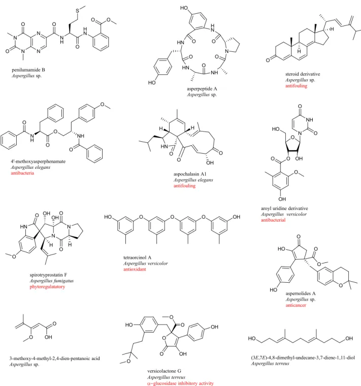
Three new lumazine peptides, penilumamides B-D and cyclic pentapeptide, asperpeptide A were isolated from the gorgonian coral-associated Aspergillus sp. [99]. Steroid derivative, (22E)-ergosta-4,6,8(14),22,24(28)-pentaen-3-one was obtained from the culture broth of Aspergillus sp. isolated from a gorgonian coral Muricella abnormalis [100]. This compound inhibited the larval settlement of barnacle Balanus amphitrite with EC50 value of 18.40 µg/mL. Phenylalanine derivative, 4’-O-methyl-asperphenamate and two cytochalasins, aspochalasin A1 and cytochalasin Z24 were isolated from the culture broth of Aspergillus elegans obtained from a soft coral Sarcophyton sp. [101]. 4’-O-Methyl-asperphenamate showed selective antibacterial activity against Staphylococcus epidermidis with the MIC value of 10 μM. Nucleoside aroyl uridine derivative was isolated from fermentation broth of Aspergillus versicolor collected from the gorgonian coral Dichotella gemmacea [102]. This compound showed antibacterial activity against Staphylococcus epidermidis with MIC value of 12.5 µM. Spirocyclic diketopiperazine alkaloid, spirotryprostatin F was isolated from Aspergillus fumigatus collected from soft coral Sinularia sp. [103]. Spirotryprostatin F in low and ultralow doses (10-6-10-17 M) exhibited stimulating action on the growth of sprout roots of soy (Glycine max) buckwheat (Fagopyrum esculentum), and corn (Zea mays). Three new cyclopentapeptides, versicoloritides A-C, a new orcinol tetramer, tetraorcinol A, and two new lactones, versicolactones A and B were isolated from the fermentation broth of Aspergillus versicolor associated with the coral Cladiella sp. [104]. Tetraorcinol A exhibited weak radical-scavenging activity against the DPPH radical with an IC50 value of 67 µM. Two aromatic butenolides, aspernolides A and B were isolated from the fermentation broth of Aspergillus terreus isolated from soft coral Sinularia kavarattiensis. Aspernolide A showed mild cytotoxicity against cancer cell lines [105]. 3-Methoxy-4-methyl-2,4-dien-pentanoic acid, was isolated from EtOAc extract of Aspergillus sp. collected from soft coral Sarcophyton tortuosum [106]. Chemical investigation on a coral-associated fungus Aspergillus terreus, led to the discovery three new compounds including a prenylated tryptophan derivative, luteoride E, a butenolide derivative, versicolactone G, and a linear aliphatic alcohol, (3E,7E)-4,8-dimethyl-undecane-3,7-diene-1,11-diol [107]. Versicolactone G showed potent α-glucosidase inhibitory activity (Figure 1-12).
N N N N N H O H N O O O O O S penilumamide B Aspergillus sp. N NH HN HN H N O HO O O O O HO asperpeptide A Aspergillus sp. O H H steroid derivative Aspergillus sp. antifouling N H O O O NH O O 4'-methoxyasperphenamate Aspergillus elegans antibacteria
aroyl uridine derivative Aspergillus versicolor antibacterial O N NH O O OH O O O OH O O OH O O versicolactone G Aspergillus terreus
glucosidase inhibitory activity
OH O N O N HN O OH H H O OH spirotryprostatin F Aspergillus fumigatus phytoregulatatory HN O O O OH H H aspochalasin A1 Aspergillus elegans antifouling O O O HO OH tetraorcinol A Aspergillus versicolor antioxidant O HO O HO O O aspernolides A Aspergillus sp. anticancer O OH O 3-methoxy-4-methyl-2,4-dien-pentanoic acid Aspergillus sp. HO HO O HO OH (3E,7E)-4,8-dimethyl-undecane-3,7-diene-1,11-diol Aspergillus terreus
Figure 1-12. Chemical structures of new compounds isolated from Aspergillus sp. associated with soft corals.
Seven aromatic polyketides, communols A-G, were isolated from the fermentation broth of Penicillium commune associated with the gorgonian coral Muricella abnormalis [108]. Communol A showed weak antimicrobial activities against Escherichia coli and Enterobacter aerogenes with MIC values of 4.1 and 16.4 µM, respectively. Two polyketides, 6,8,5’,6’-tetrahydroxy-3’-methylflavone and paecilin C were obtained from a fermentation broth of Penicillium sp. associated with gorgonian coral [109].
6,8,5’,6’-Tetrahydroxy-3’-methylflavone showed significant antifouling activity against Balanus amphitrite larvae settlement with EC50 value of 6.7 µg/mL. Dihydrothiophene-condensed chromones, oxalicumones A and B and a new natural chromone, oxalicumone C were obtained from a fermentation broth of Penicillium oxalicum associated with gorgonian coral Muricella flexuosa [110]. Oxalicumones A and B showed moderate cytotoxicity against A375 and SW-620 cell lines. Three azaphilone derivatives, pinophilins D-F and one diphenyl ether derivative, hydroxypenicillide were isolated from Penicillium pinophilum associated with gorgonian coral [111]. Five polyketides including two benzopyranones, one isochroman and two anthraquinone-citrinin derivatives were isolated from the culture broth of Penicillium citrinum associated with gorgonian coral [112]. Penicillanthranin A showed displayed moderate antibacterial activity against both Staphylococcus aureus and methicillin-resistant S. aureus with equal MIC value of 16 µg/mL. Two 2,5-piperazinedione derivatives, janthinolides A and B were isolated from the fermentation broth of Penicillium janthinellum collected from soft coral Dendronephthya sp. [113] (Figure 1-13).
communol A Penicillium commune antibacterial O OH HO O OH OH 6,8,5'6'-tetrahydroxy-3'-methylflavone Penicillium sp. antifouling O S OH O oxalicumone A Penicillium oxalicum anticancer O O O OHH O pinophilins D Penicillium pinophilum O OHOH O OH OH HO O O OH penicillanthranin A Penicillium citrinum antibacterial N NH O OH O O HO janthinolide B Penicillium janthinellum OH HO OH O O HO OH O O O HO
Figure 1-13. Chemical structures of new compounds isolated from Penicillium sp. associated with soft corals.
Chlorinated benzophenone derivative, (±)-pestalachloride D was isolated from Pestalotiopsis sp. isolated from a soft coral Sarcophyton sp. [114]. (±)-Pestalachloride D exhibited moderate antibacterial activity against Escherichia coli, Vibrio anguillarum and
Vibrio parahaemolyticus with MIC values of 5.0, 10.0 and 20.0 μM, respectively. Chemical investigation of culture broth of Cladosproium sp. collected from a gorgonian coral Anthogorgia ochracea led to the discovery of a bicyclic lactam, cladosporilactam A with promising cytotoxic activity against cervical cancer HeLa cell line with an IC50 value of 0.76 μM [115]. Five triquinane-type sesquiterpenoids, chondrosterins A-E and the known sesquiterpenoid hirsutanol C were isolated from fermentation broth of Chondrostereum sp. collected from soft coral Sarcophyton tortuosum [116]. Chondrosterin A displayed significant cytotoxic activities against cancer lines A549, CNE2, and LoVo with IC50 values of 2.45, 4.95, and 5.47 μM, respectively. Chemical investigation of Eurotium rubrum associated with soft coral Sarcophyton sp. led to the discovery of two new sulfur-containing benzofuran derivatives, eurothiocins A and B. Both compounds showed potent inhibitory effects against α-glucosidase
activity [117]. Anthraquinone alterporriol Q and the hydroanthraquinone
tetrahydroaltersolanol C were isolated from a static culture broth of Alternaria sp. retrieved from a soft coral Sarcophyton sp., showed antiviral activity against the porcine reproductive and respiratory syndrome virus (PRRSV) [118]. Two C12 polyketides, cladospolides E and F were isolated from Cladosporium sp. associated with a soft coral [119]. Cladospolide E showed potent lipid-lowering activity in HepG2 hepatocytes. Nigrospoxydons A-C and nigrosporapyrone, were obtained from Nigrospora sp. associated with a sea fan coral Annella sp [120]. Nigrospoxydon A displayed antibacterial activity against S. aureus and MRSA with MIC values of 64 and 128 µM, respectively. Four pyrone derivatives, nigrosporapyrones A-D, were isolated from the culture broth of Nigrospora sp. associated with sea fan coral Annella sp. [121]. Nigrosporapyrone A showed antibacterial activity against Staphylococcus aureus with MIC value of 128 µM. A cytochalasin derivative, xylarisin was isolated from Xylaria sp. associated with the gorgonian coral Annella sp. [122]. Xylarisin showed weak antibacterial activity. Hirsutane sesquiterpenoid, hirsutanol E was isolated from EtOAc extract of Chondrostereum sp. collected from soft coral Sarcophyton tortuosum. Hirsutanol E displayed potent cytotoxicity toward several cell lines [100] (Figure 1-14).
Figure 1-14. Chemical structures of new compounds isolated from other fungi associated with soft corals.
1-3-3 Bioactive Compounds from Stony Coral-Associated Microorganisms
Soft-coral associated microorganisms are attracting attention of researchers for discovering novel bioactive compounds. Meanwhile, stony coral-associated microorganisms are not exploited for screening of new bioactive compounds. Although stony coral-associated bacteria showed higher bioactivity than soft coral-associated bacteria, their secondary metabolites are not studied extensively [124]. This is maybe because only limited compounds were reported from stony corals and researchers speculate that the presence of calcium carbonate skeleton and very few amount of organic compounds hinders to get novel bioactive compounds from them. In addition, sustainable conservation of coral reefs, stony corals (reef
building corals) cannot be taken for the drug discovery. However, several studies revealed the presence of diverse and abundant microbial community in stony corals [125-130].
Stony coral-associated bacteria were selected for the screening of new bioactive compounds in this study because stony corals and its associated microorganisms are largely neglected for drug discovery and using new source, which has not been exploited for isolation of microorganism, ensure the isolation of new microbes with genome containing different biosynthetic pathway. To the best of our knowledge, no studies have been published with respect to new compounds from microorganisms residing stony corals before this work. So this study was conducted to estimate the ability of bacteria associated with stony corals to produce new bioactive compounds.
Literature reviews divulged that coral-associated microorganisms are the source of structurally diverse metabolites with various bioactivities. These compounds include polyketones, pyrones, cyclic peptides, lactones, alkaloids and so on, and show promising activities such as cytotoxicity, antibacterial, antifouling, phytoregulatory, and antiviral etc., which suggested that coral-associated microorganisms are promising source for finding new compounds with diverse activities. Most of the research focused on fungi associated with soft corals and, but only limited research has done with bacteria associated with soft corals. Many cultivable associated microorganisms have not been thoroughly exploited in terms of natural product biosynthesis abilities. Natural products derived from coral-associated microorganisms are still in their infancy.
References
1 Boysen RI, Hearn MTW. Comprehensive Natural Products II Chemistry and Biology.
Elsevier Science, 2010. Vol. 9, pp 5-49.
2 Dias DA, Urban S, Roessner U. Metabolites. 2012, 2, 303-336.
3 Ganjehei L, Becker RC. J. Thromb. Thrombolysis. 2015, 40, 499-511.
4 Norn S, Kruse PR, Kruse E. Dan. Medicinhist. Arbog. 2005, 33, 171-184.
5 Andrade-Neto VF, Brandão MG, Stehmann JR, Oliveira LA, Krettli AU. J.
Ethnopharmacol. 2003, 87, 253-256.
6 Giddings LA, Newman DJ. Bioactive Compounds from Extremophiles, Genomic Studies,
Biosynthetic Gene Clusters, and New Dereplication Methods. Springer International Publishing, 2015.
7 Lobanovska M, Pilla G. Yale J. Biol. Med. 2017, 29, 135-145. 8 Wainwright M. Hist. Philos. Life Sci. 1991, 13, 97-124.
9 Brock TD. Bacteriol. Rev. 1961, 25, 32-48.
10 Grossman TH. Cold Spring Harb. Perspect Med. 2016, 6, a025387.
11 Jelić D, Antolović R. Antibiotics. 2016, 5, 29. 12 Dasgupta A. Adv. Clin. Chem. 2012, 56, 75-104.
13 Unverferth DV, Magorien RD, Leier CV, Balcerzak SP. Cancer Treat. Rev. 1982, 9,
149-164.
14 Franco YL, Vaidya TR, Ait-Oudhia S. Breast Cancer (Dove Med Press). 2018, 10,
131-141.
15 Molyneux D, Taylor HR. Trends Parasitol. 2015, 31, 1.
16 Davies J. Can. J. Infect. Dis. Med. Microbiol. 2006, 17, 287-290. 17 Nunez-montero K, Barrientos L. Antibiotics. 2018, 7, 90.
18 Sayed AM, Hassan MHA, Alhadrami HA, Hassan HM, Goodfellow M, Rateb ME. J. Appl.
Microbiol. 2019.
19 Monciardini P, Iorio M, Maffioli, S, Sosio M, Donadio S. Microb. Biotechnol. 2014, 7, 209-220.
20 Leal MC, Hilario A, Munro MHG, Blunt JW, Calado R. Nat. Prod. Rep. 2016, 33, 747-750.
21 Georgiev MI. Phytochem. Rev. 2014, 13, 339-341.
22 Williams DH, Stone MJ, Hauck PR, Rahman SK. J. Nat. Prod. 1989, 53, 1189-1208.
23 Pyne ME, Narcross L, Marti VJJ. Plant Physiol.2019, 179, 844-861. 24 Theobald S, Vesth TC, Rendsvig JK. Sci. Rep.2018, 8, 17957
25 Baral B, Akhgari A, Metsä-Ketelä M. Synth. Syst. Biotechnol. 2018, 3, 163-178.
26 Newman, DJ, Cragg GM. J. Nat. Prod. 2016, 79, 629-661.
27 Snelgrove PV. Planta Med. 2016, 82, 790-799.
28 Haefner B. Drug Discov. Today. 2003, 8, 536-544.
29 Pye CR, Bertin MJ, Lokey RS, Gerwick WH, Linington RG. Proc. Natl. Acad. Sci. 2017,
114, 5601-5606.
30 Altmann KH. Chimia. 2017, 71, 646-652.
31 Jiménez C1. ACS Med. Chem. Lett. 2018, 13, 959-961.
32 Newman DJ, Cragg GM. J. Nat. Prod. 2012, 75, 311-335.
33 Hu Y, Chen J, Hu G, Yu J, Zhu X, Lin Y, Chen S, Yuan J. Mar. Drugs. 2015, 13, 202-221. 34 Núñez-Montero K, Barrientos L. Antibiotics. 2018, 7, 90.
35 Bergmann W, Burke DC. J. Org. Chem. 1955, 20, 1501-1507.
36 Mayer A. Marine Pharmaceutical: the Clinical Pipeline. Available online:
https://www.midwestern.edu/departments/marinepharmacology/clinical-pipeline.xml
(accessed on 27 October, 2019).
37 Harrison SJ, Mainwaring P, Price T, Millward MJ, Padrik P, Underhill CR, Cannell PK, Reich SD, Trikha M, Spencer A. Clin. Cancer Res. 2016, 15, 4559-4566.
38 Sun MK, Alkon DL. CNS Drug Rev. 2006, 12, 1-8.
39 Cimino PJ, Huang L, Du L, Wu Y, Bishop J, Dalsing-Hernandez J, Kotlarczyk K, Gonzales
P, Carew J, Nawrocki S, Jordan MA, Wilson L, Lloyd GK, Wirsching HG. Biomed. Rep. 2019, 10, 218-224.
40 Costales-Carrera A, Fernández-Barral A, Bustamante-Madrid P, Guerra L, Cantero R, Barbáchano A, Muñoz A. Mar. Drugs. 2019, 17, 648.
41 Ito MK. PT. 2015, 40, 826-857.
42 Chen H, Lin Z, Arnst KE, Miller DD, Li W. Molecules. 2017, 22, 1281.
43 Shang J, Hu B, Wang J, Zhu F, Kang Y, Li D, Sun H, Kong DX, Hou T. J. Chem. Inf.
Model. 2018, 58,1182-1193.
44 Romano S, Jackson SA, Patry S, Dobson ADW. Mar Drugs. 2018, 16, 244.
45 Li G, Li P, Tang X. Symbiotic Microbiomes of Coral Reefs Sponges and Corals. Springer, 2019.
46 Raimundo I, Silva SG, Costa R, Keller-Costa T. Mar Drugs. 2018, 16, 485.
48 Cooper EL, Hirabayashi K, Strychar KB, Sammarco PW. Evid. Based Complement Alternat. Med. 2014, 184959.
49 McFadden CS, Sánchez JA, France SC. Integr. Comp. Biol. 2010, 50, 389-410.
50 Aratake S, Tomura T, Saitoh S, Yokokura R, Kawanishi Y, Shinjo R, Reimer JD, Tanaka J, Maekawa H. PLoS One. 2012, 7, e30410.
51 D’Ambrosio M, Guerriero A, Pietra F. Helv. Chim. Acta. 1987, 70, 2019-2027.
52 Reddy BSG, Rao VD, Rao BCh, Dhananjaya N, Kuttan R, Babu TD. Chem. Pharm. Bull.
1999, 47, 1214-1220.
53 Look SA, Fenical W, Jacobs RS, Clardy J. Proc. Natl. Acad. Sci. U. S. A. 1985, 83, 6238-6240.
54 Bordeleau ME, Mori A, Oberer M, Lindqvist L, Chard LS, Higa T, Belsham GJ, Wagner G,
Tanaka J, Pelletier J. Nat. Chem. Biol. 2006, 2, 213-220.
55 Mayer AM, Glaser KB, Cuevas C, Jacobs RS, Kem W, Little RD, McIntosh JM, Newman
DJ, Potts BC, Shuster DE. Pharmacol. Sci. 2010, 31, 255-265.
56 Chang CH, Wen ZH, Wang SK, Duh CY. J. Nat. Prod. 2008, 71, 619-621.
57 Sata NU, Sugano M, Matsunaga S, Fusetani N. Sinulamide. Tetrahedron Lett. 1999, 40, 719-722.
58 Edrada RA, Proksch P, Wray V, Witte L, van Ofwegen L. J. Nat. Prod. 1998, 61, 358-361.
59 Shen YC, Cheng YB, Lin YC, Guh JH, Teng CM, Ko CL. J. Nat. Prod. 2004, 67, 542-546.
60 Ryuichi S, Tatsuo H. Chem. Lett. 1987, 16, 127-128.
61 Alam N, Hong J, Lee CO, Im KS, Son BW, Choi JS, Choi WC, Jung JH. J Nat Prod. 2001,
64, 956-957.
62 Fusetani N, Asano M, Matsunaga S, Hashimoto K. Comp. Biochem. Physiol. 1986, 85B,
845-846.
63 Alam M, Sanduja R, Wellington GM. Heterocycles. 1988, 27, 1998-1923.
64 Rashid MA, Gustafson KR, Cardeilina II JH, Boyd MR. J. Nat. Prod. 1995, 58, 1120-1125.
65 Fusetani N, Toyoda T, Asai N, Matsunaga S, Maruyama T. J. Nat. Prod. 1996, 59,
796-797.
66 Bae BH, Im KS, Choi WC, Hong J, Lee CO, Choi JS, Son BW, Song JI, Jung JH. J. Nat.
Prod. 2000, 63, 1511-1514.
67 Sang VT, Dat TTH, Vinh LB, Cuong LCV, Oanh PTT, Ha H, Kim YH, Anh HLT, Yang
SY. Mar. Drugs. 2019, 17, 468.
69 Lo Giudice A, Rizzo C. Diversity. 2018, 10, 80.
70 van Oppen MJH, Blackall LL. Nat. Rev. Microbiol. 2019, 17, 557-567.
71 Wooldridge SA. Biogeosciences. 2013, 10, 1647-1658.
72 Yellowlees D, Rees TA, Leggat W. Plant Cell Environ. 2008, 31, 679-694.
73 Mies M, Sumida PYG, R ̈adecker N, Voolstra CR. Front. Ecol. Evol. 2017, 5, 1-11.
74 Zhang YY, Ling J, Yang QS, Wang YS, Sun CC, Sun HY, Feng JB, Jiang YF, Zhang YZ,
Wu ML, Dong JD. Ecotoxicology. 2015, 24, 1467-1477.
75 Krediet CJ, Ritchie KB, Paul VJ, Teplitski M. Proc. Biol. Sci. B. 2013, 280, 20122328.
76 McDevitt-Irwin JM, Baum, JK, Garren M, Vega Thurber RL. Front. Mar. Sci. 2017, 4, 262.
77 Rosenberg E, Koren O, Reshef L, Efrony R, Zilber-Rosenberg I. Nat. Rev. Microbiol. 2007, 5, 355-362.
78 Bourne DG, Munn CB. Environ. Microbiol. 2005, 7, 1162-1174.
79 Lampert Y, Kelman D, Nitzan Y, Dubinsky Z, Behar A, Hill RT. FEMS Microbiol. Ecol.
2008, 64, 187-198.
80 Li J, Chen Q, Long LJ, Dong JD, Yang J, Zhang S. Sci. Rep. 2014, 4, 7320. 81 Koren O, Rosenberg E. Appl. Environ. Microbiol. 2006, 72, 5254-5259.
82 Liang J, Yu K, Wang Y, Huang X, Huang W, Qin Z, Pan Z, Yao Q, Wang W, Wu Z. Front
Microbiol. 2017, 8, 979.
83 Mendola D. In: Fusetani N (ed) Drugs from the sea. Karger, Basel, 2000, 120-133.
84 Chen YH, Lu MC, Chang YC, Hwang TL, Wang WH, Weng CF, Kuo J, Sung.
Tetrahedron Lett. 2012, 53, 1675-1677.
85 Chen YH, Kuo J, Su JH, Hwang TL, Chen YH, Lee CH, Weng CF, Sung PJ. Mar. Drugs.
2012, 10, 1566-1571.
86 Gao CH, Tian XP, Qi SH, Luo XM, Wang P, Zhang S. J. Antibiot. 2010, 63, 191-193.
87 Al-Zereini W, Fotso Fondja Yao CB, Laatsch H, Anke H. J. Antibiot. 2010, 63, 297-301.
88 Nong XH, Wei XY, Qi SH. J. Antibiot. 2017, 70, 1047-1052.
89 Nong XH, Zhang XY, Xu XY, Wang J, Qi SH. J. Nat. Prod. 2016, 79, 141-148.
90 Fu P, Kong F, Wang Y, Wang Y, Liu P, Zuo G, Zhu W. Chin. J. Chem. 2013, 31, 100-104.
91 Tapiolas DM, Roman M, Fenical W. J. Am. Chem. Soc. 1991, 113, 4682-4683.
92 Shao CL, Wang CY, Wei MY, Gu YC, She ZG, Qian PY, Lin YC. Bioorg. Med. Chem.
Lett. 2011, 21, 690-693.
94 Trisuwan K, Rukachaisirikul V, Kaewpet M, Phongpaichit S, Hutadilok-Towatana N, Preedanon S, Sakayaroj J. J. Nat. Prod. 2011, 74, 1663-1667.
95 Bao J, Zhang XY, Xu XY, He F, Nong XH, Qi SH. Tetrahedron. 2013, 69, 2113-2117.
96 Xu X, He F, Zhang X, Bao J, Qi S. Food Chem. Toxicol. 2013, 53, 46-51.
97 Chen M, Shao CL, Fu XM, Xu RF, Zheng JJ, Zhao DL, She ZG, Wang CY. J. Nat. Prod.
2013, 76, 547-553.
98 Chen M, Shao CL, Kong CJ, She ZG, Wang CY. Chem. Nat. Compd. 2014, 50, 617-620.
99 Chen M, Shao CL, Fu XM, Kong CJ, She ZG, Wang CY. J. Nat. Prod. 2014, 77,
1601-1606.
100 Chen M, Wang KL, Liu M, She ZG, Wang CY. Chem. Biodivers. 2015, 12, 1398-1406. 101 Zheng CJ, Shao CL, Wu LY, Chen M, Wang KL, Zhao DL, Sun XP, Chen GY, Wang CY.
Mar. Drugs. 2013, 11, 2054-2068.
102 Chen M, Fu XM, Kong CJ, Wang CY. Nat. Prod. Res. 2014, 28, 895-900.
103 Afiyatullov SS, Zhuravleva OI, Chaikina EL, Anisimov MM. Chem. Nat. Compd. 2012, 48, 95-98.
104 Zhuang Y, Teng X, Wang Y, Liu P, Wang H, Li J, Li G, Zhu W. Tetrahedron. 2011, 67, 7085-7089.
105 Parvatkar RR, D'Souza C, Tripathi A, Naik CG. Phytochemistry. 2009, 70, 128-132. 106 Li HJ, Cai YT, Chen YY, Lam CK, Lan WJ. Chem. Res. Chinese Universities. 2010, 26,
415-419.
107 Liua M, Suna W, Wanga J, Heb Y, Zhangb J, Lia F, Qia C, Zhua H, Xuea Y, Hua Z, Zhanga Y. Bioorganic Chemistry. 2018, 80, 525-530.
108 Wang J, Liu P, Wang Y, Wang H, Li J, Zhuang Y, Zhu W. Chin. J. Chem. 2012, 30, 1236-1242.
109 Bao J, Sun YL, Zhang XY, Han Z, Gao HC, He F, Qian PY, Qi SH. J. Antibiot. 2013, 66, 219-223.
110 Shi S, Guo K, Wang X, Chen H, Min J, Qi S, Zhao W, Li W. Mol. Med. Rep. 2017, 15, 2611-2619.
111 Zhao DL, Shao CL, Zhang Q1, Wang KL, Guan FF, Shi T, Wang CY. J. Nat. Prod. 2015, 78, 2310-2314.
112 Khamthong N, Rukachaisirikul V, Phongpaichit S, Preedanon S, Sakayaroj J. Tetrahedron. 2012, 68, 8245-8250.
114 Wei MY, Li D, Shao CL, Deng DS, Wang CY. Mar. Drugs. 2013, 11, 1050-1060. 115 Cao F, Yang Q, Shao CL, Kong CJ, Zheng JJ, Liu YF, Wang CY. Mar. Drugs. 2015, 13,
4171-4178.
116 Li HJ, Xie YL, Xie ZL, Chen Y, Lam CK, Lan WJ. Mar. Drugs. 2012, 10, 627-638. 117 Liu Z, Xia G, Chen S, Liu Y, Li H, She Z. Mar. Drugs. 2014, 12, 3669-3680.
118 Zheng CJ, Shao CL, Guo ZY, Chen JF, Deng DS, Yang KL, Chen YY, Fu XM, She ZG, Lin YC, Wang CY. J. Nat. Prod. 2012, 75, 189-197.
119 Zhu M, Gao H, Wu C, Zhu T, Che Q, Gu Q, Guo P, Li D. Bioorg. Med. Chem. Lett. 2015, 25, 3606-3609.
120 Trisuwan K, Rukachaisirikul V, Sukpondma Y, Preedanon S, Phongpaichit S, Rungjindamai N, Sakayaroj J. J. Nat. Prod. 2008, 71, 1323-1326.
121 Trisuwan K, Rukachaisirikul V, Sukpondma Y, Preedanon S, Phongpaichit S, Sakayaroj J. Phytochemistry. 2009, 70, 554-557.
122 Rukachaisirikul V, Khamthong N, Sukpondma Y, Pakawatchai C, Phongpaichit S, Sakayaroj J, Kirtikara K. Chem. Pharm. Bull. 2009, 57, 1409-1411.
123 Li HJ, Lan WJ, Lam CK, Yang F, Zhu XF. Chem. Biodivers. 2011, 8, 317-324. 124 Shnit-Orland M, Kushmaro, A. FEMS Microbiol. Ecol. 2009, 67, 371-380. 125 Huggett MJ, Apprill A. Environ. Microbiol. Rep. 2019, 11, 372-385. 126 Salerno JL, Bowen BW, Rappé MS. FEMS Microbiol. Ecol. 2016, 92.
127 Lee OO, Yang J, Bougouffa S, Wang Y, Batang Z, Tian R, Al-Suwailem A, Qian PY. Appl. Environ. Microbiol. 2012, 78, 7173-7184.
128 Rohwer F, Seguritan V, Azam F, Knowlton N. Mar. Ecol. Prog. Ser. 2002, 243, 1-10. 129 Apprill A, Weber LG, Santoro AE. mSystems. 2016, 1, e00143-00116.
130 McKew BA, Dumbrell AJ, Daud SD, Hepburn L, Thorpe E, Mogensen L, Whitby C. Appl. Environ. Microbiol. 2012, 78, 5229-5237.
CHAPTER 2
2-1 Collection of Stony Corals
Stony corals were obtained from aquarium vendors in Nagasaki and Osaka, Japan. Upon arrival of corals in our laboratory, they were kept alive in an aquarium tank until use for the isolation of bacteria.
2-2 Isolation and Identification of Coral-Associated Bacteria
In this study, aquacultured, healthy stony corals were used for isolation of bacteria. Bacteria were isolated by serial dilution and spread plate technique as follows: a small fragment of coral was taken and washed with sterile natural seawater to remove dirty particles and transiently attached microorganisms, then the surface was sterilized by using 70% ethanol to kill transiently associated microorganisms, and then washed with sterile natural seawater to remove the toxic effect of ethanol. A fragment of coral (ca. 1 g) was homogenized with mortar and pestle with an equal volume of natural seawater (1 mL), then 10-fold serial dilution was made up to 10-5, and 0.1 ml of each dilution liquid was plated onto Marine Agar 2216 (Difco). The plates were incubated at room temperature for 2 to 5 days and after two days of incubation at ambient temperature, single colonies were transferred onto Marine Agar and cultures were further purified using the same agar until to get axenic cultures (Scheme 2-1). Pure bacterial cultures were preserved by cryopreservation at -85oC in 20% glycerol in Marine Broth.
Bacterial strains were identified by 16S rRNA gene sequencing. The 16S rRNA gene of strains were amplified from a single colony by PCR with GoTaq Green Master Mix (Promega) with two universal primers (27F and 1492R). The PCR reaction was started with denaturation 98oC for 2 min followed by total 40 cycles that consisted of denaturation at 98oC for 10 sec, annealing at 55oC for 30 sec, and extension at 68oC for 10 sec. PCR products were assessed by 1% agarose gel electrophoresis (1X TAE, 1% agarose w/v). Purification of the PCR products were carried out according to the protocol of the Fast Gene Gel/PCR Extraction Kit (Nippon Genetics Co. Ltd.). The amplified 16S rRNA gene products were sequenced by Eurofins Genomics K. K., Tokyo, Japan.
2-3 Fermentation, Extraction, Isolation and Purification of Natural Products from Bacteria Associated with Stony Corals
Bacterial strains were cultured on Marine Agar was inoculated into 500 mL K-1 flask containing 100 mL of Marine Broth (Difco) or V-22 as seed medium. The flask was placed on a rotary shaker (200 rpm) at 30oC for 2 days. The seed culture (3 mL) was transferred into 500 mL K-1 flasks each containing 100 mL of A3M, A11M and A16 production medium (Table 2-1). All fermentation media except Marine Broth were prepared in natural seawater collected in Toyama Bay, Toyama, Japan. In order to increase the productivity of bacterial strains, HP-20 was added to fermentation broth, which adsorbs metabolites and minimize the toxic effects of produced metabolites [1]. The pH of the medium was adjusted to 7.0 before sterilization. The inoculated flasks were placed on a rotary shaker (200 rpm) at 30oC for 5 days. n-butanol was selected as a solvent for the extraction, because n-butanol has a wide solubility range from hydrophilic to hydrophobic compounds, but remove high molecular compounds such as polysaccharides and proteins in order to separate into organic and water layer [2]. After fermentation, 100 mL of 1-butanol was added to each flask, and the flasks were allowed to shake for 1 h. The mixture was centrifuged at 6000 rpm for 10 min, and the organic layer was separated from the aqueous layer. Purification of crude extract was carried out by mainly three steps. Firstly, crude extract was purified by silica gel column chromatography or solvent/solvent extraction, followed by ODS column chromatography and final purification was achieved by semi-preparative HPLC.
Table 2-1. Composition of seed culture and production culture media.
Media Composition Marine
Broth
peptone 0.5%, yeast extract 0.1%, and major consitutents of inorganic salts present in seawater
V22 soluble starch 1%, glucose 0.5%, NZ-case 0.3%, yeast extract 0.2%, Tryptone (Difco Laboratories) 0.5%, K2HPO4 0.1%, MgSO4·7H2O 0.05% and CaCO3 0.3%
A3M glucose 0.5%, glycerol 2%, soluble starch 2%, Pharmamedia (Traders Protein) 1.5%, yeast extract 0.3% and Diaion HP-20 (Mitsubishi Chemical, Japan) 1%
A11M glucose 0.2%, soluble starch 2.5%, yeast extract 0.5%, polypeptone (Wako Pure Chemical Industries, Ltd.) 0.5%, NZ-amine (Wako Pure Chemical Industries, Ltd.) 0.5%, CaCO3 0.3%, and Diaion HP-20 (Mitsubishi Chemical Co.) 1%
A16 glucose 2%, Pharmamedia (Traders Protein) 1%, CaCO3 0.5%, and Diaion HP-20
(Mitsubishi Chemical, Japan) 1%
2-4 HPLC-UV Screening of Metabolites of Bacteria Associated with Stony Corals
Chemical screening was employed to discover new compounds from bacteria associated with stony corals. HPLC-UV was used for chemical screening of compounds. Two milliliter of 1-butanol extract was put into vial and dried by vacuum centrifugation. Dried extract was resupeneded in 200 µL DMSO and filtered using 0.45 µm membrane filter before HPLC-DAD analysis. The extract metabolites were separated using reversed-phase HPLC and photodiode array detector (200-600 nm) was used to record their characteristic UV spectra. The conditions for the HPLC analysis were as follows: Varian Microsorb C18 column (4.6 x 100 mm) on an Agilent HP1100 system with a binary pump equipped with DAD for 200 to 600 nm. The mobile phase used the following stepwise gradient of acetonitrile (MeCN) and 0.1% formic acid at pH 2.7: 15% MeCN for 0-3 min, 15-85% MeCN for 3-25 min, 85% MeCN for 25-29 min and 85-15% MeCN for 29-32 min, with a flow-rate of 1.2 mL/min (Scheme 2-2).
Normally peaks with similar UV absorbance with in house-UV database were not considered for further study. However, this was a preliminary study conducted to assess the biosynthetic ability of associated bacteria to produce new compounds and our in-house database mainly contain UV of actinomycetes and fungi and lacking UV data for bacteria. UV only represent the chromophore present in the molecules but not the complete structure of molecules. So I considered to work with UV have hit from in-house database. Peaks with selected UV absorbance were considered for further purification and structure determination.
Scheme 2-2. Screening of metabolites of bacteria isolated from stony corals. 2-5 Structure Determination
Structure of pure compounds were determined by high resolution-mass spectrometer (HR-MS) and one dimensional and two dimensional NMR techniques and corroborated with optical rotation, UV spectrometry and IR spectrometry. Bioassays for new compounds were performed. 2-6 Dereplication of Natural Products Using HPLC-UV
Screening of natural products for novel bioactive compounds becomes increasingly a challenge. The chance of finding new bioactive compounds have become more arduous due to astronomical number of known compounds already reported in literatures. A major problem of finding new bioactive compounds from natural products is reisolation of known compounds. Reisolation of known compounds wastes time, money and resources, and easily distracts natural product chemists for more promising leads for drug discovery, and to obviate such reisolation and recharacterization of known compounds, dereplication process should be
considered before isolation of target compounds. The rapid characterization of known compounds, a process known as dereplication, has become strategically important for natural product chemists involved in screening program [3]. Nowadays, several commercial databases have been developed that can assist the natural products chemists by avoiding unnecessary structure elucidation of known compounds. Several databases are available such as CAS Registry [4-7], CSLS [8], ChemSpider [9], PubChem [10], Dictionary of Natural Products (DNP) [11], Dictionary of Marine Natural Products (DMNP) [12], AntiBase [13], MarinLit [14], Reaxys [15], and NAPRALERT [16].
Several combinations of LC hyphenated techniques such as LC-UV, LC-MS, LC-MS/MS, LC-ELSD-MS, LC-UVSPE-NMR, LC-NMR, and LC-MS/MS-NMR have been developed over the last three decades to obtain structural information of natural products [17]. UV is the simplest and most widely used detector among HPLC. Most of natural products absorb UV light in the range of 200-600 nm, including all substances having one or more double bonds and all substances that have unshared electron. Thus, even compounds with weak chromophores, such as triterpene glycosides, can be efficiently detected by UV at short wavelengths (203 nm) [18]. The major drawback of this detector is that not all natural products possess UV chromophores. UV spectra are easily acquired using a HPLC-UV examination of a crude extract or fraction. The UV profile and the maxima (λmax) of the natural products provide information on the chromophores and these distinctive spectral properties can be used for database search. For UV spectra data including, λmax and ϵ, are available in DNP and DMNP. However, partial UV spectral data available database are NAPRALERT, AntiMarin and AntiBase [19]. The UV spectra profile, including the λmax are suggestive of a chromophore present in a structure, which does not necessarily give the structure of the entire molecule. However, it offers clues for the potential substructures that can be searched for during dereplication process [19]. The UV spectrum is more informative other than just the λmax data. Matching spectra is a superior and more effective approach of UV spectra in the dereplication approach for natural products [19]. Such approaches are carried out by matching UV spectra with in-house UV spectra database [20]. Most of HPLC have the essential software for the capturing and comparing acquired UV spectral data. However, UV spectral data acquired from other HPLC cannot be compared [19]. Unfortunately, there is no UV spectral database available which contain other information required for the dereplication process [19]. UV data with mass data is a powerful and efficient tool for dereplication.
Fixed wavelength, multiple wavelength, and diode array detector (DAD) are the three main types of UV detectors which are currently available. The fixed wavelength detector has a higher instrinsic sensitivity and low cost because the light emitted at specific wavelengths with given lamp. The multiple wavelength detector is more versatile, but lower sensitivity compared to the fixed wavelength detector. The diode array detector (DAD) also known as photodiode array and provides several advantages such as peak measurement at all wavelengths, determination of the correct wavelengths in one run, detection of multiple wavelengths, peak purity analysis, scan spectrum very quickly and so on.
In this study, HPLC-UV screening of natural products from bacteria associated with stony corals was used. Having some severe drawbacks, UV is still widely used in natural products screening because of easy to use, cheap, and adapted to a relatively large numbers of natural products with detection at low wavelengths. In addition, UV spectrum is only indicative of a chromophore present in a molecule, but not the entire structure. So there is still a chance of getting new molecules even after matching UV spectra profile with an in-house database if we are using unexplored sources or organisms from different taxonomic group for screening (Figure 2-1).
References
1 Phillips T, Chase M, Wagner S, Renzi C, Powell M, DeAngelo J, Michels P. J. Ind. Microbiol. Biotechnol. 2013, 40, 411-425.
2 Takagi M, Shin-ya K. J. Antibiot. 2011, 64, 699-701.
3 Alvi KA. A strategy for rapid identification of novel therapeutic leads from natural products, Biologically active natural products: Pharmaceuticals, CRC Press, 2000. 4 CAS Available online: https://www.cas.org (accessed on 18 December 2019).
5 CAS registery. Available online:
https://www.cas.org/support/documentation/chemical-substances (accessed on 18 December 2019).
6 STN. Available online: http://www.stn-international.de/index.php?id=123 (accessed on 18 December 2019).
7 SciFinder. Available online:
https://scifinder.cas.org/scifinder/view/scifinder/scifinder-Explore.jsf (accessed on 18 December 2019).
8 CSLS. Available online: https://cactus.nci.nih.gov/cgi-bin/lookup/search (accessed on 18 December 2019).
9 ChemSpider. Available online: https://www.chemspider.com (accessed on 18 December
2019).
10 PubChem. Available online: https://pubchem.ncbi.nlm.nih.gov/ (accessed on 18 December 2019).
11 Buckingham J (ed) (2011) Dictionary of Natural Products on DVD. Chapman & Hall/CRC, Boca Raton.
12 Blunt JW, Munro MHG (eds) (2008) Dictionary of Marine Natural Products. Chapman & Hall/CRC, Boca Raton.
13 AntiBase. Available online:
https://www.wiley.com/enus/AntiBase%3A+The+Natural+-Compound+Identifier-p9783527343591(accessed on 18 December 2019).
14 MarinLit. Available online: http://pubs.rsc.org/marinlit/ (accessed on 18 December 2019). 15 Reaxys. Available online: https://www.reaxys.com/#/login (accessed on 18 December
2019).
16 NAPALERT. Available online: https://www.napralert.org/ (accessed on 18 December 2019).
17 Giddings LA, Newman DJ. Bioactive Compounds from Extremophiles, Genomic Studies,
Biosynthetic Gene Clusters, and New Dereplication Methods. Springer International Publishing, 2015.
18 Wolfender JL. Planta Med. 2009, 75, 719-734.
19 Blunt J, Munro M, Upjohn M. Handbook of Marine Natural Products. Springer
Netherlands, 2012.


![Figure 1-7. Symbiodinium cells in the host tissues [73]. a) Stony coral with brown coloration due to the presence of Symbiodinium, b) Symbiodinium cells in the gastrovascular cavity of larva (under a microscope) of the stony coral Mussismilia hispida](https://thumb-ap.123doks.com/thumbv2/123deta/7672893.1212181/21.892.243.667.112.273/symbiodinium-coloration-presence-symbiodinium-symbiodinium-gastrovascular-microscope-mussismilia.webp)