A novel Siglec-F+ neutrophil subset in the
mouse nasal mucosa exhibits an activated
phenotype and is increased in an allergic
rhinitis model.
著者
MATSUI Makoto, NAGAKUBO Daisuke, SATOOKA
Hiroki, HIRATA Takako
journal or
publication title
Biochemical and biophysical research
communications
page range
S0006-291X(20)30613-6
year
2020-04-01
URL
http://hdl.handle.net/10422/00012650
A novel Siglec-F+ neutrophil subset in the mouse nasal mucosa exhibits an activated phenotype and is increased in an allergic rhinitis model
Makoto Matsui 1, Daisuke Nagakubo 1, 2, Hiroki Satooka, Takako Hirata *
Department of Fundamental Biosciences, Shiga University of Medical Science, Otsu, Shiga 520-2192, Japan
Email address: [email protected] (M. Matsui), [email protected] (D. Nagakubo), [email protected] (H. Satooka), [email protected] (T. Hirata)
* Corresponding author. Department of Fundamental Biosciences, Shiga University of Medical Science, Seta-Tsukinowa-cho, Otsu, Shiga 520-2192, Japan. E-mail address: [email protected] (T. Hirata). Phone & Fax: +81-77-548-2122.
Abstract
Neutrophils are important phagocytic cells for host defense against pathogens. They are rapidly recruited to the site of infection, release antimicrobial peptides and cytokines, and engulf and kill microbes. Neutrophils also accumulate in allergic inflammatory sites. Here we characterized neutrophil accumulation in the nasal mucosa using a mouse model of allergic rhinitis, in which mice were sensitized by intraperitoneal injection of ovalbumin (OVA) and then challenged by intranasal administration of OVA or PBS. In the nasal mucosa of both PBS- and OVA-challenged mice, we found a cell subset expressing the eosinophil marker Siglec-F in the Ly-6G+ neutrophil population. Morphological analysis of the sorted
Ly-6G+Siglec-F+ cells revealed that they were devoid of eosinophilic granules in the cytosol and were apparently neutrophils, but compared to conventional Ly-6G+Siglec-F− neutrophils, they had a more lobulated, “botryoid” nucleus. Siglec-F+ neutrophils were barely found in the nasopharynx-associated lymphoid tissue, cervical lymph nodes, the spleen, or blood. Both Siglec-F+ neutrophils and conventional neutrophils showed increased numbers in the nasal mucosa of OVA-challenged mice. Compared to conventional Siglec-F− neutrophils, Siglec-F+ neutrophils exhibited an activated phenotype and enhanced effector functions. Taken together, our findings identify Siglec-F+ neutrophils as a novel neutrophil subset with an activated phenotype that resides specifically in the nasal mucosa.
Keywords: Allergic rhinitis Inflammation Nasal mucosa Neutrophil subset Siglec-F
Introduction
The nasal tract is an important site in host defense against inhaled pathogens; it is also the site where allergic reactions occur to airborne allergens, resulting in diseases such as allergic rhinitis (AR), which is one of the most common inflammatory diseases in both
children and adults. During the initial sensitization process in AR, allergen-specific T cells are activated, which orchestrate the production of allergen-specific IgE. Subsequent exposure to the same allergen triggers an immediate IgE-mediated mast cell response and a late-phase response characterized by an influx of inflammatory cells into the nasal mucosa, causing AR symptoms such as congestion, rhinorrhea, and sneezing [1]. The influx typically contains eosinophils, which play a critical role in the pathophysiology of AR [2]. They produce
reactive oxygen species (ROS) and release their secretory granule contents, which damage the epithelium and promote inflammatory reactions. Although neutrophils also accumulate in the nasal mucosa in AR, their pathophysiological role in AR has been less established [3,4].
Neutrophils are important phagocytic cells for host defense against pathogens. They are rapidly recruited to the site of infection, release antimicrobial peptides and cytokines, and engulf and kill microbes through ROS production and degranulation [5]. Given their short lifespan and limited protein synthesis machinery, neutrophils have long been considered a homogenous population that does not phenotypically change. However, it is now clear that activation of neutrophils by cytokines such as TNF-α and bacterial compounds such as LPS prolongs their lifespan and induces phenotype changes. In addition, neutrophil subpopulations with different actions during homeostasis, infection, inflammation, and cancer have been reported in both humans and mice [6]. Notably, a distinct subset of neutrophils that is defined by its high expression of Siglec-F, a lectin normally expressed on mouse eosinophils, has been identified in lung tumor models [7]. Compared to conventional neutrophils, Siglec-Fhi
We previously reported that the nasal mucosa was infiltrated with inflammatory cells, including eosinophils and lymphocytes, in a mouse AR model [8]. We also observed
neutrophil infiltration in the nasal mucosa in this model. We found that these neutrophils were not a homogenous population but were comprised of the following two different subsets: a conventional subset and a novel subset defined by the expression of Siglec-F and CD11c. This Siglec-F+ neutrophil subset exhibited a characteristic appearance containing a “botryoid” nucleus, an activated phenotype, and enhanced effector functions.
2. Materials and methods
2.1. Mouse AR model
The mouse AR model was prepared as described previously [8]. Briefly, on days 0, 7, and 14, female C57BL/6 mice (Japan SLC, Hamamatsu, Japan) were sensitized by an intraperitoneal injection of 25 µg of ovalbumin (OVA; Sigma-Aldrich, St. Louis, MO) and 1 mg of alum (Imject Alum; Thermo Fischer Scientific, Waltham, MA) in 300 µl of PBS. Starting on day 21, the mice received a series of seven daily intranasal OVA (500 µg in PBS) or control PBS challenges. The mice were sacrificed 24 h after the last intranasal challenge for further analyses. All experimental procedures were approved by the Animal Care and Use Committee of Shiga University of Medical Science.
2.2. Cell preparation
Single-cell suspensions were prepared from nasopharynx-associated lymphoid tissue (NALT), cervical lymph nodes (CLN), and the spleen by mechanical disruption between the frosted ends of glass slides followed by filtration twice through a 100-µm nylon mesh. Nasal mucosal cells were prepared as described previously [9]. Briefly, after isolating and removing the NALT, nasal tissues were removed from bones and cartilages and incubated in RPMI 1640
containing 1 mg/ml collagenase D (Roche Diagnostics, Basel, Switzerland) for 30 min at 37°C in a shaker incubator. The cells were washed, filtered through a 100-µm nylon mesh, and counted using a hemocytometer.
2.3. Flow cytometry
The monoclonal antibodies (mAbs) used for the flow cytometry analyses were purchased from BD Biosciences (San Jose, CA), eBioscience (San Diego, CA), or BioLegend (San Diego, CA) and included antibodies to CD11a (M17/4), CD11b (M1/70), CD11c (N418), CD18 (M18/2), CD29 (HMβ1-1), CD45 (30-F11), CD49d (R1-2), CD54 (YN1/1.7.4), CD62L (MEL-14), CCR1 (S15040E), CCR2 (SA203G11), CCR3 (J073E5), CXCR2 (SA044G4), Ly-6G (1A8), and Siglec-F (E50-2440). Single-cell suspensions were incubated with
anti-CD16/CD32 (2.4G2) for 10 min, followed by staining with mAbs for 30 min on ice and washing. Data were acquired on a FACSCantoII (BD Biosciences) and analyzed using the FlowJo software. Conventional neutrophils (CD45+Ly-6G+Siglec-F−CD11c−), Siglec-F+ neutrophils (CD45+Ly-6G+Siglec-F+CD11c+), and eosinophils
(CD45+Ly-6GloSiglec-F+CD11c−) were sorted using a FACSAria (BD Biosciences). Sorted cells were subjected to cytospin followed by May–Grünwald–Giemsa staining.
2.4. Histology
Tissue samples were prepared as described previously [10]. Briefly, the maxilla and nasal cavities were dissected, fixed overnight in 1% paraformaldehyde and 10% EDTA in PBS, and decalcified in 10% EDTA in PBS for 10–14 days. Tissues were then washed with 20% sucrose in PBS, embedded in OCT compound (Sakura Finetek Japan, Tokyo, Japan), and frozen. Sections were cut, rehydrated in PBS, and blocked with donkey serum in 2% BSA in PBS followed by a Streptavidin/Biotin Blocking Kit (Vector Laboratories, Burlingame, CA)
for 15 min each. Sections were incubated overnight with goat anti–Siglec-F (R&D systems, Minneapolis, MN) at 4°C followed by incubation with Alexa Fluor 488–labeled donkey anti-goat IgG for 1 h, and blocked again with goat IgG in 2% BSA in PBS. Sections were then incubated overnight with Alexa Fluor 647–labeled anti–Ly-6G followed by incubation with Alexa Fluor 647–labeled goat anti-rat IgG, and then incubated with DAPI. Sections were mounted with ProLong Gold Antifade Reagent (Thermo Fisher Scientific), and observed under a spectral confocal microscope (C1si; Nikon, Tokyo, Japan).
2.5. Quantification of ROS and phagocytosis
For ROS measurement, sorted cells were stimulated with or without 300 ng/ml LPS (Sigma-Aldrich) for 4 h. Stimulated cells were incubated with 25 µM CM-H2DCFDA
(Invitrogen, Carlsbad, CA) for 30 min at 37°C and analyzed by flow cytometry. A phagocytosis assay was performed using pHrodo Green E. coli BioParticles Conjugate (Invitrogen) according to the manufacturer’s instructions. Briefly, single-cell suspensions prepared from the nasal mucosa were incubated with pH-sensitive fluorescent particles resuspended at 1 mg/ml in PBS. After 30 min incubation at 37°C or on ice, phagocytosis was stopped by the addition of ice-cold PBS and cells were placed on ice. Cells were then stained for CD45, Ly-6G, Siglec-F, and CD11c. Data were acquired on a FACSCantoII and analyzed using the FlowJo software.
2.6. Stimulation of conventional neutrophils
Spleen cells were incubated with or without 300 ng/ml LPS for 4 h. The cells were then stained for Siglec-F, CD11c, and CD54 and analyzed by flow cytometry.
Statistical analysis was performed using the two-tailed Student’s t test.
3. Results
3.1. Siglec-F+ neutrophils accumulate in the nasal mucosa
We previously showed that the nasal mucosa was massively infiltrated by inflammatory cells, including eosinophils and lymphocytes, in a mouse AR model [8]. As neutrophils also accumulated, we further characterized granulocyte populations infiltrating the nasal mucosa. For the AR model, C57BL/6 mice were systemically immunized with intraperitoneal
injections of OVA and then challenged for seven consecutive days with intranasal
administrations of OVA or control PBS. We prepared single-cell suspensions from the nasal mucosa of the mice sacrificed 24 h after the last intranasal challenge, and analyzed them by flow cytometry using the neutrophil marker Ly-6G and the eosinophil marker Siglec-F. Among the CD45+ leukocytes, a side scatter (SSC)hiLy-6Glo population was markedly increased in frequency in OVA-challenged mice (Fig. 1A). These cells were mostly
Siglec-F+CD11clo and likely eosinophils [11]. Indeed, May–Grünwald–Giemsa staining of the sorted cells in this gate (gate I) confirmed that they were eosinophils with ring-shaped nuclei and eosinophilic granules in the cytoplasm (Fig. 1B). Ly-6Ghi cells in the nasal mucosa contained the following two populations: Siglec-F−CD11c− (gate II) and Siglec-F+CD11c+ (gate III). Siglec-F is an eosinophil marker that is not normally expressed on neutrophils [12]. The morphology of sorted Ly-6GhiSiglec-F−CD11c− cells is consistent with that of
conventional mouse neutrophils with ring-shaped nuclei (Fig. 1B). Although
Ly-6GhiSiglec-F+CD11c+ cells are also morphologically neutrophils with a neutrally stained cytoplasm, they had segmented nuclei (Fig. 1B). These two neutrophil populations are referred here as Siglec-F− and Siglec-F+ neutrophils. Notably, the segmented nuclei of Siglec-F+ neutrophils were radially arranged with spokelike filaments, morphologically
resembling those of “botryoid” neutrophils observed in human blood during heat stroke [13,14]. Siglec-F+ neutrophils from OVA-challenged mice were larger and had more prominent blebs than those from PBS-challenged mice. The peripheral blood of PBS- or OVA-challenged mice contained eosinophils and Siglec-F− neutrophils, both of which
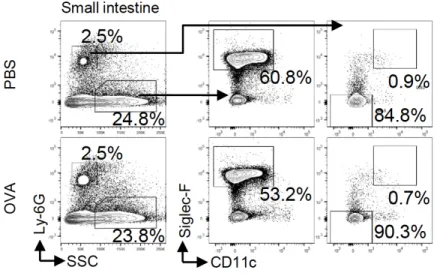
exhibited a similar morphology to the corresponding populations in the nasal mucosa (Fig. 1C, D). In contrast, Siglec-F+ neutrophils were barely found in the blood from either PBS- or OVA-challenged mice (Fig. 1C). They were also barely present in lymphoid tissues, including the NALT, CLN, and spleen (Fig. 1E). To examine whether this subset resides in mucosal tissue other than the nasal mucosa, we isolated cells from the small intestinal mucosa and analyzed them by flow cytometry. Ly-6Ghi neutrophils were much less frequent than
eosinophils in the intestinal mucosa, and most of them were Siglec-F−CD11c− (Fig. S1). Thus, Siglec-F+ neutrophils are a subset that specifically resides in the nasal mucosa.
As expected, the number of eosinophils in the nasal mucosa was remarkably increased in OVA-challenged mice compared to PBS-challenged mice (Fig. 1F). Albeit less abundant than eosinophils, both Siglec-F− and Siglec-F+ neutrophils were also increased in the nasal mucosa upon OVA challenge (Fig. 1F).
Immunofluorescence staining of nasal mucosa sections revealed the presence of cells expressing both Ly-6Gand Siglec-F in both PBS- and OVA-challenged mice, which were likely Siglec-F+ neutrophils (Fig. 2A). Ly-6G+Siglec-F+ cells accumulated in the same region where Ly-6G+Siglec-F− cells (Siglec-F− neutrophils) and Ly-6G−Siglec-F+ cells (eosinophils) accumulated. In agreement with the flow cytometry results, Ly-6G+Siglec-F+ cells were more abundant in the nasal mucosa from OVA-challenged mice than from PBS-challenged mice. In contrast, Ly-6G+Siglec-F+ cells were hardly found in the NALT and spleen sections from either PBS- or OVA-challenged mice (Fig. 2B), again confirming that Siglec-F+ neutrophils are a unique subset that resides in the nasal mucosa.
3.2. Siglec-F+ neutrophils exhibit an activated phenotype
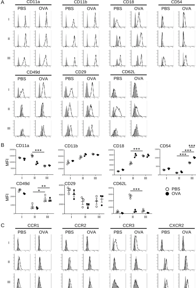
To characterize Siglec-F+ neutrophils, we examined the cell surface expression of adhesion molecules known to be involved in neutrophil activation and migration. Neutrophils express β2 integrins such as LFA-1 (αLβ2; CD11a/CD18) and Mac-1 (αMβ2; CD11b/CD18). In
PBS-challenged mice, the expression of CD11a on Siglec-F+ neutrophils was lower than that on conventional Siglec-F− neutrophils, while CD18 expression was higher on Siglec-F+ neutrophils (Fig. 3A, B). In addition to β2 integrins, the α4 integrin VLA-4 (α4β1;
CD49d/CD29) is also expressed on neutrophils. The expression of CD49d was higher on Siglec-F+ neutrophils than on Siglec-F− neutrophils in both PBS- and OVA-challenged mice. Most notably, ICAM-1 (CD54), a member of the Ig-like gene superfamily, which is generally absent on circulating neutrophils, was considerably expressed on Siglec-F+ neutrophils but only minimally expressed on Siglec-F− neutrophils. Furthermore, CD54 expression on Siglec-F+ neutrophils was upregulated upon OVA challenge. Another remarkable feature of Siglec-F+ neutrophils is the downregulation of L-selectin (CD62L), which is known to be shed in response to activation. These results suggested that Siglec-F+ neutrophils in the nasal mucosa exhibit adhesion molecule expression profiles typical of activated neutrophils.
We next investigated chemokine receptor expression profiles. Siglec-F+ neutrophils did not express CCR3, a chemokine receptor selectively expressed on eosinophils, but expressed CCR1, CCR2, and CXCR2, which mediate neutrophil migration (Fig. 3C). CXCR2 was apparently upregulated in Siglec-F+ neutrophils compared to Siglec-F− neutrophils in the nasal mucosa of OVA-challenged mice.
3.3. Siglec-F+ neutrophils have increased phagocytic activity
the three granulocyte subsets to produce ROS. Eosinophils, Siglec-F− neutrophils, and Siglec-F+ neutrophils were isolated from the nasal mucosa of OVA-challenged mice and stimulated with or without LPS. LPS-induced ROS generation was measured using the ROS-sensitive fluorescent probe CM-H2DCFDA by flow cytometry. Siglec-F+ neutrophils
showed a tendency toward increased ROS generation, although there were no significant differences among the three subsets (Fig. 4A).
We next measured the phagocytic activity of the three granulocyte subsets in the nasal mucosa of PBS- and OVA-challenged mice using fluorescent E. coli particles. As expected, compared to the two neutrophil subsets, eosinophils showed only minimal phagocytic activity (Fig. 4B, C). Compared to Siglec-F− neutrophils, Siglec-F+ neutrophils from both PBS- and OVA-challenged mice showed significantly higher phagocytosis activity (Fig. 4B, C). Collectively, these results demonstrate that Siglec-F+ neutrophils had enhanced effector functions.
3.4. Stimulation of Siglec-F− neutrophils in vitro induces only a slight increase in Siglec-F expression
Having identified an activated phenotype of Siglec-F+ neutrophils, we evaluated whether Siglec-F− neutrophils could be converted to Siglec-F+ neutrophils by stimulating these cells in vitro. LPS stimulation of Siglec-F− neutrophils from the spleen induced the upregulation of cell surface expression of Siglec-F, CD11c, and CD54 (Fig. 4D). However, the increase in Siglec-F expression was only minimal, suggesting that Siglec-F+ neutrophils cannot be generated simply by stimulating conventional neutrophils.
4. Discussion
mucosa and accumulates during nasal inflammation in a mouse AR model. These neutrophils expressed Siglec-F and exhibited an activated phenotype and enhanced effector functions.
Conventional mouse neutrophils in the bone marrow and blood have ring-shaped nuclei rather than segmented nuclei [15]. These neutrophils were also constitutively present in the nasal mucosa and accumulated under inflammatory conditions. In addition to these
conventional neutrophils, the nasal mucosa contained a distinct neutrophil population defined by the expression of Siglec-F. Remarkably, Siglec-F+ neutrophils had radially segmented, “botryoid” nuclei. Radial segmentation is caused by contractile microfilaments and
microtubules radiating from the centriole [16] and appears to be associated with inflammatory conditions [17]. In humans, botryoid neutrophils have been reported in patients with heat stroke [13,14], encephalitis [18], and cocaine abuse [19]. Although it is not known whether botryoid changes were induced in situ in the nasal mucosa, nuclear segmentation implies increased maturation of Siglec-F+ neutrophils.
The subset is characterized by unique cell surface expression profiles of adhesion molecules (CD11c+CD54+CD62Llo), indicating an activated phenotype. Indeed, compared to conventional neutrophils, Siglec-F+ neutrophils exhibited enhanced phagocytosis and a tendency toward increased ROS generation. It has been reported that in a mouse model of endotoxemia, CD54-expressing neutrophils exhibit enhanced phagocytosis and ROS
generation and that CD54 expression correlates with these effector functions [20]. Engblom et al. [7] reported that Siglec-Fhi neutrophils infiltrating lung tumors in a mouse tumor model showed increased ROS activity. Furthermore, gene expression profiles showed that CD11c (Itgax) and CD54 (Icam1) were upregulated while CD11a (Itgal) was downregulated in Siglec-Fhi neutrophils compared to Siglec-Flo neutrophils, suggesting that nasal Siglec-F+ neutrophils and lung tumor–infiltrating Siglec-Fhi neutrophils have overlapping phenotypes and functions. In humans, the following three neutrophil subsets are found in the blood during
acute inflammation: neutrophils with a conventional segmented nucleus, neutrophils with a banded nucleus, and CD62Llo neutrophils with a hypersegmented nucleus [21,22]. Siglec-F+ neutrophils phenotypically resemble human CD62Llo neutrophils, which are also
CD11c+CD54+. Functionally, human CD62Llo neutrophils inhibit T cell responses and thereby maintain tolerance [21]. Conversely, CD62Llo neutrophils reported in the nasal lavage fluid from allergic patients exhibit a T cell priming capacity and enhance eosinophil migration [23]. It remains to be determined whether Siglec-F+ neutrophils in the nasal mucosa have any interactions with T cells and promote or inhibit allergic inflammation. In this regard, during intestinal mucosal inflammation, a functionally activated neutrophil subset negatively regulates the pathogenesis of inflammatory bowel disease [24].
The origin of Siglec-F+ neutrophils also remains elusive. We showed that stimulation of Siglec-F− neutrophils with LPS induced only a slight increase in the expression of Siglec-F. It may thus be less likely that Siglec-F− neutrophils simply convert into the Siglec-F+ neutrophil subset upon stimulation. It has been proposed that human CD62Llo neutrophils are a separate neutrophil subset based on proteome profiling and in vivo pulse-chase labeling [22]. Lung tumor–infiltrating Siglec-Fhi neutrophils acquire their molecular features before arrival at the tumor site [7]. Whether the Siglec-F+ subset is generated from conventional neutrophils in situ in the nasal environment or elsewhere before arrival at the nasal mucosa will be determined.
In conclusion, our study shows a distinct subset of neutrophils residing specifically in the mouse nasal mucosa. They exhibit an activate phenotype and enhanced effector functions. Further investigation will be required to clarify the functional role and origin of this subset.
Acknowledgments
This work was supported by the JSPS KAKENHI [grant numbers 16K08831 and 17K08785].
Footnotes
Abbreviations: AR, allergic rhinitis; CLN, cervical lymph node: mAb, monoclonal antibody;
mean fluorescence intensity, MFI; NALT, nasopharynx-associated lymphoid tissue; OVA, ovalbumin; ROS, reactive oxygen species; SSC, side scatter.
1 Equally contributed.
2 Present address: Division of Health and Hygienic Sciences, Faculty of Pharmaceutical
Sciences, Himeji Dokkyo University, 7-2-1 Kamiohno, Himeji, Hyogo, 670-8524, Japan.
References
[1] L. Rosenwasser, New insights into the pathophysiology of allergic rhinitis, Allergy Asthma Proc. 28 (2007) 10-15. https://doi.org/10.2500/aap.2007.28.2977.
[2] D. Adamko, P. Lacy, R. Moqbel, Eosinophil function in allergic inflammation: from bone marrow to tissue response, Curr. Allergy Asthma Rep. 4 (2004) 149-158.
https://doi.org/10.1007/s11882-004-0061-x.
[3] K. Hosoki, I. Boldogh, S. Sur, Innate responses to pollen allergens, Curr. Opin. Allergy Clin. Immunol. 15 (2015) 79-88. https://doi.org/10.1097/ACI.0000000000000136.
[4] K. Hosoki, T. Itazawa, I. Boldogh, et al., Neutrophil recruitment by allergens contribute to allergic sensitization and allergic inflammation, Curr. Opin. Allergy Clin. Immunol. 16 (2016) 45-50. https://doi.org/10.1097/ACI.0000000000000231.
[5] T.N. Mayadas, X. Cullere, C.A. Lowell, The multifaceted functions of neutrophils, Annu. Rev. Pathol. 9 (2014) 181-218. https://doi.org/10.1146/annurev-pathol-020712-164023. [6] G. Christoffersson, M. Phillipson, The neutrophil: one cell on many missions or many cells with different agendas?, Cell Tissue Res. 371 (2018) 415-423.
[7] C. Engblom, C. Pfirschke, R. Zilionis, et al., Osteoblasts remotely supply lung tumors with cancer-promoting SiglecFhigh neutrophils, Science 358 (2017).
https://doi.org/10.1126/science.aal5081.
[8] D. Nagakubo, O. Yoshie, T. Hirata, Upregulated CCL28 expression in the nasal mucosa in experimental allergic rhinitis: Implication for CD4+ memory T cell recruitment, Cell.
Immunol. 302 (2016) 58-62. https://doi.org/10.1016/j.cellimm.2016.02.001.
[9] M.A. Rodriguez-Monroy, S. Rojas-Hernandez, L. Moreno-Fierros, Phenotypic and
functional differences between lymphocytes from NALT and nasal passages of mice, Scand. J. Immunol. 65 (2007) 276-288. https://doi.org/10.1111/j.1365-3083.2006.01898.x.
[10] M. Costalonga, P.P. Cleary, L.A. Fischer, et al., Intranasal bacteria induce Th1 but not Treg or Th2, Mucosal Immunol. 2 (2009) 85-95. https://doi.org/10.1038/mi.2008.67. [11] W.W. Stevens, T.S. Kim, L.M. Pujanauski, et al., Detection and quantitation of eosinophils in the murine respiratory tract by flow cytometry, J. Immunol. Methods 327 (2007) 63-74. https://doi.org/10.1016/j.jim.2007.07.011.
[12] J.Q. Zhang, B. Biedermann, L. Nitschke, et al., The murine inhibitory receptor
mSiglec-E is expressed broadly on cells of the innate immune system whereas mSiglec-F is restricted to eosinophils, Eur. J. Immunol. 34 (2004) 1175-1184.
https://doi.org/10.1002/eji.200324723.
[13] J.A. Hernandez, S.W. Aldred, J.R. Bruce, et al., "Botryoid" nuclei in neutrophils of patients with heatstroke, Lancet 2 (1980) 642-643.
https://doi.org/10.1016/s0140-6736(80)90307-4.
[14] M.B. Boutilier, N.M. Hardy, R.O. Saffos, Botryoid nuclei in neutrophils of patients with heatstroke, Lancet 1 (1981) 53. https://doi.org/10.1016/s0140-6736(81)90168-9.
[15] H. Biermann, B. Pietz, R. Dreier, et al., Murine leukocytes with ring-shaped nuclei include granulocytes, monocytes, and their precursors, J. Leukoc. Biol. 65 (1999) 217-231.
https://doi.org/10.1002/jlb.65.2.217.
[16] B. Norberg, Cytoplasmic microtubules and radial-segmented nuclei (Rieder cells). Ultrastructural studies, Scand. J. Haematol. 7 (1970) 445-454.
https://doi.org/10.1111/j.1600-0609.1970.tb01930.x.
[17] K.A. Neftel, O.M. Muller, Heat-induced radial segmentation of leucocyte nuclei: a non-specific phenomenon accompanying inflammatory and necrotizing diseases, Br. J. Haematol. 48 (1981) 377-382. https://doi.org/10.1111/j.1365-2141.1981.tb02729.x. [18] A. Fujimi, Y. Ikeda, K. Ono, et al., Botryoid nuclei of neutrophils and monocytes in autoimmune limbic encephalitis, Int. J. Hematol. 95 (2012) 329-330.
https://doi.org/10.1007/s12185-012-1042-9.
[19] M. Fumi, Y. Pancione, S. Sale, et al., Botryoid nuclei resulting from cocaine abuse, Am. J. Hematol. 92 (2017) 1260-1261. https://doi.org/10.1002/ajh.24769.
[20] A. Woodfin, M. Beyrau, M.B. Voisin, et al., ICAM-1-expressing neutrophils exhibit enhanced effector functions in murine models of endotoxemia, Blood 127 (2016) 898-907.
https://doi.org/10.1182/blood-2015-08-664995.
[21] J. Pillay, V.M. Kamp, E. van Hoffen, et al., A subset of neutrophils in human systemic inflammation inhibits T cell responses through Mac-1, J. Clin. Invest. 122 (2012) 327-336.
https://doi.org/10.1172/JCI57990.
[22] T. Tak, P. Wijten, M. Heeres, et al., Human CD62L(dim) neutrophils identified as a separate subset by proteome profiling and in vivo pulse-chase labeling, Blood 129 (2017) 3476-3485. https://doi.org/10.1182/blood-2016-07-727669.
[23] J. Arebro, S. Ekstedt, E. Hjalmarsson, et al., A possible role for neutrophils in allergic rhinitis revealed after cellular subclassification, Sci. Rep. 7 (2017) 43568.
https://doi.org/10.1038/srep43568.
negatively regulate IBD, Gut 67 (2018) 1052-1063.
https://doi.org/10.1136/gutjnl-2016-313535.
Figure legends
Fig. 1. Two neutrophil subsets are observed in the nasal mucosa. (A) Expression of Ly-6G,
Siglec-F, and CD11c in CD45+ cells in the nasal mucosa from PBS- or OVA-challenged mice. Gate I, SSChiLy-6GloSiglec-F+CD11clo cells (eosinophils); gate II, Ly-6GhiSiglec-F−CD11c− cells (conventional Siglec-F− neutrophils); and gate III, Ly-6GhiSiglec-F+CD11c+ cells
(Siglec-F+ neutrophils). (B) May–Grünwald–Giemsa staining of sorted cells in gates I, II, and III shown in (A). Scale bar, 10 µm. (C) Expression of Ly-6G, Siglec-F, and CD11c in CD45+ cells in the blood from PBS- or OVA-challenged mice. (D) May–Grünwald–Giemsa staining of sorted cells in gates I and II shown in (C). Scale bar, 10 µm. (E) Expression of Siglec-F and CD11c in Ly-6Ghi neutrophils in the NALT, CLN, and spleen from PBS- or OVA-challenged mice. (F) The number of eosinophils (I), Siglec-F− neutrophils (II), and Siglec-F+ neutrophils (III) in the nasal mucosa from PBS- or OVA-challenged mice. Data (mean ± SEM) represent the results from three mice per group. *p < 0.05, **p < 0.01.
Fig. 2. Siglec-F+ neutrophils are located in the nasal lamina propria. Immunofluorescence staining of nasal mucosa sections (A) and NALT and spleen sections (B) from PBS- or OVA-challenged mice. Frozen sections were stained for Ly-6G (red), Siglec-F (green), and DAPI (blue). Arrowheads indicate cells co-expressing Ly-6G and Siglec-F. Squares in the images on the left in each panel correspond to the images on the right. Scale bars, 50 µm (left images) and 10 µm (right images).
representative adhesion molecules in eosinophils (I), Siglec-F− neutrophils (II), and Siglec-F+ neutrophils (III) in the nasal mucosa from PBS- or OVA-challenged mice. Representative staining results with mAbs to indicated adhesion molecules (open graphs) and isotype
controls (shaded graphs) are shown. (B) Quantitative analysis of expression of representative adhesion molecules. Data (mean ± SEM) are expressed as the mean fluorescence intensity (MFI) and represent the results from three mice per group. *p < 0.05, **p < 0.01, ***p < 0.001. (C) Expression of representative chemokine receptors in eosinophils (I), Siglec-F− neutrophils (II), and Siglec-F+ neutrophils (III) in the nasal mucosa from PBS- or
OVA-challenged mice. Representative staining results with mAbs to indicated chemokine receptors (open graphs) and isotype controls (shaded graphs) are shown.
Fig. 4. Siglec-F+ neutrophils exhibit enhanced effector functions. (A) ROS production in eosinophils (I), Siglec-F− neutrophils (II), and Siglec-F+ neutrophils (III) in the nasal mucosa from OVA-challenged mice. Data (mean ± SEM) are expressed as the MFI increase calculated by subtracting the average of MFI of unstimulated cells from the MFI of LPS-stimulated cells and represent the results from four mice. (B) Phagocytic activity of eosinophils (I), Siglec-F− neutrophils (II), and Siglec-F+ neutrophils (III) in the nasal mucosa from PBS- or
OVA-challenged mice. Representative flow cytometry histograms of E. coli uptake are shown. (C) Quantitative analysis of phagocytic activity. Data (mean ± SEM) are expressed as the MFI and represent the results from three mice per group. *p < 0.05. (D) Expression of Siglec-F, CD11c, and CD54 in Siglec-F– neutrophils stimulated with or without LPS. Data (mean ± SEM) are expressed as the MFI and represent the results from three mice per group. *p < 0.05, **p < 0.01, ***p < 0.001.
Figure 1
A E I II B PBS O VA III C I II PBS O VA I 98.5% I II II III III 75.9% 60.2% 73.6% 20.6% 14.2% 27.3% 12.3% 7.0% 52.3% Ly -6G SSC S igle c -F CD11c PBS OVA Nasal mucosa I 74.4% I II II III III 70.4% 91.0% 95.5% 0.9% 0.06% 8.6% 3.8% 3.4% 5.5% Ly -6G SSC S igle c -F CD11c PBS OVA Blood S igle c -F CD11c PBS OVA II II III III 85.1% 94.7% 0.0% 2.6% NALT CLN II II III III 82.5% 75.7% 1.8% 4.0% Spleen II II III III 93.2% 93.4% 0.11% 0.05% D F C e ll n u m b e r 5000 10000 15000 20000 III 50000 100000 150000 200000 II*
*
100000 200000 300000 400000 I**
Nasal mucosaFigure 2
B A PBS OVA Nasal mucosa Ly -6G / S igle c -F D IC Ly -6G / S igle c -F D IC NALT PBS OVA Ly -6G / S igle c -F D IC Ly -6G / S igle c -F D IC Spleen Ly -6G / S igle c -F D IC Ly -6G / S igle c -F D ICFigure 3
A PBS OVA CD62L PBS OVA CD11b PBS OVA CD18 PBS OVA CD54 PBS OVA CD11a I II III PBS OVA CD49d I II III PBS OVA CD29 B CD11a CD11b CD18 0 10000 20000 30000 40000 I II III PBS OVA CD62L CD49d 0 2000 4000 6000 I II III CD29 0 1000 2000 3000 *** I II III MFI CXCR2 PBS OVA CCR2 PBS OVA CCR3 PBS OVA PBS OVA CCR1 I II C 0 5000 10000 15000 20000 *** MFI I II III 0 500 1000 1500 ****** *** I II III CD54 0 1000 2000 3000 4000 *** I II III 0 2000 4000 6000 8000 10000 I II III ***Figure 4
-2000 -1000 0 1000 2000 3000 M FI I II III A D LPS− LPS+ CD54 1500 2000 2500 *** FI Siglec-F 1000 1500 * FI CD11c 0 5000 10000 15000 20000 20000 30000 40000 3 7 ºC o n i c e 3 7 ºC o n i c e 3 7 ºC o n i c e*
*
MFI PBS OVA I II III B C III II I PBS OVA 37ºC PBS OVA On ice 600 800 FI **Supplementary Figure
Fig. S1. Siglec-F+ neutrophils are barely observed in the small intestine. Expression of
Ly-6G, Siglec-F, and CD11c in CD45+ cells in the small intestine from PBS- or OVA-challenged mice is shown.