はじめに
Portal-systemic encephalopathy(PSE)は門脈-大循環系シャ
ントによる代謝性脳症を意味する用語1)で,本邦では猪瀬型
肝性脳症とも呼ばれる.今回我々は,肝硬変や腹部手術歴を 含めた基礎疾患がないにもかかわらず,90 歳という超高齢で 亜急性に発症した PSE を経験した.視床・内包に MRI- 拡散 強調像(diffusion-weighted image; DWI)で高信号を呈した超 高齢発症 PSE の報告は稀であり,ここに報告する. 症 例 患者:90 歳,女性 主訴:物忘れ 既往歴:高血圧・糖尿病(10 年前),脳挫傷(2 年前),腹 部手術歴なし. 現病歴:2016 年 5 月から急に同じ言葉を繰り返す,意思疎 通が出来ない,尿失禁などが出現したため,近医に入院した. その際,脳波で前頭葉に高振幅徐波を認め,脳挫傷後遺症に よる二次性てんかんが疑われた.その後,疎通性は徐々に改 善した.10 日後に某院物忘れ外来を受診し,アルツハイマー 型認知症が疑われ,ドネペジル内服(5 mg/day)が開始され た.6 月の MRI で左頭頂葉に亜急性期脳病変を認め,発熱, 貧血もあったため精査目的に当院内科に入院した. 入院時現症:身長 160 cm,体重 57 kg,血圧 106/50 mmHg, 脈拍 56 回 / 分,胸腹部に異常は認めなかった.意識は軽度見 当識障害があり,mini mental state examination(MMSE)は
10/30点であった.脳神経,四肢運動系に明らかな異常は認
めなかった.Asterixis の有無は記載がなかった.
入院時検査所見:血液検査では CRP 2.2 mg/dl と上昇し,PR3-ANCA 10.8 IU/ml(正常 3.5 IU/ml <)が陽性,D-dimer 14.7 μg/ml であったが,その他,末梢血,肝・腎機能,凝固系に異常は 認めなかった.頭部 MRI では,T1強調像(T1-weighted image; T1WI)に異常はなかったが(Fig. 1A),T2*強調像では右前頭 葉,左頭頂葉に低信号域(Fig. 1B),DWI では左頭頂葉に高信 号域(Fig. 1C),頭部 CT では同部位に高吸収域を認め(Fig. 1D), 左頭頂葉は亜急性期の出血性梗塞,右前頭葉は陳旧性出血と 推定した.Magnetic resonance angiography(MRA)では頭蓋 内動脈の壁不整が高度であった.脳波ではシータ波が全般性 に見られた.99m Tc-ECD脳血流シンチグラフィ(SPECT)の eazy-Z-score imaging system(eZIS)では左優位に前頭葉,頭 頂葉,後頭葉の血流低下を認めた. 入院後経過:血管炎などを念頭に置きながら検査を進めてい たところ,ある日の午後から急に意思疎通が困難になり,そ
症例報告
MRI
拡散強調像で両側視床・内包病変を認めた
超高齢発症 portal-systemic encephalopathy の 1 例
松田 倫明
1)*
竹迫 慎平
2)中 満浩
2)南立 亮
3)梅原 藤雄
1) 要旨: 症例は 90 歳女性である.入院中,急激に意識レベルが低下し深昏睡に至った.頭部 MRI−拡散強調像 (diffusion-weighted image; DWI)で両側視床,内包に高信号を認め,血中アンモニア値の上昇,脳波で前頭葉優 位のデルタ波・三相波を認めた.肝性脳症に準じて治療を行い,意識レベルの改善,血中アンモニア値低下,DWI 高信号の消失を認めた.腹部 CT で肝自体に異常は認めなかったが,回結腸静脈から下大静脈に連続する異常血管を 認め,portal-systemic encephalopathy と診断した.深昏睡時,視床・内包に DWI 高信号を認めた場合には,急性 高アンモニア血症に起因する脳症も鑑別上重要である.(臨床神経 2017;57:759-763)
Key words: portal-systemic encephalopathy,高アンモニア血症,視床・内包,MRI 拡散強調像,超高齢者
*Corresponding author: 公益社団法人鹿児島共済会南風病院神経内科〔〒 892-8512 鹿児島県鹿児島市長田町 14-3〕
1)公益社団法人鹿児島共済会南風病院神経内科
2)公益社団法人鹿児島共済会南風病院糖尿病・内分泌内科
3)公益社団法人鹿児島共済会南風病院放射線科
(Received June 6, 2017; Accepted September 7, 2017; Published online in J-STAGE on November 28, 2017) doi: 10.5692/clinicalneurol.cn-001068
臨床神経学 57 巻 12 号(2017:12) 57:760
の日の深夜には Japan Coma Scale III-300 程度の深昏睡に至っ た.頭部 MRI DWI で両側中脳背側・視床・内包後脚付近に高 信号(Fig. 2A~D),fluid-attenuated inversion recovery(FLAIR) 像では同部位に淡い高信号を認め(Fig. 2E~H),apparent diffusion coefficient(ADC)map では同部位の低信号を認めた (Fig. 2I, J).意識障害の精査目的で,翌日神経内科に紹介され た.神経学的に,開眼はしているが呼びかけに反応なく,意 思疎通は困難であった.瞳孔は正円同大,対光反射は迅速, 発語はなかった.四肢の自発運動はなく,asterixis を含める 不随意運動は認めなかった.腱反射は正常で,Babinski 徴候 は疑陽性であった.症状,画像所見から代謝性脳症を疑い採 血を追加したところ,血中アンモニア値が 327 μg/dl と高値で あることが判明した.動脈血ガス分析は,pH 7.55,pCO2
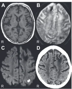
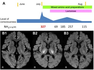
24 mmHg,pO2 85 mmHg,HCO3 21 mmol/lであり,意識障害 を来しうるような他の電解質,血糖(Na 138 mEq/l,K 4.7 mEq/l, Cl 109 mEq/l,Ca 7.8 mg/dl,P 3.3 mg/dl,Mg 2.7 mg/dl,血糖 143 mg/dl)に関しては異常を認めなかった.脳波では前頭葉 優位にデルタ波,三相波を認め(Fig. 3),トポグラフィーで は両側前頭葉にデルタ波を認めた.肝性脳症に準じて蛋白制 限食,混合アミノ酸製剤の点滴,ラクツロース内服で治療を 開始したところ,数日で意識は著明に改善した.その後も血 中アンモニア値は変動を示し,それに対応して意識レベルも 変動した(Fig. 4A).意識レベルが安定した 1 ヶ月後の頭部 MRI では DWI の高信号は消失していた(Fig. 4B).肝性脳症の精 Fig. 1 Brain MRI findings on admission (A–D; axial view).
T1-weighted image (1.5 Tesla, TR 597 ms, TE 15 ms) showed no
abnormality (A), but T2*-weighted image (1.5 Tesla, TR 675 ms, TE
23 ms) showed hypointense lesions in the right frontal lobe and left parietal lobe (B). Diffusion-weighted MRI (DWI) (1.5 Tesla, TR 5,000 ms, TE 70 ms) showed hyperintensity in the left parietal lobe (C). Brain CT scan showed a high-density legion in the left parietal lobe (D).
Fig. 2 Brain MRI findings during deep coma.
Diffusion-weighted MRI (DWI) (1.5 Tesla, TR 6,500 ms, TE 97 ms) showed hyperintense lesions in the dorsal midbrain, thalamus, and posterior limb of internal capsule bilaterally (A–D). Apparent diffusion coefficient (ADC) map (1.5 Tesla, TR 6,500 ms, TE 97 ms) showed hypointensity in the same lesions (I, J). Fluid-attenuated inversion recovery (FLAIR) (1.5 Tesla, TR 10,002 ms, TE 2,300 ms) showed slight hyperintensity (E–H) similar to DWI.
査目的で腹部 CT を施行したところ,肝自体には異常はな かったが,回結腸静脈から下大静脈に連続する異常血管を認 め(Fig. 5),PSE と診断した.その後の検査で,直腸に進行 期の大腸癌が発見され手術を施行した.PSE に対しては内服 薬療法を継続しており,新たな脳症の出現は認めていない. 考 察 本例は,90 歳で急性脳症を発症し,特徴的な MRI 所見か ら,直ちに急性高アンモニア血症に気づき,その原因として PSEが見いだされた症例である. 高アンモニア血症を伴う脳症は,慢性肝障害の急性増悪2), 先天性尿素サイクル異常症3),急性薬物中毒4),PSE5)6),感 染症7)などで発症することがあり,高アンモニア血症性脳症 (hyperammnonemic encephalopathy)と呼ばれる8).特徴的な MRI所見としては,DWI で左右対称性の帯状回,島回皮質, 視床,中脳の高信号が報告されている8). 高アンモニア血症性脳症の原因として,最も頻度の高い肝 性脳症の MRI 所見としては,以前から T1WIでの両側淡蒼球 の高信号がよく知られている9)が,近年,急性肝性脳症(acute
Fig. 4 Clinical course on diffusion-weighted MRI (DWI).
Treatment for hepatic encephalopathy was administered, which led to an improvement in consciousness level, a decrease in plasma ammonia levels (A), and a normalization in the DWI scan (B1: On admission, B2: Deep coma, and B3: After the treatment).
Fig. 5 Three-dimensional computed tomographic angiography (3DCTA).
Abdominal 3DCTA showed an abnormal blood vessel leading from the ileocolic vein to the inferior vena cava. Fig. 3 Electroencephalogram during deep coma.
臨床神経学 57 巻 12 号(2017:12) 57:762 hepatic encephalopathy)では,異なる所見を呈することが報 告されている.McKinney らは,20 例の急性肝性脳症(慢性 肝障害を伴う急性肝機能障害患者 14 例,アセトアミノフェン 過量内服などによる薬剤性急性肝不全患者 6 例)の MRI 所見 を検討し,FLAIR では視床 17/20(85%),内包後脚 15/20 (75%),脳室周囲白質 16/20(80%),脳幹背側 14/20(70%), 大脳灰白質 6/20(30%),小脳白質 3/20(15%)に病変を認 め,DWI で視床 14/20(70%),内包後脚 16/20(80%),脳室 周囲白質 17/20(85%),脳幹背側 7/20(35%),大脳皮質 5/20 (25%)などに高信号を認めることを報告した10).このうち 16例では血中アンモニアレベルが上昇しており,高アンモニ ア血症が脳症に関連していると推定している.実際,これら の急性肝性脳症として報告された所見は,高アンモニア血症 性脳症の報告とほぼ類似したものを示している11).本例の MRIでも,深昏睡期に DWI で両側脳幹背側・視床・内包に 高信号を認め,著明な高アンモニア血症を伴っていた.これ らの所見は血中アンモニアの正常化に伴い消失したことか ら,アンモニアが脳症及び MRI 異常に直接的に関与したもの と推定された. アンモニアは血液脳関門を通過し,血管周囲のアストロサ イト突起から細胞内に拡散し,細胞内グルタミンを増加させ る.これにより細胞内浸透圧が増加し,脳浮腫が引き起こさ れる.また,細胞外スペースにも拡散し,GABA 受容体やグ ルタミン輸送体にも作用し,アストロサイトの形態や機能の 変容を生じることが報告されている12).高アンモニア血症を 伴う急性脳症に対しては,速やかな治療により不可逆的な脳 障害を食い止めることが可能であり,迅速な診断が重要であ る.しかしながら,高アンモニア血症性脳症の MRI 所見は, 低酸素性脳症13),低血糖性脳症14)15),脳炎,Creutzfeldt-Jakob disease(CJD)等にも類似しているため,注意を要する. 本例で,次に特筆すべき点は,超高齢で PSE を発症した 点にある.国内の PSE 報告を調べえた範囲では,発症年齢は 50~80 歳であり,本例は最高齢であった.高齢発症 PSE は, 認知症や精神疾患などと診断される場合が少なくない16).本 例でも,半年程前から繰り返す意識障害に関して,てんかん・ アルツハイマー型認知症と診断されていたが,結果的には PSEによるものであったと推定される. 門脈-大循環シャント形成の原因には,先天性17),または 後天性(肝生検や腹部手術後による肝実質の変性,交通事故 などによる腹部外傷後に生じたもの)が挙げられる18).本例 は肝硬変や入院以前の腹部手術歴はなく,大腸癌手術の際に 癌との血行も認めなかったことから,先天性の可能性が高い. 超高齢で PSE を発症した原因は不明であるが,誘因として は,1)腹腔内血行動態の変化によるシャント血流の増加, 2)アンモニアに対する中枢神経脆弱性の亢進,3)大腸癌に よる便秘によって生じた腸内細菌叢の変化によるアンモニア 産生の増加,などが推定される. PSEの治療は,急性期には蛋白制限食,混合アミノ酸製剤 の点滴,ラクツロース投与などの保存的加療を行い,症状が 持続する場合は短絡結紮術・短絡切除術,バルーン閉塞下逆 行性経静脈的塞栓術19)~21)などによる根治術も選択される.本 例は,超高齢であり,保存的加療のみで速やかに症状が改善 し,かつ再発防止が可能であったため,根治的な治療は行な わなかった. 本報告の要旨は,第 215 回日本神経学会九州地方会で発表し,会長 推薦演題に選ばれた. ※本論文に関連し,開示すべき COI 状態にある企業,組織,団体 はいずれも有りません. 文 献
1) Sherlock S, Summerskill WH, White LP, et al. Portal-systemic encephalopathy; neurological complications of liver disease. Lancet 1954;267:454-457.
2) Morgan MY. Cerebral magnetic resonance imaging in patients with chronic liver disease. Metab Brain Dis 1998;13:273-290. 3) Gropman AL, Summar M, Leonard JV. Neurological implications
of urea cycle disorders. J Inherit Metab Dis 2007;30:865-879. 4) Twilla JD, Pierce AS. Hyperammonemic encephalopathy due to
valproic acid and topiramate interaction. Case Rep Psychiatry 2014;2014:410403.
5) Akahoshi T, Nishizaki T, Wakasugi K, et al. Portal-systemic encephalopathy due to a congenital extrahepatic portosystemic shunt: three cases and literature review. Hepatogastroenterology 2000;47:1113-1116.
6) Toru S, Matumura K, Kawaguchi R, et al. Widespread cortical lesions on diffusion-weighted imaging in acute portal systemic shunt encephalopathy caused by primary biliary cirrhosis. AJNR Am J Neuroradiol 2011;32:E55-E56.
7) Samtoy B, DeBeukelaer MM. Ammonia encephalopathy secondary to urinary tract infection with Proteus mirabilis. Pediatrics 1980;65:294-297.
8) U-King-Im JM, Yu E, Bartlett E, et al. Acute hyperammonemic encephalopathy in adults: imaging findings. AJNR Am J Neuroradiol 2011;32:413-418.
9) Rovira A, Alonso J, Cordoba J. MR imaging findings in hepatic encephalopathy. AJNR Am J Neuroradiol 2008;29:1612-1621. 10) McKinney AM, Lohman BD, Sarikaya B, et al. Acute hepatic
encephalopathy: diffusion-weighted and fluid-attenuated inversion recovery findings, and correlation with plasma ammonia level and clinical outcome. AJNR Am J Neuroradiol 2010;31:1471-1479. 11) McKinney AM, Sarikaya B, Spanbauer J, et al. Acute hepatic (or
hyperammonemic) encephalopathy: diffuse cortical injury and the significance of ammonia. AJNR Am J Neuroradiol 2011;32: E142.
12) Braissant O, McLin VA, Cudalbu C. Ammonia toxicity to the brain. J Inherit Metab Dis 2013;36:595-612.
13) 後藤泰伸,綿谷崇史,荒川芳輝ら.蘇生後脳症の MRI―時間 的変化と予後の分析―.脳神経 2001;53:535-540.
14) Kang EG, Jeon SJ, Choi SS, et al. Diffusion MR imaging of hypoglycemic encephalopathy. AJNR Am J Neuroradiol 2010;31: 559-564.
15) Albayram S, Ozer H, Gokdemir S, et al. Reversible reduction of apparent diffusion coefficient values in bilateral internal capsules in transient hypoglycemia-induced hemiparesis. AJNR Am J
Neuroradiol 2006;27:1760-1762.
16) Miyata K, Tamai H, Uno A, et al. Congenital portal systemic encephalopathy misdiagnosed as senile dementia. Intern Med 2009;48:321-324.
17) Zhang DY, Weng SQ, Dong L, et al. Portal hypertension induced by congenital hepatic arterioportal fistula: report of four clinical cases and review of the literature. World J Gastroenterol 2015; 21:2229-2235.
18) Alonso-Gamarra E, Parron M, Perez A, et al. Clinical and radiologic manifestations of congenital extrahepatic portosystemic shunts: a comprehensive review. Radiographics 2011;31:707-722. 19) Murakami M, Nishino K, Satou T, et al. Percutaneous
transretroperitoneal direct approach to occlude a major shunt in a patient with extrahepatic portal-systemic encephalopathy. Hepatol Res 2009;39:313-317.
20) Tanaka O, Ishihara K, Oyamada H, et al. Successful portal-systemic shunt occlusion with balloon-occluded retrograde transvenous obliteration for portosystemic encephalopathy without liver cirrhosis. J Vasc Interv Radiol 2006;17:1951-1955. 21) Kato T, Uematsu T, Nishigaki Y, et al. Therapeutic effect of
balloon-occluded retrograde transvenous obliteration on portal-systemic encephalopathy in patients with liver cirrhosis. Intern Med 2001;40:688-691.
Abstract
Portal-systemic encephalopathy with bilateral thalamic and internal capsule lesions
using diffusion-weighted MRI in a super-aged patient
Michiaki Matsuda, M.D.
1), Shinpei Takesako, M.D.
2), Mitsuhiro Nakazaki, M.D., Ph.D.
2),
Toru Nandate, M.D.
3)and Fujio Umehara, M.D., Ph.D.
1)1)Department of Neurology, Nanpuh Hospital
2)Department of Diabetology and Endocrinology, Nanpuh Hospital 3)Department of Radiology, Nanpuh Hospital
We describe the case of a 90-year-old woman who was hospitalized in July 2016 and subsequently experienced a
sudden decline in consciousness level resulting in a state of deep coma. Involuntary movements were not observed, and
bilateral Babinski signs were inconclusive. Diffusion-weighted MRI (DWI) of the brain showed bilateral hyperintensity in
the thalamus and internal capsule, laboratory testing detected high levels of plasma ammonia, and an electroencephalogram
showed delta waves and triphasic waves predominantly in the frontal lobe. Based on these results, treatment for hepatic
encephalopathy was administered, which led to an improvement in consciousness level, a decrease in plasma ammonia
levels, and a normalization in the DWI scan. Abdominal computed tomography scan showed no abnormality in the liver,
but revealed an abnormal blood vessel leading from the ileocolic vein to the inferior vena cava; the patient was diagnosed
with portal-systemic encephalopathy. In deep coma patients, acute encephalopathy with hyperammonemia is important
for differential diagnosis when DWI shows high-density legions in the thalamus and internal capsule.
(Rinsho Shinkeigaku (Clin Neurol) 2017;57:759-763)
Key words: portal-systemic encephalopathy, hyperammonemia, thalamus and capsula interna, diffusion-weighted MRI, super-aged patient