Japan Advanced Institute of Science and Technology Title
Effect of dual‐drug‐releasing micelle‒hydrogel composite on wound healing in vivo in
full‐thickness excision wound rat model Author(s) Patel, Monika; Nakaji‐Hirabayashi, Tadashi;
Matsumura, Kazuaki
Citation Journal of Biomedical Materials Research Part A, 107(5): 1094-1106
Issue Date 2019-01-31
Type Journal Article
Text version author
URL http://hdl.handle.net/10119/17043
Rights
(c) 2019 Wiley Periodicals, Inc. This is the peer reviewed version of the following article: Monika Patel, Tadashi Nakaji‐Hirabayashi, Kazuaki Matsumura, Journal of Biomedical Materials Research Part A, 107(5), 2019, 1094-1106, which has been published in final form at
https://doi.org/10.1002/jbm.a.36639. This article may be used for non-commercial purposes in
accordance with Wiley Terms and Conditions for Use of Self-Archived Versions.
1
Effect of dual-drug–releasing micelle-hydrogel composite on wound
1healing in vivo in full-thickness excision wound rat model
23
Monika Patel1, Tadashi Nakaji-Hirabayashi2, 3, Kazuaki Matsumura1* 4
1 School of Materials Science, Japan Advanced Institute of Science and Technology, Ishikawa, 5
923-1292, Japan 6
2 Graduate School of Science and Engineering ,University of Toyama, Toyama, 930-8555, Japan 7
3Graduate School of Innovative Life Science, University of Toyama, Toyama, 930-8555, Japan 8
9
* Corresponding author: [Kazuaki Matsumura e-mail: mkazuaki@jaist.ac.jp] 10
2
Abstract
11
Wound healing is a complex process involving an intricate cascade of body responses. A 12
composite dressing that would effectively target different stages of wound healing and 13
regeneration is urgently needed. In the current study, we tested the efficacy of a previously 14
prepared micelle-hydrogel composite loaded with two drugs, in full-thickness excision wound 15
model in rat. We found that the composite elicited almost no inflammation and effectively 16
enhanced healing at all stages of the healing process. An initial burst of the first drug, amphotericin 17
B, eliminated any preliminary infection. This burst was followed by a gradual release of curcumin 18
as the healing and anti-inflammatory agent. Better healing was observed in rats treated with the 19
drug-loaded composites than in blank and control groups. Wounds showed up to 80% closure in 20
the treated group, with high collagen deposition. Re-epithelialization and granulation were also 21
better in the treated group than in the non-treated control and blank groups. Histopathological 22
examination revealed that drug-loaded composites improved cutaneous wound healing and 23
regeneration. In conclusion, the micelle-hydrogel composite is an effective dressing and might 24
have major applications in wound healing. 25
26
Keywords: Micelle-hydrogel composite, dermal wound healing, pH-sensitive release, dual-drug
27
release, polypeptide hydrogel 28
29 30 31 32
3
INTRODUCTION
33
In the last few decades, development of new dressing material to aid wound healing has received 34
great attention.1-3 Although conventional (non-occlusive) wound dressings, which generate dry 35
wound healing conditions, continue to constitute the largest type of dressing materials, the use of 36
occlusive dressings,4-6 hydrocolloid,7, 8 and hydrogel dressings,9-11 which offer hydrated wound 37
healing conditions, is currently increasing. The next vital phase in the development of new dressing 38
material is the development of material capable of delivering active molecules and/or drugs 39
directly at the wound site. Indeed, dressings loaded with active factors and/or drugs are becoming 40
increasingly popular because of the well-known fact that topical or exogenous application of active 41
substances directly at the wound site improves healing. 42
Wound healing involves a series of complex and well-orchestrated events occurring after 43
an injury or physical trauma to the skin,12-13 that aims to completely restore the integrity of 44
damaged tissue and reinstate it as a functional barrier.14-16 However, in some extreme situations 45
(i.e., trauma with large full-depth skin damage),17 complete re-epithelialization takes a long time.18 46
Therefore, extensive studies are focusing on wound dressing systems to promote better wound 47
healing and to reduce scar formation.19 48
Wound dehydration perturbs the healing process,20-22 compromising the optimal 49
environment required by that process. Therefore, maintenance of the moisture of the wound is of 50
prime importance for effective and fast wound healing. In such cases, hydrogels are a promising 51
candidate material, with the ability to absorb wound exudates,23-24 control wound dehydration, and 52
allow oxygen access. Furthermore, in addition to the hydrated environment that hydrogels provide, 53
they can serve an additional purpose, delivering bioactive substances directly to the wound in a 54
sustained manner. 55
4 Curcumin25-26 is the principle curcuminoid and active component of Curcuma longa. 56
Chemically, it is diferuloylmethane, or 1,7-bis(4-hydroxy-3-methoxyphenyl)-1,6-heptadiene-3,5-57
dione, a naturally occurring low-molecular weight polyphenolic phytoconstituent. Curcumin, in a 58
form of turmeric (powder of dried rhizome of Curcuma longa), has been widely and predominantly 59
used in Asian countries, especially India27 and China, as a dyeing material,28 flavoring agent,29 and 60
in many forms of customary medical practices to treat a range of inflammatory and chronic 61
ailments. Various studies involving curcumin present evidence in support of its numerous 62
pharmacological benefits, such as anti-oxidant,30, 31 anti-inflammatory,32, 33 anti-bacterial,34 anti-63
viral,35 anti-tumor,36 and hyperlipidemic activities. It has been reported that administration of 64
curcumin, both topically and orally, results in rapid wound healing. Yet, the therapeutic efficacy 65
of curcumin is restricted because of its poor solubility in aqueous media, reduced oral 66
bioavailability, and high first-pass metabolism. Another disadvantage of curcumin is the means of 67
application. Curcumin is a polyphenol, which can result in toxicity if applied in a highly 68
concentrated dose. Hence, a water-soluble formulation with a controlled release would be 69
preferred for clinical application of curcumin. 70
We recently reported preparation of a new micelle-hydrogel composite.37 The composite 71
consists of polypeptide micelles cross-linked with genipin, both of which are biocompatible and 72
frequently used for medical purposes. The micelle-hydrogel composite is composed of two 73
oppositely charged polypeptide-based micelle systems, the positively charged poly(L
-lysine)-b-74
poly(phenylalanine) (PLL-PPA), and negatively charged poly(glutamic acid)-b-75
poly(phenylalanine) (PGA-PPA). Because of the presence of amphiphilic polypeptide chains, 76
these polypeptides easily self-assemble into micelles, rendering drug loading of the hydrophobic 77
core effortless and facile. In a previous study, we showed that these micelle systems release drugs 78
5 under various conditions.37 Because of the opposite charge of the micelles in the composite, the 79
two micellar systems behave differently at varying pH values, hence enabling various drug release 80
rates. This phenomenon makes it easy to tune the release rate of different drugs from these different 81
micelle types in the composite, making it an ideal candidate for dual-drug release studies, 82
especially for wound healing studies. 83
The aim of the current study was to evaluate the in vivo biocompatibility and efficacy of the 84
micelle-hydrogel composite37 as a wound dressing, serving as a reservoir for sustained delivery of 85
curcumin (Figure 1). We evaluated the activity of the prepared composite in wound healing in vivo, 86
in a full-thickness excision wound model in rat. Biomechanical tests, biochemical analysis, and 87
histopathological examinations were also conducted to investigate the therapeutic effects of 88
curcumin-loaded micelle hydrogel composites in the model. 89
90 91
6
MATERIALS AND METHODS
92
Preparation of dual-drug–loaded micelle-hydrogel composites
93
The dual-drug–loaded micelle-hydrogel composites were generated by using poly(L
-lysine-b-94
phenylalanine) and poly(glutamic acid-b-phenylalanine) (Scheme S1) polymers, as previously 95
described37 (Supporting Information). The polymers were synthesized using the common N-96
carboxyanhydride (NCA) method. NCA were prepared using protected amino acids (Scheme S2). 97
The generated polymers (PLL-PPA and PGA-PPA) were dialyzed in solutions containing 98
curcumin and amphotericin B (respectively) to form drug-loaded micelles and were then gelled 99
using genipin (Scheme S3) to form a micelle-hydrogel composite. 100
101
Wound model
102
Wound generation. Adult (9-week-old, 290−310 g, n = 25 male Sprague−Dawley rats (Japan SLC,
103
Inc. Shizuoka, Japan) were housed under a 12-h light/12-h dark cycle with ad libitum access to 104
food and water. All animals were in quarantine for a week before the study. All manipulations 105
were performed under aseptic conditions. NIH guidelines (or for non-U.S. residents similar 106
national regulations) for the care and use of laboratory animals (NIH Publication #85-23 Rev. 107
1985) have been observed. Further, all animal procedures were performed following the protocol 108
approved by the ethical committee in University of Toyama (Toyama, Japan). All rats were treated 109
humanely throughout the experimental period. Transplantation experiments with dual-drug– 110
loaded micelle-hydrogel composites and control samples were carried out under anesthesia with 111
isoflurane gas (250–350 mL/min, isoflurane: 1.5–2.5%) using the UNIVENTOR 400 anesthesia 112
unit (Univentor, Zejtun, Malta) and according to the guidelines of the Animal Welfare Committee 113
of University of Toyama and Ministry of Education, Culture, Sports, Science and Technology 114
7 (MEXT). A standard full-thickness excision wound was created for the purpose of the study. 115
Briefly, on day 0, rats were anaesthetized, and the dorsum shaved and cleaned using saline-soaked 116
gauze, and then swabbed with 70% ethanol. A single full-thickness wound (20 mm × 20 mm) was 117
created in the left dorsal flank skin of each rat to the depth of the loose subcutaneous tissues, and 118
was left open (Figure 2). 119
Treatments. Animals were divided into four groups (6 rats per group). The wounds were topically
120
treated with a single application of blank hydrogels (without drugs); low-concentration hydrogels 121
(LC; hydrogels loaded with low concentration, 0.5 mg, of curcumin); or high-concentration 122
hydrogels (HC; hydrogels loaded with high concentration, 1.5 mg, of curcumin). Both LC and HC 123
groups were loaded with low concentration (50 µg) of amphotericin B to demonstrate dual-drug 124
release as well as prevent any infections of the wound. The wounds in the final group of animals 125
(the control group) were dressed using medical gauze. A piece of Tegaderm (3M, Maplewood, 126
MN, USA) was placed on top of all wounds to prevent the rats from removing the treatment 127
material. Upon experimental wounding, animals were housed in individual cages, and maintained at 128
an ambient temperature (23°C), with 12-h light/12-h dark cycles, with ad libitum access to food and 129
water. 130
For biochemical studies, histopathological examinations, and antioxidant enzyme analysis, 131
animals (3 rats per group) were sacrificed under anesthesia on days 4 and 8 after surgery, because 132
the most pronounced changes in tissue occur during the first week after wounding. Wound collagen 133
content, granulation tissue formation, wound maturity, and superoxide dismutase (SOD) and 134
catalase activity were investigated in detail as described below. 135
8 136
Histopathological examination
137
Adjacent skin fragments were removed together with the wound area to evaluate any 138
histopathological alterations. The collected specimens were fixed in 10% buffered formalin, 139
processed, embedded in paraffin, and then sectioned perpendicular to the wound surface into thin 140
sections following standard protocols. Tissue sections were stained with hematoxylin and eosin, 141
and analyzed using light microscopy (Biozero Keyence BZ 8000, Osaka, Japan). Tissue sections 142
were also stained with rabbit anti-Iba1 IgG antibodies (Wako Pure Chemical Corp., Osaka, Japan) 143
and Alexa488-conjugated anti-rabbit IgG antibodies (ThermoFisher Scientific, Waltham, MA, 144
USA) to visualize macrophages, and counter-stained with Hoechst 33258 (DOJINDO Laboratories, 145
Kumamoto, Japan) following the manufacturers’ instructions. 146
147
Wound healing and wound closure evaluation
148
Wounds were digitally photographed together with an identity plate and calibration bar 149
immediately after wounding, and subsequently after dressing removal and cleansing with sterile 150
saline on days 4 and 8 (following re-anaesthetization, as above). Wound closure was determined 151
based on scaled digital images of each wound using Image J image analysis software. Wound 152
closure was calculated by measuring the open wound area in each digital image, at each time point. 153
Open wound area was calculated as % of the original area immediately after wounding on day 0, 154
by using the following formula: 155
% 𝑤𝑤𝑤𝑤𝑤𝑤𝑤𝑤𝑤𝑤 𝑐𝑐𝑐𝑐𝑤𝑤𝑐𝑐𝑤𝑤𝑐𝑐𝑐𝑐 =[𝑤𝑤𝑤𝑤𝑤𝑤𝑤𝑤𝑤𝑤 𝑎𝑎𝑐𝑐𝑐𝑐𝑎𝑎 𝑤𝑤𝑤𝑤 𝑤𝑤𝑎𝑎𝑑𝑑 0 − 𝑤𝑤𝑤𝑤𝑤𝑤𝑤𝑤𝑤𝑤 𝑎𝑎𝑐𝑐𝑐𝑐𝑎𝑎 𝑤𝑤𝑤𝑤 𝑤𝑤𝑎𝑎𝑑𝑑 𝑋𝑋]𝑤𝑤𝑤𝑤𝑤𝑤𝑤𝑤𝑤𝑤 𝑎𝑎𝑐𝑐𝑐𝑐𝑎𝑎 𝑤𝑤𝑤𝑤 𝑤𝑤𝑎𝑎𝑑𝑑 0 × 100 156
9 157
Evaluation of granulation
158
Granulation tissue deposition in wounds was semi-quantitatively scored based on panoramic 159
photomicrographs of hematoxylin- and eosin-stained sections in the center of each wound. The 160
granulation was estimated as the depth of granulated tissue at the site of scarring, by two 161
experienced observers who were unaware of the treatment group allocation. 162
163
Evaluation of craniocaudal wound contraction (re-epithelialization)
164
Percentage craniocaudal contraction (a histological measure of central wound contraction, in a 165
craniocaudal dimension) was determined in hematoxylin- and eosin-stained sections in the center 166
of the wound. Wound width was expressed as the percentage of the original central wound width 167
based on wound images taken on day 0. 168
169
Evaluation of tissue inflammation
170
The extent of inflammation in the wound was evaluated in each group of animals by Hoechst 171
33258 and anti-Iba1 antibody staining of tissue samples. 172
173
Evaluation of enzyme activity
174
Tissue samples were washed with phosphate-buffered saline to remove adhering red blood cells. 175
The samples were homogenized in ice-cold 0.1 M Tris-HCl, pH 7.4, containing 0.5% Triton X-176
100, and 5 mM β-mercaptoethanol. The obtained crude mixture was centrifuged for 25 min at 177
8000× g and 4°C, and the pellet containing cell debris was discarded. The supernatant contained 178
the total tissue enzyme activity (cytosolic and mitochondrial). SOD activity was determined in the 179
10 supernatant using a method based on the reduction of nitro blue tetrazolium, with sample 180
absorbance measured at 560 nm.38 To determine the catalase activity, the supernatant was mixed 181
with H2O2 and decrease in sample absorbance was recorded at 240 nm, as previously described.39 182
183
Evaluation of collagen content
184
Wounded tissue samples were frozen in liquid nitrogen and then freeze-dried by lyophilization. 185
The lyophilized samples were then incubated overnight in 0.5 M acetic acid and homogenized. 186
The homogenate was centrifuged at 12000g for 15min at 4°C and total collagen content determined 187
using a total collagen assay kit (BVN K218-100; Biovision, CA,USA) as per manufacturer’s 188
recommendations. 189
190
Determination of the mechanical properties of hydrogels
191
Rheological properties of the gels were evaluated using a rheometer equipped with a 24.99-mm 192
2.069° cone (Rheosol G5000, UBM Co., Ltd., Kyoto, Japan). Hydrogels were prepared as for the 193
wound-healing test. The dynamic storage (G′) and loss (G′′) moduli of the hydrogels were 194
determined by a frequency dispersion mode, between 0.01 and 10 Hz. All analyses were carried 195
out at 37°C. For the analysis, mineral oil was placed around the sample circumference to prevent 196
evaporation of water from the micelle-hydrogel composite. 197
198
Statistical analysis
199
All the variables were tested in independent experiments repeated three times. Values are reported 200
as the mean ± standard error of the mean. Experimental data from different groups were compared 201
11 using one-way analysis of variance (ANOVA). A p-value < 0.05 in a two-tailed test was 202
considered statistically significant. 203
12
RESULTS
204
Rationale for the study
205
Our group has recently designed a polypeptide-based system that enabled a highly efficient control 206
of the rate of drug release by varying a range of parameters, including pH.37 Since wound healing 207
is highly impacted by the pH of healthy tissue surrounding the wounded tissue, the observation 208
had a valid implication for testing the developed system in vivo. Previous studies indicated that the 209
pH of tissue in the vicinity of a wound is acidic during healing and that this acidic environment 210
(approximately pH 4.5)40 is automatically created around the wounded tissue by the body. This 211
intrigued us as the developed composite system could be exploited in response to pH, thus 212
potentially improving the healing environment. Further, to improve wound retraction and healing, 213
infection at the early stages of healing would ideally be prevented. This prompted us to use a dual-214
drug release system to controllably release an anti-bacterial drug (amphotericin B) during early 215
stages of healing, followed by a slow release of the healing drug (curcumin). Indeed, an in vitro 216
assay (Figure 3) indicated a controlled and desired release profile of these drugs at pH 4.5, which 217
strengthened the hypothesis that the polypeptide-based system could be used as a superior wound 218
healing system. 219
220
Evaluation of the novel micelle-hydrogel composite in vivo
221
Macroscopic observations. The bio-efficacy of the newly formulated micelle-hydrogel composite
222
as a wound dressing was evaluated in vivo in a subcutaneous implantation study in the rat model. 223
Dorsal wounds were generated and dressed with hydrogel or gauze, as required, covered by 224
Tegaderm, and various wound parameters were monitored over 8 d (Figure 2). 225
13 Wound healing progression in the control, blank, LC, and HC groups is shown in Figure 4. 226
Wounds treated with LC and HC micelle-hydrogel composites exhibited noticeable dryness and 227
no indication of pathological fluid oozing out. In addition, no signs of inflammation or infection 228
were apparent in these groups compared with the control and blank groups. Wound closure was 229
analyzed in each group as a percentage of the reduction in wounded area on days 4 and 8 [Figure 230
5(a)]. Animals treated with micelles containing high concentration of curcumin showed more 231
substantial wound closure (53.04 ± 4.26% on day 4; 87.32 ± 3.11% on day 8) than those treated 232
with gels loaded with low concentration of curcumin (22.23 ± 3.86% on day 4; 73.39 ± 4.03% on 233
day 8), blank (15.12 ± 2.92% on day 4; 32.67 ± 3.81% on day 8), or in the control groups 234
(7.31 ± 3.64% on day 4; 18.73 ± 6.21% on day 8). 235
The residual wound area was determined in each group, by measuring the open wound area 236
on days 4 and 8 [Figure 5 (b)]. Wounds began to close on day 4 and residual wound sizes were 237
reduced in all rat groups by the end of day 8. A drastic reduction in the residual wound area was 238
observed after 8 d of treatment with HC gels. By contrast, the largest residual wound area was 239
noted in the control group, indicating slow wound healing. Decrease of the wounded area is an 240
important parameter in wound healing, indicative of reduced infection and inflammation. Overall, 241
on days 4 and 8, wound contraction in HC group was significantly greater than that in other groups. 242
243
Microscopic observations. To evaluate wound closure in more detail, the effect of the treatments
244
on the process of granulation41 and re-epithelialization42, 43 was studied. Thickness of granulation 245
tissue and extent of re-epithelization were evaluated in hematoxylin- and eosin-stained tissue 246
samples. As shown in Figure 6, the granulation was significantly enhanced in wounds after 8-d 247
treatment with HC gels. However, no significant improvement in the granulation was apparent in 248
14 the control samples, which exhibited minimum or almost no granulation. In the blank group, 249
granulation was moderate, and better than that in the control but significantly lower than of the LC 250
and HC treated groups. 251
Re-epithelialization was analyzed in all test groups on days 4 and 8. As shown in Figure 7, 252
no pronounced epithelial regeneration was apparent in blank and control groups on day 4. 253
Conversely, in the LC and HC groups, enhanced formation of the epithelial lining was apparent as 254
early as 4 d after wounding. Re-epithelialization was improved in all samples by day 8. These 255
results were consistent with the analysis of the residual wound area. As shown in Figure 8, wounds 256
treated with HC exhibited a well-defined regenerated and differentiated epidermal layer on day 8, 257
with a fairly higher cell number and a relatively thicker dermis than wounds in other samples. 258
Wounds in the LC group also exhibited an enhanced re-epithelialization but the effect was not as 259
pronounced as in the HC group. Samples from other groups showed an early, on-going epithelial 260
layer formation with poor granulation and traces of edema. 261
262
Effect on tissue inflammation. Hematoxylin and eosin staining supported the notion of enhanced
263
wound healing in groups treated with HC and LC gels. To better understand the effect of the 264
implanted gels on tissue and contribution to wound healing, the inflammatory response at 265
implantation site was evaluated.44-46 Wound tissue sections from different groups after 4-d and 8-266
d treatment were stained with Hoechst 33258 and anti-Iba1 antibodies. 267
And shown in Figure 9, on day 4 after surgery, an extremely high inflammatory response 268
was noted in the control group, with a massive accumulation of macrophages at the wound site 269
(green dots marking the cytosol of macrophages stained with anti-Iba1 antibodies). The 270
accumulation of macrophages in the control group was reduced on day 8 after wounding but 271
15 remained appreciably higher than that in other groups. The second highest inflammatory response 272
on day 4 was evident in the blank group. The response visibly declined by day 8. By contrast, in 273
the remaining two groups (LC and HC groups), no accumulation of macrophages was apparent on 274
day 4, indicating enhanced wound healing, with the cell proliferation phase already started. That 275
was also suggested by the large number of accumulated cells in LC and HC samples (blue dots in 276
Figure 9, stained by Hoechst 33258). On day 4, clear granulation was apparent in HC samples, 277
indicative of accumulation of non-inflammatory cells, which by day 8 turned into a well-defined 278
regenerated epithelium. Similarly, no visible signs of enhanced inflammation were apparent on 279
day 4 in LC samples, with a clear onset of re-epithelialization by day 8, supporting the notion that 280
the hydrogels improved wound healing in the LC and HC treatment groups. 281
282
Effect on tissue enzyme activity, collagen content, and angiogenesis. In addition to histological
283
analysis, other biochemical wound parameters were evaluated to assess the efficiency of wound 284
healing. Previous studies indicated that wounding induces oxidative stress in the injured tissue, 285
enhancing the expression of SOD-encoding gene.47 SOD activity was determined in injured tissues, 286
and a clear reduction in the net SOD activity was observed. As shown in Figure 10, SOD levels in 287
the HC and LC groups were reduced on days 4 and 8 in comparison with those in blank and control 288
groups, where an increment in the level of SOD activity on day 8 was apparent. A contrasting trend 289
was observed for the activity of catalase, another antioxidant enzyme (Figure 11). Accordingly, 290
catalase activity on day 4 in the control and blank groups was similar to or higher than that in the 291
LC and HC groups, whereas it was significantly increased by day 8. By day 8, catalase activity in 292
HC group was almost double that in the control group. 293
16 The net collagen content48-51 of the wounded tissues on days 4 and 8 after the surgery was 294
next examined (Figure 12). As shown, the total collagen deposition was highest in the HC group 295
on days 4 and 8, strongly indicating enhanced wound healing in comparison with other samples. 296
Since angiogenesis is a crucial parameter of the wound healing process, tissue sections 297
were stained with anti-CD31 antibodies to evaluate the effect of treatments on the formation of 298
blood vessels. As shown in Figure 13, wounds in the LC and HC groups contained more CD31-299
positive cells than those in the blank and control groups. 300
301
Rheological properties of the hydrogels
302
Finally, rheological properties of the hydrogels were evaluated to better understand hydrogel 303
behavior. As shown in Figure 14, a composite lacking the PGA-PPA micelles showed a very low 304
storage modulus (G′), in the range of 102 Pa, and a low loss modulus (G′′), in the order of 101 Pa, 305
in comparison with the composite with both micelles present, where the storage and loss moduli 306
were in the range of 104 and 103 Pa, respectively. This suggested the role and importance of PGA-307
PPA micelles in the maintenance of gel structure and strength. The values of storage and loss 308
moduli of the hydrogel steadily decreased over 48 h (Figure 15). This supported the notion of 309
controlled drug release from the hydrogels. 310
311 312
17
DISCUSSION
313
In the current study, we evaluated the effectiveness of a novel dual-drug–releasing micelle-314
hydrogel composite in wound healing in vivo, in the full-thickness excision wound rat model. 315
The process of wound healing follows a distinct timeline of physical events (phases), 316
including post-trauma repair in the case of an injury. In intact skin, the epidermis (upper skin layer) 317
and dermis (deep skin layer) act as a defensive barrier against the external environment. When the 318
barrier is broken, i.e., when the skin is injured, a coordinated cascade of biochemical reactions is 319
brought into motion to heal the damage. The sequence of events includes blood clotting, 320
inflammation, cell proliferation, and maturation (remodeling). 321
In the initial moments following the injury, platelets in the blood begin to accumulate at 322
the site of injury.52 The platelets become activated and release chemical cues to promote clotting. 323
The resultant clot facilitates the closing of the opening in the blood vessel, preventing further 324
bleeding. Inflammation is an important phase of wound healing.53, 54 Cells that had been damaged 325
or are dead as a result of the injury are cleared out. Inflammation also facilitates the removal of 326
bacteria and other infectious pathogens. Proliferation marks the growth of new tissue at the injury 327
site.55, 56 The beginning of this phase accompanies the start of granulation, with new cells migrating 328
to the site of injury and proliferating. Angiogenesis, connective tissue deposition, re-329
epithelialization, and wound contraction are the key events of the proliferation phase. Finally, 330
tissue repair is completed in the maturation (remodeling) phase.57 Then, the connective tissue is 331
rearranged along tension lines, and cells that have served their purpose are strategically removed 332
by programmed cell death (apoptosis). 333
To determine the effect of the micelle-hydrogel composite on different stages of wound 334
healing, we performed various analyses, and reported strikingly positive results. The specific 335
18 composite was used because of its ability to release drugs in response to the need of the 336
environment in the vicinity of the wound. At acidic pH (ca. 4.5), PGA chains in the PGA-PPA 337
micelles become relatively un-charged and acquire a helical conformation, which strains the core 338
of the micelle and results in faster release of the drug. This is required for the initial prevention of 339
infection at the site of wounding.37 On the other hand; PLL-PPA micelles in the composite exist 340
in charged random-coil state. The micellar organization and drug release remain stable, releasing 341
the drug slowly over a period of time, aiding wound healing (Figure S1). 342
We observed that in the LC- and HC-treated groups, wound size decreased with time in the 343
absence of oozing or visible signs of infection. This supported the notion that the micelle-hydrogel 344
composite accelerated wound healing. The blank and LC treatment groups showed an intermediate 345
response between that of the control and HC groups. Granulation in the LC group was improved 346
because of the regular supply of curcumin to the tissue by the implanted gels. Quantitative analysis 347
of wound closure revealed a significant improvement in the LC and HC groups in comparison with 348
the blank and control groups. The implanted micelle hydrogel composites prevented drying out of 349
the wounds. 350
Several previous studies demonstrated the consequences of the innate immune response 351
of resident cells and incoming inflammatory cells (such as monocytes and granulocytes) during 352
skin wound repair.58 These cells fight the invading microbes, contribute to scavenging of dead and 353
decaying cells, and also (crucially) support the repair process by releasing a spectrum of growth 354
factors. However, because of the release of pro-inflammatory and cytotoxic mediators, 355
uncontrolled activity of macrophages may become detrimental to tissue repair. Indeed, imbalanced 356
inflammation characterized by increased numbers of macrophages is a hallmark of attenuated 357
repair response in human diseases, including diabetes mellitus,59 vascular disease, and aging. Data 358
19 presented in the current study (Figure 6) indicated that the initial migration of cells was faster in 359
the HC and LC groups than in the blank and control groups. This might be a consequence of the 360
constant release of curcumin in the HC and LC groups, in agreement with published observations 361
that curcumin considerably improves granulation in non-ischemic wounds.60 362
A series of important events takes place at the edge of the wound, accompanying 363
granulation. Epidermal cells in the direct vicinity of the edge of the wound begin to thicken within 364
the first 24–48 h post injury.61 Basal cells at the edge start to flatten towards the wound, eventually 365
covering the wound. The newly formed epithelium, however, is thinner than the normal 366
(uninjured) epithelium. In large and open wounds, epithelialization proceeds over the bed of 367
granulized tissue, involving the activity of proteolytic enzymes. The re-epithelialization process is 368
evident in Figure 8, with a steady migration of cells towards wound closure (marked by a dotted 369
line), proceeding over the course of few days. In typical wounded tissues, inflammation onsets and 370
subsides by 2–3 d of wound creation, however, the exact time line depends on the type and location 371
of the wound.58, 62 372
As the wound progresses through the inflammation phase, cell debris and necrotic tissues 373
are cleared off, creating room for proliferation. Early onset of inflammation is essentially a sign of 374
improved wound healing, indicating that the wound is rapidly going through the proliferation 375
phase, in which fibroblasts migrate to the wound bed. Fibrin strands that facilitate fibroblast 376
migration to the wound site are deposited in the inflammatory phase. As shown in Figure 9 wounds 377
in the HC and LC groups progressed through the inflammatory phase by day 4, in contrast with 378
the blank and control group, where the wounds contained very high numbers of macrophages at 379
that time point (marking the inflammatory phase). The early onset and completion of inflammatory 380
phase in the HC and LC groups may be attributed to curcumin, a strong anti-inflammatory drug.63 381
20 Analysis of the biochemical aspects of wound healing, including SOD and catalase 382
activities, and the amount of collagen in wounded tissue, yielded interesting results. Wounding is 383
a stressful event for any organism, not only causing discomfort and pain, but also initiating a 384
cascade of events at the wound site. Oxidative stress is one of such of events, and is marked by the 385
presence of superoxide radicals at the site of injury. As the radical concentration increases, so does 386
the expression of SOD, a radical-scavenging enzyme.64 Considering the antioxidant activity of 387
curcumin, a model drug in the current study, we anticipated that oxidative stress in the wound 388
should show a decreasing trend over the period of wound healing (Figure 10). This trend could be 389
easily attributed to the radical-scavenging (antioxidant) activity of curcumin, resulting in lower 390
SOD levels in cells at the wound site, as indeed was apparent (Figure 10). This indicated an 391
improvement in the wound-healing environment and also supported the notion of a controlled 392
release of curcumin from the micelle-hydrogel composite, slowly over a period of time, keeping 393
the oxidative stress in check. High SOD activity in the control and blank groups confirmed these 394
conclusions (Figure 10). 395
Upon scavenging, superoxide radicals in the tissue are converted to hydrogen peroxide. 396
Hydrogen peroxide is toxic to cells and hampers the wound healing process, by causing oxidative 397
stress, albeit one that is milder than the oxidative stress associated with superoxide radicals.65, 66 398
This, in turn, stimulates the expression of the peroxide-scavenging enzyme catalase. Indeed, 399
catalase activity generally increased in the wounded tissue, maintaining a low oxidative stress in 400
the surrounding therein (Figure 11). Consequently, in the LC and HC groups, SOD activity was 401
low, and catalase activity was high. Even though SOD activity was significantly lower in the HC 402
group than that in the blank or control groups (Figure 10), catalase activity in the HC group was 403
slightly higher than that in the LC group, and significantly higher than that in the blank and control 404
21 groups. Considering the low SOD activity and high catalase activity in the granulation tissues in 405
the HC group, wound-healing efficacy was the highest in that group among all groups examined. 406
Combination of various histopathological analysis of wounds in the HC, LC, blank, and 407
control groups on days 4 and 8 after surgery revealed that they indeed were in different stages of 408
wound healing. As discussed earlier, the proliferative and maturation phases mark improved 409
wound healing, with angiogenesis and connective tissue (collagen) deposition taking place in those 410
phases. The presented results unambiguously supported the notion that the developed dual-drug– 411
loaded micelle-hydrogel composites improved wound healing. Namely, in agreement with 412
advanced granulation and re-epithelialization, and reduced inflammation, HC-treated wounds 413
attained the late proliferative phase, with enhanced accumulation of collagen fibers in the 414
extracellular matrix (Figure 12). Similarly, in the LC group, the total collagen content of the wound 415
was higher than that in the blank and control groups, indicating improved wound healing. New 416
collagen is observed in tissue as early as on the day of scarring. However, the newly formed 417
collagen is not strong and as the wound matures, the amount and deposition of collagen changes, 418
strengthening the tissue bed and increasing the tensile strength of the new formed tissue. 419
Consequently, high level of collagen is an optimistic indicator of improved wound healing. 420
Since the pre-existing vascular network around the wound is not sufficient to provide ample 421
nutrients and oxygen to the injury site, vessel damage at the wound site leads to ischemia.67, 68 422
Therefore, the maintenance of cell viability in the wound and continuation of rapid healing 423
essentially requires the formation of new vasculature, i.e., angiogenesis.69 Angiogenesis involves 424
the synthesis of new blood vessels from dividing differentiated endothelial cells of the local 425
vascular system, mononuclear cells, and bone marrow-derived circulating endothelial cells.70 426
While it remains debatable whether circulating cells escalate the formation of the luminal 427
22 endothelium layer, many studies demonstrated that circulating CD31+ endothelial cells can indeed 428
form new blood vessels.71 Consequently, we investigated the presence of circulating CD31+ cells 429
at the wound site. The experiment revealed angiogenesis in the vicinity of the wounded area in the 430
LC- and HC-treated groups, which confirmed the notion of improved wound healing in the treated 431
groups (Figure 13). However, further studies are required to unequivocally verify this, since 432
circulating macrophages also show CD31-positivity.72 Collectively, the presented data were in 433
agreement with the original hypothesis that the micelle-hydrogel composite would facilitate wound 434
healing in case of trauma or skin patch excision. 435
Although the micelle-hydrogel composite performed well in the in vivo wound-healing 436
model, amphotericin B was added only in trace amounts. Hence, an obvious question arises about 437
whether loading the composite with one drug only would facilitate healing, and why two micelle 438
types or two drugs in the composite were required. The composite system was used because the 439
wounding was done in a controlled environment, which is not always the case out of the laboratory, 440
and the second drug (at high concentration and defined dosage) is likely to be always required to 441
accelerate healing. The drug can be a broad-spectrum antibiotic or a growth factor. In addition, the 442
second micelle in the composite is required to maintain the structural integrity of the composite by 443
electrostatic interactions between the micelles. As shown in Figure 14, the storage and loss moduli 444
were substantially reduced in the absence of PGA-PPA micelles. That is because the two micelles 445
types in the composite are oppositely charged, and during mixing and cross-linking they are 446
involved in electrostatic interactions, stabilizing the system even in the absence of drug, and 447
maintaining the integrity of the micelle-hydrogel composite. Furthermore, the hydrophobic core 448
of the micelle in the composite acts as the drug reservoir. We hypothesized that the (hydrophobic) 449
drug is involved in some kind of hydrophobic interactions with the core chains of the micelle. 450
23 Should that be so, the overall mechanical strength of the composite should change with drug 451
release, as the core becomes looser with the diffusion of the drug. To evaluate this, we undertook 452
a time-dependent rheological evaluation of the composite. Indeed, we observed a clear decreasing 453
trend in the mechanical modulus of the composites at different time points of drug release (Figure 454
15). The gradual reduction in the modulus might indirectly reflect a slow and gradual drug release. 455
That was important for the current study, as a sudden or burst-type release of curcumin can have 456
several adverse effects. As shown in previous studies, a burst or high-dose release of curcumin at 457
a wound site can cause DNA damage or chromosomal alterations (in rare cases), and delay wound 458
healing.73, 74 Further, the mechanical evaluation confirmed that the storage modulus of the devised 459
micelle-hydrogel system was within the limits for gel systems used in wound healing and, hence, 460
was an ideal candidate for such a gel. 461
In summary, the reported experiments and their implications indicate that the novel 462
micelle-hydrogel composite can serve as effective would-healing material for enhanced skin repair 463
and regeneration, aided by controlled release of encapsulated drugs. The composite positively 464
impacted each stage of wound repair and healing, resulting in enhanced wound contraction, 465
granulation, and re-epithelialization, and with a minimal inflammatory response. This suggests 466
that the composite is extremely biocompatible and non-toxic for animal use. The exact mechanistic 467
effect on wound healing remains unknown. However, even in the absence of encapsulated drug, 468
no detrimental effects on the process of wound healing were observed (in the blank group in 469
comparison with the control group). Consequently, this type of material could be optimized to 470
enhance wound healing and developed as dressing material for clinical use. 471
472
Acknowledgments
24 The authors have no conflicts of interest to declare.
474 475
Monika Patel 476
School of Materials Science 477
Japan Advanced Institute of Science and Technology, 1-1, Asahidai, Nomi, Ishikawa, 923-1292, 478 Japan 479 480 Tadashi Nakaji-Hirabayashi 481
Graduate School of Science and Engineering, University of Toyama, 3190, Gofuku, Toyama, 482
Japan 930-8555 483
Graduate School of Innovative Life Science, University of Toyama, 3190 Gofuku, Toyama, Japan 484 930-8555 485 Kazuaki Matsumura 486 E-mail: mkazuaki@jaist.ac.jp 487
School of Materials Science 488
Japan Advanced Institute of Science and Technology, 1-1, Asahidai, Nomi, Ishikawa, 923-1292, 489
Japan 490
25
References
492
1. Gelinsky E. Regarding wound healing and dressing material problems once from a different 493
point. Bruns' Beitrage zur klinischen Chirurgie.1957;194:51-73. 494
2. Patrulea V, Ostafe V, Borchard G, Jordan O. Chitosan as a starting material for wound healing 495
applications. European journal of pharmaceutics and biopharmaceutics : official journal of 496
Arbeitsgemeinschaft fur Pharmazeutische Verfahrenstechnik eV. 2015;97:417-26. 497
3. Wharram SE, Zhang X, Kaplan DL, McCarthy SP. Electrospun silk material systems for wound 498
healing. Macromol. Biosci. 2010;10:246-57. 499
4. Saraceno R, Chiricozzi A, Nistico SP, Tiberti S, Chimenti S. An occlusive dressing containing 500
betamethasone valerate 0.1% for the treatment of prurigo nodularis. J Dermatolog Treat. 501
2010;21:363-6. 502
5. Yamamoto N, Kiyosawa T. Histological effects of occlusive dressing on healing of incisional 503
skin wounds. Int Wound J. 2014;11:616-21. 504
6. Zadeh Farahani RM, Shahidi A. Occlusive dressing of wounds: old tradition, new concepts. J 505
Tissue Viability. 2009;18:57-8. 506
7. Khan MI, Islam JM, Kabir W, Rahman A, Mizan M, Rahman MF, et al. Development of 507
hydrocolloid Bi-layer dressing with bio-adhesive and non-adhesive properties. Mater. Sci. Eng. C. 508
2016;69:609-15. 509
8. Yanagibayashi S, Kishimoto S, Ishihara M, Murakami K, Aoki H, Takikawa M, et al. Novel 510
hydrocolloid-sheet as wound dressing to stimulate healing-impaired wound healing in diabetic 511
db/db mice. Biomed Mater Eng. 2012;22:301-10. 512
26 9. Rakhshaei R, Namazi H. A potential bioactive wound dressing based on carboxymethyl 513
cellulose/ZnO impregnated MCM-41 nanocomposite hydrogel. Mater. Sci. Eng. C. 2017;73:456-514
64. 515
10. Rezvanian M, Ahmad N, Mohd Amin MC, Ng SF. Optimization, characterization, and in vitro 516
assessment of alginate-pectin ionic cross-linked hydrogel film for wound dressing applications. 517
Int J Biol Macromol 2017;97:131-40. 518
11. Wathoni N, Motoyama K, Higashi T, Okajima M, Kaneko T, Arima H. Physically crosslinked-519
sacran hydrogel films for wound dressing application. Int J Biol Macromol 2016;89:465-70. 520
12. Brooks MP. Wound healing: a review. : J Miss State Med Assoc. 1973;14:385-90. 521
13. Kirker KR, James GA. In vitro studies evaluating the effects of biofilms on wound-healing 522
cells: a review. APMIS. 2017;125:344-52. 523
14. Atiyeh BS, Costagliola M, Hayek SN, Dibo SA. Effect of silver on burn wound infection 524
control and healing: review of the literature. Burns. 2007;33:139-48. 525
15. Babavalian H, Latifi AM, Shokrgozar MA, Bonakdar S, Mohammadi S, Moosazadeh 526
Moghaddam M. Analysis of Healing Effect of Alginate Sulfate Hydrogel Dressing Containing 527
Antimicrobial Peptide on Wound Infection Caused by Methicillin-Resistant Staphylococcus 528
aureus. Jundishapur J.Microbiol. 2015;8:e28320. 529
16. Betts J. Review: wound cleansing with water does not differ from no cleansing or cleansing 530
with other solutions for rates of wound infection or healing. Evid Based Nurs. 2003;6:81 531
17. Jagetia GC, Rajanikant GK. Acceleration of wound repair by curcumin in the excision wound 532
of mice exposed to different doses of fractionated gamma radiation. Int Wound J 2012;9:76-92. 533
27 18. Laplante AF, Germain L, Auger FA, Moulin V. Mechanisms of wound reepithelialization: 534
hints from a tissue-engineered reconstructed skin to long-standing questions. FASEB. 535
2001;15:2377-89. 536
19. Hunt DL. Review: debridement using hydrogel seems to be better than standard wound care 537
for healing diabetic foot ulcer. ACP J Club. 2003;139:16 538
20. Boury-Jamot M, Daraspe J, Bonte F, Perrier E, Schnebert S, Dumas M, et al. Skin aquaporins: 539
function in hydration, wound healing, and skin epidermis homeostasis. Handbook Exp Pharmacol. 540
2009:205-17. 541
21. Posthauer ME. Hydration: does it play a role in wound healing? Adv Skin Wound Care. 542
2006;19:74-6. 543
22. Rippon MG, Ousey K, Cutting KF. Wound healing and hyper-hydration: a counterintuitive 544
model. J Wound Care. 2016;25:68, 70-5. 545
23. Ishihara M, Ono K, Sato M, Nakanishi K, Saito Y, Yura H, et al. Acceleration of wound 546
contraction and healing with a photocrosslinkable chitosan hydrogel. Wound Repair Regen. 547
2001;9:513-21. 548
24. Luo Y, Diao H, Xia S, Dong L, Chen J, Zhang J. A physiologically active polysaccharide 549
hydrogel promotes wound healing. J Biomed Mater Res A. 2010;94:193-204. 550
25. Singh U, Barik A, Singh BG, Priyadarsini KI. Reactions of reactive oxygen species (ROS) 551
with curcumin analogues: Structure-activity relationship. Free Radic Res. 2011;45:317-25. 552
26. Zheng XH, Shao YX, Li Z, Liu M, Bu X, Luo HB, et al. Quantitative structure-retention 553
relationship of curcumin and its analogues. J Sep Sci. 2012;35:505-12. 554
28 27. Roy M, Sinha D, Mukherjee S, Biswas J. Curcumin prevents DNA damage and enhances the 555
repair potential in a chronically arsenic-exposed human population in West Bengal, India. Eur J 556
Cancer Prev. 2011;20:123-31. 557
28. Hashem MM, Atta AH, Arbid MS, Nada SA, Asaad GF. Immunological studies on Amaranth, 558
Sunset Yellow and Curcumin as food colouring agents in albino rats. Food Chem Toxicol. 559
2010;48:1581-6. 560
29. Ukil A, Maity S, Karmakar S, Datta N, Vedasiromoni JR, Das PK. Curcumin, the major 561
component of food flavour turmeric, reduces mucosal injury in trinitrobenzene sulphonic acid-562
induced colitis. Br J Pharmacol. 2003;139:209-18. 563
30. Choudhury AK, Raja S, Mahapatra S, Nagabhushanam K, Majeed M. Synthesis and Evaluation 564
of the Anti-Oxidant Capacity of Curcumin Glucuronides, the Major Curcumin Metabolites. 565
Antioxidants. 2015;4:750-67. 566
31. Weber WM, Hunsaker LA, Abcouwer SF, Deck LM, Vander Jagt DL. Anti-oxidant activities 567
of curcumin and related enones. Bioorg Med Chem. 2005;13:3811-20. 568
32. Menon VP, Sudheer AR. Antioxidant and anti-inflammatory properties of curcumin. Adv Exp 569
Med Bio. 2007;595:105-25. 570
33. Wessler S, Muenzner P, Meyer TF, Naumann M. The anti-inflammatory compound curcumin 571
inhibits Neisseria gonorrhoeae-induced NF-kappaB signaling, release of pro-inflammatory 572
cytokines/chemokines and attenuates adhesion in late infection. J Biol Chem. 2005;386:481-90. 573
34. Xie M, Fan D, Zhao Z, Li Z, Li G, Chen Y, et al. Nano-curcumin prepared via supercritical: 574
Improved anti-bacterial, anti-oxidant and anti-cancer efficacy. Int. J. Pharm. 2015;496:732-40. 575
29 35. Umar S, Shah MA, Munir MT, Yaqoob M, Fiaz M, Anjum S, et al. Synergistic effects of 576
thymoquinone and curcumin on immune response and anti-viral activity against avian influenza 577
virus (H9N2) in turkeys. Poult Sci. 2016;95:1513-20. 578
36. Zhang W, Cui T, Liu L, Wu Q, Sun L, Li L, et al. Improving Anti-Tumor Activity of Curcumin 579
by Polymeric Micelles in Thermosensitive Hydrogel System in Colorectal Peritoneal 580
Carcinomatosis Model. J Biomed Nanotechnol. 2015;11:1173-82. 581
37. Patel M, Kaneko T, Matsumura K. Switchable release nano-reservoirs for co-delivery of drugs 582
via a facile micelle-hydrogel composite. J Mater Chem B. 2017;5:3488-97. 583
38. Freeman R, King B. Technique for the performance of the nitro-blue tetrazolium (NBT) test. 584
J Clin Pathol. 1972;25:912-4. 585
39. Hugo A. Catalase In vitro. Methods Enzymol. 1984;105C:121-6. 586
40. Nagoba BS, Suryawanshi NM, Wadher B, Selkar S. Acidic Environment and Wound Healing: 587
A Review. Wounds. 2015;27:5-11. 588
41. Mori HM, Kawanami H, Kawahata H, Aoki M. Wound healing potential of lavender oil by 589
acceleration of granulation and wound contraction through induction of TGF-beta in a rat model. 590
BMC Complement Altern Med. 2016;16:144. 591
42. Moulin V, Auger FA, Garrel D, Germain L. Role of wound healing myofibroblasts on re-592
epithelialization of human skin. Burns. 2000;26:3-12. 593
43. Raja, Sivamani K, Garcia MS, Isseroff RR. Wound re-epithelialization: modulating 594
keratinocyte migration in wound healing. Front Biosci. 2007;12:2849-68. 595
44. King DF, King LA. A brief historical note on staining by hematoxylin and eosin. The Am J 596
Dermatopathol. 1986;8:168. 597
30 45. Oschman JL, Chevalier G, Brown R. The effects of grounding (earthing) on inflammation, the 598
immune response, wound healing, and prevention and treatment of chronic inflammatory and 599
autoimmune diseases. J Inflamm Res. 2015;8:83-96. 600
46. Resan M, Vukosavljevic M, Vojvodic D, Pajic-Eggspuehler B, Pajic B. The acute phase of 601
inflammatory response involved in the wound-healing process after excimer laser treatment. Clin 602
Ophthalmol. 2016;10:993-1000. 603
47. Wang Y, Fu X, Ma N. Relationship between wound healing and TNF, MDA and SOD contents 604
in granulation tissues of rats in the first week. Chinese journal of plastic surgery and burns. 605
1996;12:45-7. 606
48. Douglas DM, Forester JC, Ogilvie RR. Physical characteristics of collagen in the later stages 607
of wound healing. Br J Surg. 1969;56:219-22. 608
49. Grabska-Liberek I, Galus R, Owczarek W, Wlodarsk K, Zabielski S, Malejczyk J, et al. 609
Collagen based dressings in the treatment of wound healing. Pol Merkur Lekarsk. 2013;35:51-4. 610
50. Kamma-Lorger CS, Boote C, Hayes S, Albon J, Boulton ME, Meek KM. Collagen 611
ultrastructural changes during stromal wound healing in organ cultured bovine corneas. Exp Eye 612
Res. 2009;88:953-9. 613
51. Mussini E, Hutton JJ, Jr., Udenfriend S. Collagen proline hydroxylase in wound healing, 614
granuloma formation, scurvy, and growth. Science. 1967;157:927-9. 615
52. Van Waes, C. Cell adhesion and regulatory molecules involved in tumor formation, hemostasis, 616
and wound healing. Head Neck. 1995;17:140-147. 617
53. Koh T, DiPietro L. Inflammation and wound healing: The role of the macrophage. Expert Rev 618
Mol Med. 2011;13:E23. 619
54. Wilson HJ. Inflammation and wound healing. J Artif Organs. 2005;7:71-76. 620
31 55. Chikuma HM, Verkman, AS. Aquaporin-3 facilitates epidermal cell migration and 621
proliferation during wound healing. Int J Mol Med. 2008;86:221. 622
56. Reinders Y, Felthaus O, Brockhoff G, Pohl F, et al. Impact of Platelet-Rich Plasma on Viability 623
and Proliferation in Wound Healing Processes after External Radiation. Int J Mol Sci. 624
2017;18:1819. 625
57. Meir M, Flemming S, Burkard N, Bergauer L, Metzger M, et al. Glial cell line-derived 626
neurotrophic factor promotes barrier maturation and wound healing in intestinal epithelial cells in 627
vitro. Am J Physiol Gastrointest Liver Physiol. 2015;309(8): G613-24. 628
58. Ud‐Din S, Bayat A. Non‐invasive objective devices for monitoring the inflammatory, 629
proliferative and remodelling phases of cutaneous wound healing and skin scarring. Exp Dermatol. 630
2016;25:579-585. 631
59. Okizaki S, Ito Y, Hosono K, Oba K, Ohkubo H, Kojo K, et al. Vascular Endothelial Growth 632
Factor Receptor Type 1 Signaling Prevents Delayed Wound Healing in Diabetes by Attenuating 633
the Production of IL-1beta by Recruited Macrophages. Am J Pathol. 2016;186:1481-98. 634
60. Jia S, Xie P, Hong S J, Galiano R, Singer A, Clark R, Mustoe T. Intravenous curcumin efficacy 635
on healing and scar formation in rabbit ear wounds under non-ischemic, ischemic, and ischemia-636
reperfusion conditions. Wound Repair Regen. 2014;22. 637
61. E. E. Peacock Jr . Wound repair. Third edition. 1984. 526p. 638
62. Martin P. Wound Healing--Aiming for Perfect Skin Regeneration. Science. 1997;276:75-81. 639
63. Hunt T, Dunphy JE. Fundamentals of wound management. New York: Appleton-640
CenturyCrofts, 1979. 641
64. Carrasco L, María I C, Blázquez-Castro A, Vecchio D, et al. Photoactivation of ROS 642
Production In Situ Transiently Activates Cell Proliferation in Mouse Skin and in the Hair Follicle 643
32 Stem Cell Niche Promoting Hair Growth and Wound Healing. J Invest Dermatol. 644
2015;135(11):2611-22. 645
65. Gong C Y, Wu Q, Wang Y J, Zhang D D, Luo F, et al. A biodegradable hydrogel system 646
containing curcumin encapsulated in micelles for cutaneous wound healing. Biomaterials. 647
2013;34:6377-87. 648
66. Bae G U, Seo D W, Kwon H K, Lee H Y, Hong S, et al. Hydrogen Peroxide Activates p70S6k 649
Signaling Pathway. World J Biol Chem. 1999;274:32596. 650
67. Arnold F, West DC. Angiogenesis in wound healing. Pharmacol Therapeut. 1991;52:407-22. 651
68. Yoshida S, Yoshimoto H, Hirano A, Akita S. Wound Healing and Angiogenesis through 652
Combined Use of a Vascularized Tissue Flap and Adipose-Derived Stem Cells in a Rat Hindlimb 653
Irradiated Ischemia Model. Plast Reconstr Surg. 2016;137:1486-97. 654
60. Grunewald M, Avraham I, Dor Y, Bachar-Lustig E, Itin A, Jung S, et al. VEGF-induced adult 655
neovascularization: recruitment, retention, and role of accessory cells. Cell. 2006;124:175-89. 656
70. Urbich C, Dimmeler S. Endothelial progenitor cells: characterization and role in vascular 657
biology. Circ Res. 2004;95:343-53. 658
71. Galiano R D, Tepper O M, Pelo C R, Bhatt K A, Callaghan M, et al.Topical Vascular 659
Endothelial Growth Factor Accelerates Diabetic Wound Healing through Increased Angiogenesis 660
and by Mobilizing and Recruiting Bone Marrow-Derived Cells. Am J Pathol. 2004;164(6):1935-661
47. 662
72. Croix B S, Rago C, Velculescu V, Traverso1 G, Romans K E, et al. Genes Expressed in Human 663
Tumor Endothelium. Science. 2000;289:1197-1202. 664
33 73. Shang HS, Chang CH, Chou YR, Yeh MY, Au MK, Lu HF, et al. Curcumin causes DNA 665
damage and affects associated protein expression in HeLa human cervical cancer cells. Oncol Rep. 666
2016;36:2207-15. 667
74. Lu HF, Yang JS, Lai KC, Hsu SC, Hsueh SC, Chen YL, et al. Curcumin-induced DNA damage 668
and inhibited DNA repair genes expressions in mouse-rat hybrid retina ganglion cells. Neurochem 669
Res. 2009;34:1491-7. 670
671 672
34
Figure legends
673
FIGURE 1. Schematic Representation of the Formulation of Micelle-Hydrogel Composite for
674
Drug Release. Amp B, amphotericin B; DMSO, dimethyl sulfoxide. 675
FIGURE 2. Schematic Representation and Actual Images of Wound Generation.
676
FIGURE 3. In Vitro Drug Release of Curcumin and Amphotericin B at Inflammatory pH (ca. 4.5).
677
Data Are Presented as Mean ± SD (n = 3). 678
FIGURE 4. Macroscopic Appearance of Wounds in Rats from Different Experimental Groups on
679
Days 0, 4, and 8. The Images Are Representative of Three Biological Replicates. 680
FIGURE 5. (a) Wound Closure (%) in Rats in Different Groups on Days 4 and 8, and (b) Residual
681
Wound Size in Treated Rats in Comparison with Day 0. **p < 0.05. Data Are Presented as Mean 682
± SD (n = 3). 683
FIGURE 6. The Thickness of Granulation Area in the Tested Animals. (a) Histological Evaluation
684
of the Newly Formed Granulated Tissue on day 8. The Images Are Representative of Three 685
Biological Replicates. (b) Comparison of the Granulation Thickness in Samples. **p < 0.05. Data 686
Are Presented as Mean ± SD (n = 3). 687
FIGURE 7. Degree of Re-Epithelialization in Different Rat Groups on Days 4 and 8. **p < 0.05
688
When Compared with the Control. Data Are Presented as Mean ± SD (n = 3). 689
FIGURE 8. Histological Evaluation of Epithelial Tissue Regeneration in Wounds in Different Rat
690
Groups. The Arrows Indicate the Wound Edge and the Dotted Lines Trace the Path of Re-691
Epithelialization. The Images Are Representative of Three Biological Replicates. 692
FIGURE 9. Evaluation of Inflammatory Response by Hoechst 33258 and Iba1 Staining of Tissue
693
Sections from Different Rat Groups. Blue Dots Are the Nuclei of All Cells Stained by Hoechst 694
35 33258 and Green Dots Represent the Macrophage Cytosol Stained by Anti-Iba1 Antibodies. The 695
Images Are Representative of Three Biological Replicates. 696
FIGURE 10. SOD Activity in the Wounded Tissue in Different Rat Groups on Days 4 and 8 After
697
the Surgery. **p < 0.05. Data Are Presented as Mean ± SD (n = 3). 698
FIGURE 11. Catalase Activity in the Wounded Tissue in Different Rat Groups on Days 4 and 8
699
After the Surgery. **p < 0.05. Data Are Presented as Mean ± SD (n = 3). 700
FIGURE 12. The Amount of Collagen in Wounded Tissue in Different Rat Groups on Days 4 and
701
8 After the Surgery. **p < 0.05. Data Are Presented as Mean ± SD (n = 3). 702
FIGURE 13. Evaluation of Angiogenesis in Different Rat Groups on 8 Day. Thin Sections Were
703
Stained Using Anti-CD31 Antibodies. The Images Are Representative of Three Biological 704
Replicates. 705
FIGURE 14. Storage (G′) and Loss (G′′) Moduli of Micelle-Hydrogel Composites Containing
706
PGA-PPA (a) and Gels without PGA-PPA (b), at 37°C. The Graphs Are Representative of 3 707
Replicates. 708
FIGURE 15. Storage (G′) and Loss (G′′) Moduli of Micelle-Hydrogel Composites during Drug
709
Release at 37°C. The Graphs Are Representative of 3 Replicates. 710
in vivo in full-thickness excision wound rat model
Monika Patel1, Tadashi Nakaji-Hirabayashi2,3, Kazuaki Matsumura1*
1 School of Materials Science, Japan Advanced Institute of Science and Technology, Ishikawa, 923-1292, Japan 2 Graduate School of Science and Engineering ,University of Toyama, Toyama, 930-8555, Japan
3Graduate School of Innovative Life Science, University of Toyama, Toyama, 930-8555, Japan
SUPPORTING INFORMATION
Gel formation
Polymer synthesis. Two different di-block polypeptides were first prepared: poly(L-lysine)-b-poly(phenylalanine) (PLL-PPA) and poly(glutamic acid)-b-poly(phenylalanine) (PGA-PPA) (Scheme S1). The block copolymers PZLL-b-PPA and P(OBzl)GA-b-PZLL-b-PPA were synthesized in a two-step reaction using the protected amino acid precursors ε-benzyloxycarbonyl-L-lysine [H-Lys(Z)-OH], γ-benzyl-L-glutamic acid [H-Glu(OBzl)-OH], and phenylalanine (H-Phe-OH). First, the hydrophilic block (of either glutamic acid or lysine) was synthesized by ring opening polymerization of the respective N-carboxyanhydride (NCA). Upon complete consumption of the first monomer, Phe-NCA was added as the second hydrophilic block, and the reaction carried out until complete consumption of the second block. The di-block polypeptides were precipitated in diethyl ether. These polypeptides were further protected in trifluoroacetic acid and HBr to yield PLL-PPA and PGA-PPA (Scheme S2).
Formation of drug-loaded micelles. To prepare drug-loaded micelles, 2% (w/v) solution of above synthesised
amphiphilic polypeptides was prepared. This solution was then mixed with the desired amount of drug (dissolved in dimethyl sulfoxide) and dialyzed. After dialysis, the solution was lyophilized to yield drug-loaded micelles.
Preparation of hydrogel. To prepare, drug-loaded micelle-hydrogel composite, the two drug-loaded micelles
(curcumin-loaded PLL-PPA and amphotericin B-loaded PGA-PPA) were mixed in 1:1 ratio. This mixture was cross-linked using a biocompatible cross-linker genipin, utilizing the free amino group in PLL-PPA polymers (Scheme S3).
SCHEME S1. Schematic Diagrams of the Prepared Polymers. O OH NH2 R HN O O O R1 N H O R1 HN O O O N H H N R1 R2 O O R2 m m n
Amino acid NCA Monomer Homo Polypeptide Protected di-block polypeptide
Nucleophile or Base
For amphiphillic polypeptide R1 = -(CH2)4-NH2 / -(CH2)2-COOH
R 2 = -CH2-C6H5 ; X = Protection group X X X X N H H N R1 R2 O O m n Di-block polypeptide Deprotection
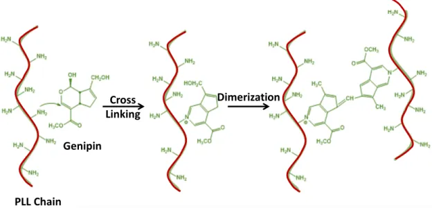
Genipin
PLL Chain
Linking
SCHEME S3. Schematic Representation of Genipin Crosslinking.
Charged (mostly) random coil state of PLL-PPA at neutral pH
(relaxed core)
Uncharged alpha helix state of PLL-PPA at pH~4.5 (wound) (Strained core = drug leaching) pH Change
Loss of charge
= Drug (Curcumin)
FIGURE S1. Schematic Representation of Effective Drug Release at Wound pH (ca. 4.5) from PLL-PPA Micelles in the