九州大学学術情報リポジトリ
Kyushu University Institutional Repository
Colorectal diffuse large B-cell lymphoma:
molecular subclassification and prognostic significance of immunoglobulin gene
translocation
保利, 喜史
http://hdl.handle.net/2324/4060045
出版情報:九州大学, 2019, 博士(医学), 課程博士 バージョン:
権利関係:© 2019 Elsevier Inc. All rights reserved.
Original contribution
Colorectal diffuse large B-cell lymphoma: molec- ular subclassi fi cation and prognostic signi fi cance of immunoglobulin gene translocation ☆ , ☆☆
Yoshifumi Hori MD
a, Hidetaka Yamamoto MD, PhD
a, Yui Nozaki MD
a,
Takehiro Torisu MD, PhD
b, Minako Fujiwara MD, PhD
a, Kenichi Taguchi MD, PhD
c, Kenichi Nishiyama MD, PhD
d, Shotaro Nakamura MD, PhD
e,
Takanari Kitazono MD, PhD
b, Yoshinao Oda MD, PhD
a,⁎
aDepartment of Anatomic Pathology, Kyushu University, Japan
bDepartment of Medicine and Clinical Science, Graduate School of Medical Sciences, Kyushu University, Japan
cDepartment of Pathology, National Kyushu Cancer Center, Fukuoka, Japan
dDepartment of Pathology, Fukuoka Red Cross Hospital, Fukuoka, Japan
eDivision of Gastroenterology, Department of Internal Medicine, School of Medicine, Iwate Medical University, Morioka, Japan
Received 21 May 2019; revised 6 September 2019; accepted 9 September 2019
Keywords:
Diffuse large B-cell lym- phoma;
Colon;
Rectum;
Translocation;
Immunoglobulin;
Prognosis
SummaryPrimary colorectal diffuse large B-cell lymphoma (DLBCL) is rare, and its clinicopatholog- ical and genetic features are poorly understood. The aim of our study was to elucidate the frequency and prognostic significance of molecular subgroups in colorectal DLBCL. We examined 25 cases of colorectal lymphoma with DLBCL-like morphology and classified them into germinal center B-cell like (GCB)/non-GCB subgroups by immunohistochemistry (IHC) for CD10, bcl-6 and MUM1, or into dou- ble-expressor (DE)/non-DE subgroups by IHC for bcl-2 and c-myc. Translocations involvingBCL2, BCL6,MYC,IGH,IGK,IGL, andMALT1were also investigated using break-apartfluorescencein situ hybridization (FISH). The 25 cases were classified into two entities—DLBCL, not otherwise specified (NOS) (n = 23; 92%) and high grade B-cell lymphoma, double hit (n = 2; 8%)—according to the recent WHO classification. None of them showed histological evidence of Epstein-Barr virus infection or high-grade transformation from low grade B-cell lymphoma. Ten cases were GCB-type and four cases were DE-type, but these subtypes did not contribute to clinicopathological differences. Translocations involvingBCL2, BCL6, MYC,IGH,IGK,IGL, andMALT1were detected in 3 (12%), 3 (12%), 10 (40%), 14 (56%), 3 (12%), 3 (12%), and 0 (0%) of 25 cases, respectively. Of note, the presence of IGHtranslocation was significantly associated with better overall survival (P= .0053) and progression free survival (P= .0259). Similarly, the translocation involving at least one of theIGs (IGH,IGK, and/
☆ Conflicts of interest/Disclosures: The authors declare that there are no conflicts of interest to disclose.
☆☆ Acknowledgements: We are grateful to Hiroaki Miyoshi, Koichi Ohshima, Shinichi Kawano, Koji Ikegami, Yuichi Matsuno, Keizou Zeze, Takahide Tanaka, Takashi Hirata, Motohiro Esaki, and Shinichi Tsuruta for scientific support, and Naoko Ieiri and Mami Nakamizo for technical assistance. The English in this article was edited by KN international (http://www.kninter.com/).
⁎ Corresponding author at: Department of Anatomic Pathology, Pathological Sciences, Graduate School of Medical Sciences, Kyushu University, Maidashi 3-1-1, Higashi-ku, Fukuoka 812-8582, Japan.
E-mail address:[email protected](Y. Oda).
www.elsevier.com/locate/humpath
https://doi.org/10.1016/j.humpath.2019.09.003 0046-8177/© 2019 Elsevier Inc. All rights reserved.
Human Pathology (xxxx)xx, xxx
YHUPA-04922; No of Pages 12
orIGL) was associated with more favorable prognosis in DLBCLs or even in DLBCL, NOS. This is thefirst report to reveal that a small subset of colorectal DLBCL corresponds to double-hit lymphoma.
In addition, translocations involving at least one of theIGs may be a favorable prognostic factor in co- lorectal DLBCL. Testing the translocation involving rearrangement ofIGs as well asMYCandBCL2/
BCL6may thus be useful for diagnosis and prognosis.
© 2019 Elsevier Inc. All rights reserved.
1. Introduction
Primary colorectal lymphoma is very rare, accounting for 0.2% to 0.6% of all colonic malignancies and 10% to 20% of gastrointestinal (GI) non-Hodgkin lymphoma[1,2]. Never- theless, diffuse large B-cell lymphoma (DLBCL) is relatively common among colorectal lymphomas[1-7]. DLBCL is of- ten difficult to treat and the continuing subject of interest among pathologists, hematologists and gastroenterologists.
According to the recent World Health Organization (WHO) classification published in 2017, colorectal B-cell lymphoma and lymphoproliferative disorder (LPD) with DLBCL-like morphology can be subdivided into the following categories:
(i) DLBCL, not otherwise specified (NOS), (ii) high-grade B- cell lymphoma withMYCandBCL2and/orBCL6rearrange- ments [so-called double-hit lymphoma (DHL): HGBL-DH], (iii) Epstein-Barr virus (EBV)-positive DLBCL, NOS (for- merly, age-related LPD), (iv) EBV-positive mucocutaneous ulcer, and (v) immunodeficiency-associated LPD (including primary immune disorder, human immunodeficiency virus infection, post-transplant and other iatrogenic conditions) [3]. These lymphomas should be distinguished from Burkitt lymphoma (BL) or morphologically BL-like lymphomas such as HGBL-DH and HGBL-NOS.
DLBCL is a clinically, immunologically and genetically heterogenous group of lymphomas [3,8-10]. As for nodal DLBCLs, HGBL-DH (DHL) is considered to have an ag- gressive biological behavior[11,12]. The double expressor, as defined by the co-expression of c-myc and bcl-2, are also an aggressive subgroup of DLBCL[13-15]. According to the gene expression profile, the germinal center B-cell like (GCB) subtype is thought to have a more favorable prognosis than the non-GCB or activated B-cell-like (ABC) subtype among DLBCLs[16]. However, the prevalence and clinico- pathological significance of these subtypings have been un- clear in primary DLBCL of the GI tract, especially of the large intestine[17].
The reported cytogenic abnormalities in DLBCL include the translocations involvingIGH(14q32),IGK(2p12),IGL (22q11),BCL2(18q21),BCL6(3q27),MYC(8q24),API2 (11q21) andMALT1(18q21)[18,19]. Nevertheless, it is not easy to examine all the above translocations at the daily prac- tice level, while the G-band of chromosomal inspection is not sufficiently sensitive, and next-generation sequencing is ex- pensive[8-10]. Therefore, it is necessary to establish bio- markers that are cost-effective and useful for diagnosis and
prognosis. We previously reported that translocations involv- ingIGHare associated with favorable prognosis in patients with gastric and small bowel DLBCLs[20,21]. In this study, we attempted to classify colorectal DLBCLs according to the recent WHO classification, and to elucidate the clinicopatho- logical significance of molecular subtyping and candidate gene rearrangements.
2. Materials and methods
2.1. SubjectsData on cases of colorectal lymphomas with DLBCL-like morphology (n = 25) were retrieved from thefiles of Kyushu University Hospital and its affiliated hospitals between 1999 and 2018. All cases satisfied the criteria for primary gastroin- testinal lymphoma as defined by Lewin et al[22]. A method for discriminating primary colorectal lymphomas from sys- temic lymphomas involving the large intestine has not been fully developed, but in the current study we considered a case to be colorectal primary if the main bulk of the lesions were located in the large intestine in the current study[2,4-6].
All cases were histologically reviewed by two pathologists (mostly Y.H. and H.Y.), and the morphological diagnosis of DLBCL was confirmed according to the criteria of WHO classification[3]. Immunodeficiency-associated LPDs with DLBCL morphology were excluded in the current study. Al- though pleomorphic mantle cell lymphoma can show DLBCL-like morphology, we confirmed that both CD5 and cyclin D1 were immunohistochemically negative in all 25 cases. Burkitt lymphoma (BL) and high-grade lymphoma with BL-like morphology were also excluded. The clinical stage was determined according to the Lugano classification [23]. Staging workup included computed tomography of the neck, chest, and abdomen, esophagogastroduodeno- scopy, colonoscopy, small bowel endoscopy (double-balloon and/or capsule endoscopy) or barium radiography, bone mar- row aspiration/biopsy, andfluorine-18fluorodeoxyglucose positron emission tomography. The international prognostic index (IPI) for aggressive lymphoma was classified as low, low-intermediate, intermediate-high or high risk[24]. The macroscopic growth pattern was classified as polypoid, ul- cerative, lymphomatous polyposis, diffuse-infiltrating, or mixed by three experienced endoscopists (S.N., T.T., and Y.H.) based on the endoscopicfindings in 8 cases and on
2 Y. Hori et al.
the surgical excision specimens in 17 cases[6,20,21]. The initial treatment modalities included surgical resection, che- motherapy with a cyclophosphamide, doxorubicin, vincris- tine and prednisolone (CHOP)-based regimen with or without rituximab, radiotherapy, or a combination of them.
Complete remission (CR) was defined as the complete disap- pearance of clinical evidence of lymphoma. Partial remission (PR) was defined as a tumor reduction of ≥50%. The resected or biopsied specimens werefixed in 10% formalin solution, embedded in paraffin and cut into 4-μm-thick slices for subsequent analysis. This retrospective study was ap- proved by the Institutional Review Board of Kyushu Univer- sity (no.29-240) and the National Kyushu Cancer Center (no.2017-56).
2.2. Immunohistochemical staining
For immunohistochemistry (IHC), we used the primary mouse monoclonal antibodies against CD10 (clone 56C6, 1:100 dilution; Leica Biosystems, Newcastle upon Tyne, UK), bcl-2 (clone 124, 1:100 dilution; Dako), bcl-6 (clone PG-B6p, 1:10 dilution; Dako), MUM1 (clone MUM1p, 1:50 dilution; Dako) and c-myc (clone Y69, 1:100 dilution;
Abcam, Cambridge, UK). Samples in which more than 30% of tumor cells were immunoreactive were considered to be positive for CD10, bcl-6 and MUM1[3,20,21]. There- after, tumors were classified as either GCB or non-GCB phe- notype according to the algorithm of Hans[25]. Moreover, the cut-off points for c-myc and bcl-2 were defined as 40%
or more and 50% or more, respectively[3,14,15,26].
2.3. Fluorescence in situ hybridization (FISH)
Gene translocation was investigated by interphasefluores- cence in situ hybridization (FISH) on FFPE with dual-color, break-apart rearrangement probes forBCL2,BCL6,MYC, IGH,MALT1(Vysis-Abbott, Des Plaines, IL),IGKandIGL (Cytocell Technologies, Cambridge, UK) as previously de- scribed[20,21]. Cases with bothIGHandBCL2rearrange- ments by break-apart FISH were further investigated by FISH withIGH/BCL2dual-color, dual fusion translocation probe (Vysis-Abbott, Des Plaines, IL). Likewise, cases with bothIGHandMYCrearrangements were examined by FISH withIGH/MYC/CEP8tri-color, dual fusion probe (Vysis).
FISH analysis was performed using direct viewing on a stan- dard fluorescence microscope (BX53; Olympus, Tokyo).
Cut-off levels were the same as those in previous studies [20,21]. If three or more gene copies were detected in tumor cells, the result was categorized as a copy number gain, as de- scribed in the previous studies[20,21].
2.4. In situ hybridization for EBV infection
To test for the presence of EBV,in situ hybridization (ISH) was performed on FFPE with an EBV-encoded
RNA (EBER)-specific peptide nucleic acid (PNA) probe and a PNA ISH detection kit (Dako Cytomation, Car- pinteria, CA) according to the manufacturer’s instruc- tion, as previously described [27]. EBV-positive gastric cancer with lymphoid stroma was used as the positive control.
2.5. Statistical analysis
Overall survival (OS) was defined as the time from the date of diagnosis to the date of death from a tumor-specific cause. Progression-free survival (PFS) was defined as the time from the date of diagnosis to the date when progressive disease (PD) wasfirst documented or relapse was confirmed clinically. The OS and PFS rates were calculated with the Kaplan-Meier method, and values were compared by using the log-rank test. Other differences were evaluated with Fish- er’s exact test, theχ2test, or the Mann-WhitneyUtest.P values ofb.05 were considered to be statistically significant.
All analyses were performed with JMP PRO 13 software (SAS Institute, Cary, NC).
3. Results
3.1. Clinical features
Clinicopathologicalfindings are shown inTable 1 and summarized in Supplemental Table S1. The patients were di- agnosed at the mean age of 66 years (range: 38-84 years) with a slight male predominance (male: female = 1.27:1). The most frequent tumor location was the cecum (n = 17, 68%).
Sixteen patients (64%) had a single tumor located in the large intestine, whereas nine (36%) had multiple tumors, with the main lesion being located in the large intestine. Among cases with multiple tumors, the smaller tumors were present in the ileum (n = 8, 32%) or stomach (n = 1, 4%). The macroscopic type was classified as the ulcerative type in 11 patients (44%), the polypoid type in 12 (48%), mixed-type in 1 (4%), and diffuse type in 1 (4%). No patients had lymphoma- tous polyposis-type tumors. The clinical stage was I in 9 pa- tients (36%), II1 in 2 patients (8%), II2 in 3 patients (12%), IIE in 4 patients (16%), and IV in 7 patients (28%). The IPI risk was low in 11 patients (44%), low-intermediate in 7 pa- tients (28%), high-intermediate in 5 patients (20%), and high in 2 patients (8%). Seventeen patients (68%) underwent sur- gical resection followed by chemotherapy with a cyclophos- phamide, doxorubicin, vincristine and prednisolone (CHOP)-based regimen with or without rituximab, 5 patients (20%) received chemotherapy without surgical resection and 2 patients (8%) received radiation therapy after chemother- apy, and 1 patient (4%) underwent surgery alone. As a result, 17 patients (71%) achieved CR, 4 (16%) achieved PR, and 3 (13%) showed PD. The follow-up duration after diagnosis ranged from 3 to 187 months (median: 54 months). During 3 Immunoglobulin gene translocation in colorectal DLBCL
Table 1 Clinicopathologicalfindings in 25 cases of colorectal large B-cell lymphoma
Case Age Sex Size
(mm)
Site Multiplicity Macroscopic type
Clinical stage (Lugano)
IPI Colectomy Initial treatment Response to initial treatment
Progression/relapse, follow-up period (months)
Tumor specific death, follow-up period (months) DLBCL, NOS (n = 23)
1 38 F 27 Cecum Single Ulcerative II2 0 (-) R-CHOP CR (-, 83 mo) (-, 83 mo)
2 56 F 135 Cecum Multiple Polypoid IV 3 (+) R-CHOP CR (+, 75 mo) (+, 92 mo)
3 64 M 47 Cecum Multiple Polypoid IV 2 (-) R-CHOP CR (-, 53 mo) (-, 53 mo)
4 64 M 40 Colon Single Ulcerative IIE 2 (+) R-CHOP CR (-, 79 mo) (-, 79 mo)
5 51 M 87 Cecum Single Ulcerative IIE 1 (+) R-CHOP CR (-, 118 mo) (-, 118 mo)
6 74 F 68 Cecum Multiple Ulcerative IV 3 (+) R-CHOP PR (-, 8 mo) (-, 8 mo)
7 71 M 31 Cecum Single Polypoid I 1 (+) (-) (-) (-, 28 mo) (-, 28 mo)
8 69 F 55 Cecum Single Ulcerative II1 1 (+) R-CHOP PR (-, 20 mo) (-, 20 mo)
9 67 M 85 Colon Multiple Polypoid II2 4 (+) ESHAP PD (+, 1 mo) (+, 3 mo)
10 74 M 90 Cecum Multiple Ulcerative IV 3 (+) EPOCH PD (+, 3 mo) (+, 8 mo)
11 84 M 80 Colon Multiple Mixed IV 2 (-) R-CHOP CR (+, 44 mo) (+, 44 mo)
12 66 F 60 Cecum Multiple Polypoid II1 2 (+) THP-COP PD (+, 4 mo) (+, 9 mo)
13 59 M 100 Rectum Single Polypoid I 2 (-) THP-COP CR (+, 12 mo) (+, 56 mo)
14 64 M 50 Cecum Single Polypoid II2 1 (+) THP-COP +
Radiation
CR (+, 13 mo) (-, 39 mo)
15 78 M 49 Cecum Multiple Polypoid I 1 (+) R-CHOP CR (-, 45 mo) (-, 45 mo)
16 53 F 48 Cecum Single Polypoid I 0 (+) R-CHOP CR (-, 187 mo) (-, 187 mo)
17 73 F 110 Cecum Single Ulcerative IIE 3 (+) CHOP CR (-, 95 mo) (-, 95 mo)
18 63 F 42 Cecum Multiple Polypoid I 1 (+) CHOP CR (-, 58 mo) (-, 58 mo)
19 79 M 103 Rectum Single Polypoid IV 4 (-) R-CHOP PR (-, 2 mo) (-, 2 mo)
20 65 F 94 Cecum Single Ulcerative I 1 (+) R-THP-COP CR (-, 82 mo) (-, 82 mo)
21 71 M 60 Cecum Single Polypoid I 3 (+) R-CHOP CR (-, 101 mo) (-, 101 mo)
22 61 M 104 Rectum Single Ulcerative I 1 (+) R-CHOP CR (-, 14 mo) (-, 14 mo)
23 60 F 100 Rectum Single Ulcerative I 0 (-) R-CHOP +
Radiation
CR (-, 8 mo) (-, 8 mo)
HGBL-DH (n = 2)
24 68 M 140 Cecum Single Ulcerative IIE 2 (+) R-ESHAP PR (+, 61 mo) (-, 85 mo)
25 77 F 93 Colon Single Diffuse IV 2 (-) R-THP-COP CR (+, 4 mo) (+, 6 mo)
Abbreviations: IPI, international prognostic index; R, rituximab; CHOP, cyclophosphamide, doxorubicin, vincristine, and prednisone; ESHAP, etoposide, methyl-prednisolone, cytarabine and cisplatin; EPOCH, etoposide, prednisone, vincristine, cyclophosphamide, and doxorubicin.; THP-COP, tetrahydropyranyl adriamycin, cyclophosphamide, vincristine, and prednisolone; CR, complete response; PR, partial re- mission; PD, progressive disease.
4Y.Horietal.
the follow-up period, 3 patients (13%) who had initially been treated with surgery plus chemotherapy showed PD. A total of 9 patients (36%) died and 7 (28%) of them died of lym- phoma. Of the 2 remaining patients, 1 patient (4%) died of esophageal cancer, and the other patient (4%) died from pneumoniae post chemotherapy. The OS and PFS rates after 5 years were 75% and 68%, respectively (Supplemental Fig.
S1).
3.2. Histopathological findings
Histopathologically, all cases showed diffuse proliferation of CD20-positive centroblast-like or immunoblast-like large atypical lymphoid cells (Fig. 1A and B). None of them re- vealed morphologically definitefindings of high grade trans- formation from low grade B-cell lymphoma, such as follicular lymphoma and MALToma. EBV-EBER was nega- tive in all cases, which excluded both EBV-positive DLBCL, NOS and EBV-positive mucocutaneous ulcer. According to the immunohistochemical and molecularfindings, our cases were classifiable into two categories; DLBCL, NOS (n = 23, 92%) (cases 1–23) and HGBL-DH (n = 2, 8%) (case 24 and 25) (see below) (Table 1).
3.3. Immunohistochemical findings
Immunohistochemically, CD10, bcl-2, bcl-6, MUM1, and c-myc were positive in 8/25 (32%), 14/25 (56%), 15/25 (60%), 17/25 (68%), and 7/25 (28%) cases, respectively (Table 2and Supplemental Table S2). According to the algo- rithm of Hans[25], 10 (40%) and 15 (60%) cases were sub- divided into the GCB and non-GCB phenotype, respectively. Among 23 cases of DLBCL, NOS, 9 (39%) were GCB type and 14 (61%) were non-GCB type (Table 2, Supplemental Fig. S2). Among cases of HGBL-DH (n = 2), 1 belonged to the GCB and 1 to the non-GCB phenotype.
Four of 25 (16%) cases were consistent with double expres- sor (DE). These cases comprised of 3/23 (13%) cases of DLBCL, NOS and 1/2 (50%) cases of HGBL-DH. In other words, 1/4 (25%) cases of DE lymphoma and 1/21 (5%) cases of non-DE lymphoma was a case of HGBL-DH.
3.4. Gene translocations by FISH
Break-apart FISH was successful in all 25 cases. A retrospec- tive split signal on FISH is shown inFig. 1C. Gene rearrange- ments suspicious for translocations involvingIGH,IGK,IGL, BCL2,BCL6, andMYCwere detected in 14 (56%), 3 (12%), 3 (12%), 3 (12%), 3 (12%), and 10 (40%) of 25 cases, respectively (Fig. 2, Supplemental Table S2). No cases harboringMALT1 translocation were observed. Among the 10 patients withMYC rearrangements, breakage ofBCL2was observed in 1 case (case 24) and breakage ofBCL6in 1 case (case 25); that is, 2 cases cor- responded to HGBL-DH (Fig. 2). One of the 2 HGBL-DHs also hadIGH-MYC translocation confirmed by dual-fusion FISH
(cases 25) (Supplemental Table S3). Among the 14 patients with IGHrearrangements, breakage ofBCL2,BCL6andMYCwas detected in 2 (14%), 3 (21%), and 5 (36%) cases, respectively (Fig. 2). Among the 5 cases (cases 1, 5, 10, 19 and 25) with both IGHandMYCrearrangements by break-apart FISH,IGH-MYC translocation was confirmed in 2 cases (cases 5 and 25) by dual-fusion FISH (Supplemental Table S3). Of the 2 cases (cases 3 and 20) with bothIGHandBCL2rearrangements by break- Fig. 1 A representative case of colorectal diffuse large B-cell lymphoma. A,A high-power histological view shows diffuse infil- trates of large, atypical lymphoid cells (hematoxylin and eosin stain). B,Immunohistochemically, the neoplastic cells are positive for CD20. C,Interphasefluorescencein situhybridization with an IGHdual-color break-apart probe shows a split of green and red sig- nals (indicated by arrows), indicating the presence of a translocation.
5 Immunoglobulin gene translocation in colorectal DLBCL
apart FISH,IGH-BCL2translocation was confirmed in one case (case 20) by dual-fusion FISH. Of all 25 cases, 16 (64%) cases showed translocations involving at least one of theIGs (IGH, IGKandIGL) (Fig. 2). Among these 16 cases, 12 hadIGHtrans- location alone, 1 hadIGKtranslocation alone (case 24), and there was 1 case each with a combination ofIGH/IGK/IGL(case 3),IGH/IGL(case 10) orIGK/IGLtranslocation (case 6). Among 9 cases withoutIGs translocations, 3 cases were associated with MYCtranslocation, but none of 9 tumors hadBCL2orBCL6 translocation (Fig. 2). Extra copies ofMALT1and/orBCL2,sug- gesting trisomy 18, were detected in 1 (4%) cases and extra cop- ies ofBCL6, suggesting trisomy 3, were detected in 3 (12%) cases (Supplemental Table S2). Extra copies ofMYCorIGs were not detected in any case. In total, 21 (84%) cases showed some structural or numerical aberrations.
3.5. Clinicopathological and prognostic comparison
The correlation between molecular factors and OS or PFS is shown inFig. 3and4, respectively.
3.5.1. DLBCL, NOS, versus HGBL-DH
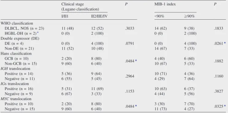
Both cases (2/2) of HGBL-DH showed higher clinical stage and higher MIB-1 index (≥90%) (Table 3 and 4).
HGBL-DH exhibited a tendency toward higher frequency of relapse (2/2, 100% versus 7/23, 30%) (Table 3) and shorter progression free survival time (P= .0696) as compared with DLBCL, NOS; however, these differences did not reach the level of statistical significance (Fig. 4A).
3.5.2. DE versus non-DE
All 4 cases of DE showed higher clinical stage and higher MIB-1 index (≥90%) (Table 4). Three of 4 (75%) cases of DE relapsed, whereas 6 of 21 (29%) cases of non-DEs re- lapsed (P= .1162); however, these 2 groups did not show significant differences in OS (P = .6304) and PFS (P = .3188) (Fig. 3B and4B).
3.5.3. GCB versus non-GCB subtype
Between GCB (n = 10, 25%) and non-GCB (n = 15, 75%) cases, there were no significant differences in clinico- pathological factors (data not shown) or prognosis (Fig. 3C and4C).
3.5.4.IGHtranslocation versus nonIGHtranslocation IGHtranslocation-positive groups showed significantly longer time of OS and PFS as compared with the negative group, respectively (P= .0053, .0259) (Fig. 3D and4D).
Table 2 Immunohistochemical results in 25 cases of colorectal large B-cell lymphoma
Case CD10 bcl-2 bcl-6 MUM1 c-myc Double expressor MIB-1 (%) Hans
DLBCL, NOS (n = 23)
1 (+) (+) (+) (+) (+) (+) N95 GCB
2 (+) (+) (+) (+) (+) (+) 90-95 GCB
3 (+) (+) (+) (+) (-) (-) 60 GCB
4 (-) (-) (+) (-) (-) (-) 64 GCB
5 (+) (-) (+) (-) (-) (-) 90 - 95 GCB
6 (+) (+) (+) (+) (-) (-) 90 GCB
7 (+) (+) (+) (+) (-) (-) 90 GCB
8 (+) (-) (-) (-) (+) (-) 85 GCB
9 (-) (+) (+) (-) (-) (-) 67 GCB
10 (-) (+) (-) (+) (+) (+) 90 Non-GCB
11 (-) (-) (-) (-) (-) (-) 85 Non-GCB
12 (-) (-) (-) (-) (-) (-) 66 Non-GCB
13 (-) (+) (-) (+) (-) (-) 90 Non-GCB
14 (-) (+) (+) (+) (-) (-) 50 Non-GCB
15 (-) (-) (-) (+) (-) (-) 60 Non-GCB
16 (-) (+) (+) (+) (-) (-) 59 Non-GCB
17 (-) (-) (+) (+) (-) (-) 80 Non-GCB
18 (-) (+) (-) (-) (-) (-) 80 Non-GCB
19 (-) (-) (+) (+) (+) (-) 75 Non-GCB
20 (-) (-) (-) (-) (-) (-) 50 Non-GCB
21 (-) (+) (-) (+) (-) (-) N95 Non-GCB
22 (-) (-) (-) (+) (+) (-) 90 Non-GCB
23 (-) (+) (+) (+) (-) (-) 89 Non-GCB
HGBL-DH (n = 2)
24 (+) (-) (+) (+) (-) (-) N95 GCB
25 (-) (+) (+) (+) (+) (+) 90 Non-GCB
Abbreviation: GCB, germinal center B-cell-like. Immunophenotype by Hans classification.
6 Y. Hori et al.
10 1 6 5 19 14 8 15 16 17 18 3 4 20 11 12 7 2 9 13 21 22 23 24 25 Case No.
Diagnosis
HL H KL H H H H H H H H HKL H H K H IGs (IGHand/or IGK and/or IGL)
translocation
2,6 6 2 2 6 BCL2/BCL6
translocation
MYC translocation
DE
GCB
Progression/relapse
DLBCL, NOS DH
Fig. 2 Histological subtype,IGs rearrangement and other molecular factors in colorectal large B-cell lymphomas. H:IGHtranslocation; K:IGLtranslocation; L:IGLtranslocation; 2:
BCL2translocation; 6:BCL6translocation
7ImmunoglobulingenetranslocationincolorectalDLBCL
3.5.5.IGs translocation versus nonIGs translocation TheIGs translocation-positive group had longer time of OS (P= .0240) and PFS (P= .0771) as compared with the negative group, although the latter did not reach the level of statistical significance (Fig. 3E and4E).
3.5.6.MYCtranslocation versus non MYCtranslocation The MYC translocation-positive group tended to have worse prognosis compared with the negative group in terms of both OS (P= .1642) and PFS (P= .2024) (Figure 3F and 4F). In addition, clinical stage (P= 0.0484) and MIB-1 index Fig. 3 Kaplan-Meier analysis for overall survival (OS) in colorectal DLBCLs. A, HGBL-DH show slightly worse OS, but without statistical significance (P= .4478). B,There is no significant difference in OS between DEs and non-DEs (P= .6304). C,There is no significant difference in OS between GCB and non-GCB subtypes (P = .9276). D,Cases involvingIGHtranslocation are correlated with significantly better OS (P= .0053). E, Cases involvingIGs translocation are correlated with significantly better OS (P= .0240). F,Cases involvingMYCtranslocation are associated with slightly worse OS, but not to a statistically significant degree (P= .2024).
8 Y. Hori et al.
(P= .0325) were significantly higher in theMYCtransloca- tion positive group (Table 4).
3.5.7. Prognostic factors in colorectal DLBCL, NOS When the cases were limited to DLBCL, NOS (n = 23), both the presence of IGH translocation and the
presence of IGs translocation were correlated with bet- ter OS (P = .0136, P = .0101, respectively) (Supple- mental Fig. S3C, S3E) and PFS (P = .0304, P = .0210, respectively) (Supplemental Fig. S3D, S3F).
Other molecular factors did not influence the prognosis.
Fig. 4 Kaplan-Meier analysis for progression free survival (PFS) in colorectal DLBCLs. A, HGBL-DH show slightly worse PFS, but the difference does not reach statistical significance (P= .0696). B,DEs show slightly worse PFS, but the difference is not significant (P= .5812). C,Non-GCB subtypes show slightly worse PFS, but statistical difference is not detected (P= .5812). D,Cases involvingIGHtrans- location are correlated with significantly better PFS (P= .0259). E, Cases involvingIGs translocation show better PFS, but the difference is not significant (P= .0771). F,Cases involvingMYCtranslocation are associated with slightly worse prognosis, but the difference is not signif- icant (P= .1642).
9 Immunoglobulin gene translocation in colorectal DLBCL
4. Discussion
In our study, we detected 2 cases of HGBL-DHs (8%) among the colorectal DLBCLs; a similar ratio of HGBL-DHs (DHL) has been reported among nodal DLBCLs[3,11,12]. Although the number of examined cases was very small in our previous studies on GI-DLBCL, the ratio of DHL was 0% in the stomach (n = 83) and 6% in the small intestine (n = 33)[20,21]. Addition- ally, in a study by Magnoli et al[26], no DHL cases were recog- nized in any GI organ, including the stomach (n = 5), small intestine (n = 5), and large intestine (n = 4). It has been reported that DHL represents a more aggressive group among nodal DLBCLs[11,12]. In this study, both cases (2/2) of DHL showed relapse; 1 case of DHL (case 25) died of tumor, while the other (case 24) experienced a relapse but remained alive for 85 months.
It is unclear why the latter case showed a relatively indolent clin- ical course. These cases had bothIGKandBCL2translocation and GCB phenotype, and thus it might be that the DHL origi- nated from follicular lymphoma[3], although it was difficult to determine the exact origin and clinicopathological significance of such a genotype of DHL. Recently, Miyaoka et al reported that follicular lymphomas withMYCtranslocation (“double-hit follic- ular lymphoma”) may have a relatively indolent clinical course [28]. Conversely, among our cases of DLBCL, NOS (n = 23), 3 patients (case 3, 4, and 20) had bothIGHandBCL2orBCL6 rearrangements by break-apart FISH (case 20 also hadIGH- BCL2translocation by dual-fusion FISH) withoutMYCtranslo- cations and they remained alive without relapse. Morphologi- cally, these 3 cases did not show the definitive findings of follicular lymphoma, but genetically at least some of them might be identical to DLBCL derived from follicular lymphoma and may be consistent with a relatively indolent clinical course.
In this study, 4 (16%) cases of colorectal DLBCL were DE, and 2 (8%) cases were DHL (Table 2and Supplemental Table S2). In previous studies, among DLBCLs of various sites, about 19% to 34% have been reported as DE [3,26,29]. However, there have been no studies focusing on the digestive tract. Patients with DE werefirst noted as a poor prognostic group after R-CHOP, and many cases of DE were later found to belong to activated B cell-like (ABC) subtype [13,14,29]. In our series, patients with the DE phenotype showed significantly higher MIB-1 index (P = .0261) (Table 4). In addition, 3 of 4 patients with DE relapsed and two died of lymphoma, but there was no statistically signifi- cant difference in prognosis between the DE and non-DE groups (Fig. 3B and4B). Thus, the prognostic significance of DE subtypes in GI DLBCL remains to be further eluci- dated in studies enrolling a larger number of cases.
According to earlier reports on nodal DLBCL, the GCB type accounts for about 35% to 56% of cases and has a more favorable prognosis than the non-GCB type[25,30-32]. As for colorectal DLBCL, there are few reports on GCB/non-GCB phenotype, due to its scarcity of colorectal DLBCL itself. As for DLBCL of the stomach and small intestine, the actual ratio of the GCB to non-GCB subtype is controversial, but all reports agree that there is no statistically significant difference in prognosis between
the two subtypes[20,21,33,34]. In our series of colorectal cases, the GCB type accounted for 40% (10/25) of DLBCLs; moreover, half of the DHL cases (1/2) and half of the DE cases (2/4) were the GCB subtype. As for prognosis, there was no significant prognostic difference between the GCB and non-GCB group. Al- though the examined number of cases examined in this study was limited, the prevalence and clinicopathological significance of the GCB subtype might be different between GI and nodal DLBCLs.
On the other hand, in GI DLBCL, the prognostic value of the GCB/non-GCB phenotype based on a system other than Hans’ criteria is still unknown[31,32].
In the present study,IGHtranslocation was confirmed in 14 cases (56%), which was the similar to the frequency in nodal DLBCL[35]. The frequency ofIGHtranslocation in other GI
Table 3 Clinicopathological features in DLBCL, NOS, and HGBL-DH
DLBCL, NOS (n = 23, 92%)
HGBL-DH (n = 2, 8%)
Male/female 13:10 1:1
Age median (range) 65 (38-84) 73 (68-77) Location
Cecum (n = 17) Colon (n = 4) Rectum (n = 4)
16 (70) 3 (13) 4 (17)
1 (50) 1 (50) 0 (0) Tumor size
b10cm (n = 18)
≥10cm (n = 7)
17 (74) 6 (26)
1 (50) 1 (50) Macroscopic type
Ulcerative (n = 11) Polypoid type (n = 12) Mixed/others (n = 2)
10 (44) 12 (52) 1 (4)
1 (50) 0 (0) 1 (50) Clinical stage
(Lugano classification) I, II1 (n = 11) II2, IIE, IV (n = 14)
11 (48) 12 (52)
0 (0) 2 (100) IPI
Low (n = 11)
Low-intermediate (n = 7) Intermediate-high (n = 5) High (n = 2)
11 (48) 5 (22) 5 (22) 2 (87)
0 (0) 2 (100) 0 (0) 0 (0) Colectomy
Yes (n = 18) No (n = 7)
17 (74) 6 (26)
1 (50) 1 (50) Responsea
CR (n = 17) PR (n = 4) NC/PD (n = 3)
16 (70) 3 (13) 3 (13)
1 (50) 1 (50) 0 (0) Progression/relapse
Yes (n = 9) No (n = 16)
7 (30) 16 (70)
2 (100) 0 (0) Tumor specific death
Yes (n = 7) No (n = 18)
6 (26) 17 (74)
1 (50) 1 (50) Abbreviations: IPI, International Prognostic Index; CR, complete re- sponse; PR, partial remission; NC, no change; PD, progressive disease.
aOne case did not receive an imaging test after initial examination, but did not relapse clinically.
10 Y. Hori et al.
organs was reported to be 27% to 70% in the small intestine and 36% in the stomach (except for DLBCL with MALToma) [20,21]. As for prognosis, in our series of colorectal DLBCLs, translocations involvingIGHwere found to be associated with favorable OS and PFS (Fig. 3D and4D). Even when considering only cases of DLBCL, NOS, the same relationship was con- firmed (Supplemental Fig. S3C, S3D). A similar prognostic value ofIGHtranslocation has been reported in DLBCLs of the stomach and small intestine[20,21], whereas it has not been confirmed in nodal DLBCL. It is well known that overexpression ofIGHpartner genes such asBCL2,BCL6andMYCplays an im- portant role in lymphomagenesis; presumably, however, addi- tional alterations are also indispensable for the development and progression of DLBCL[18]. The differences in the gene al- terations involved might influence the above-mentioned prog- nostic impact ofIGH-translocation in GI-DLBCL. In addition, it is possible that the genes responsible for the development of lymphoma are different between the GI tract and nodal DLBCLs.
Meanwhile, translocation ofIGs (IGHand/orIGKand/or IGL) was found in 16 cases (64%) of colorectal DLBCL.
Among the 16 cases, 1 case withoutIGHtranslocation had IGKtranslocation (case 24) and 1 case withoutIGHtransloca- tion had bothIGKandIGLtranslocations (case 6). In one patient withIGHtranslocation, we also found concomitant transloca- tions involving bothIGKandIGL; this is a rare phenomenon that has only been reported in nodal DLBCL[19,36]. Interest- ingly, similar toIGHtranslocation, the presence ofIGs translo- cation was associated with favorable prognosis in our patients with colorectal DLBCL (n = 25) (Fig. 3E and4E) or in those
with DLBCL, NOS (n = 23) (Supplemental Fig. S3E, S3F).
As for gastric and small intestinal DLBCL, to the best of our knowledge, there have been no investigations on the prognostic relevance ofIGs translocation. Akasaka et al[37]reported that BCL6-IGgene fusions were associated with a better prognosis thanBCL6-non-IGfusions in patients with nodal DLBCL.
We still cannot fully explain the reason for the favorable prognosis of patients withIGs translocation. Nevertheless, the examination ofIGs translocation might be helpful for the prognostic prediction of colorectal DLBCL. Further stud- ies will be needed to confirm whether these translocations truly influence the prognosis of gastrointestinal DLBCL and to elucidate the underlying molecular mechanism.
In conclusion, we comprehensively examined the immu- nophenotype and candidate gene rearrangements in cases of colorectal DLBCL and revealed that 8% of these patients had the“so-called double-hit”. We also found that translo- cation involving at least one ofIGH,IGK, andIGLwas as- sociated with more favorable behavior in our patients with DLBCL or DLBCL, NOS. Therefore, identifying the trans- locations ofIGs as well asMYCandBCL2/BCL6may be useful for the diagnosis and prognostic prediction of colo- rectal DLBCL.
Appendix A. Supplementary data
Supplementary data to this article can be found online at https://doi.org/10.1016/j.humpath.2019.09.003.
Table 4 The correlation between subclassifications and clinical stage or MIB-1 index in colorectal DLBCL Clinical stage
(Lugano classification)
P MIB-1 index P
I/II1 II2/IIE/IV b90% ≥90%
WHO classification DLBCL, NOS (n = 23) HGBL-DH (n = 2)a
11 (48) 0 (0)
12 (52) 2 (100)
.3033 14 (62)
0 (0)
9 (38) 2 (100)
.1833 Double expressor (DE)
DE (n = 4) Non-DE (n = 21)
0 (0) 11 (52)
4 (100) 10 (48)
.0791 0 (0)
14 (67)
4 (100) 7 (33)
.0261⁎ Hans classification
GCB (n = 10) Non-GCB (n = 15)
2 (20) 9 (60)
8 (80)
6 (40) .0484⁎ 4 (40)
10 (67)
6 (60)
5 (33) .1882
IGHtranslocation Positive (n = 14) Negative (n = 11)
5 (36) 6 (55)
9 (64)
5 (45) .2964 10 (71)
4 (29)
4 (36)
7 (64) .1160
IGs translocation Positive (n = 16) Negative (n = 9)
5 (31) 6 (67)
11 (69)
3 (33) .1153 10 (63)
4 (44)
6 (37)
5 (56) .3827
MYCtranslocation Positive (n = 10) Negative (n = 15)
2 (20) 9 (60)
8 (80)
6 (40) .0484⁎ 3 (30)
11 (73)
7 (70)
4 (27) .0325⁎
Abbreviation GCB, germinal center B-cell.
a MYCtranslocation andBCL2and/orBCL6translocation.
⁎ Statistically significant.
11 Immunoglobulin gene translocation in colorectal DLBCL
References
[1] Müller-Hermelink HK, Delabie J, Ko YH, et al. B-cell lymphoma of the colon and rectum. In: Bosman FT, Carneiro F, Hruban RH, et al, eds. WHO Classification of Tumours of the Digestive System, Fourth edition. Lyon: IARC Press; 2010:178–180.
[2] Nakamura S, Matsumoto T, Iida M, Yao T, Tsuneyoshi M. Primary gas- trointestinal lymphoma in Japan: a clinicopathologic analysis of 455 pa- tients with special reference to its time trends. Cancer 2003;97:2462-73.
[3] Gascoyne RD, Campo E, Jaffe ES, et al. Diffuse large B-cell lymphoma, not otherwise specified. In: Swerdlow SH, Campo E, Harris NL, et al, ed- itors. World Health Organization classification of tumours of haematopoie- tic and lymphoid tissues. Revised 4th edition. Lyon: IARC Press; 2017.
[4] Cai YB, Chen HY, He JJ, et al. The role of surgical intervention in primary colorectal lymphoma: A SEER population-based analysis. Oncotarget 2016;44:72263-75.
[5] Lai YL, Lin JK, Liang WY, Huang YC, Chang SC. Surgical resection combined with chemotherapy can help achieve better outcomes in patients with primary colonic lymphoma. J Surg Oncol 2011;104:265-8.
[6] Nakamura S, Matsumoto T, Iida M, et al. Endoscopic diagnosis of intesti- nal malignant lymphoma diagnosis and management of gastrointestinal lymphomas. Gastroenterol Endosc 2009;51:3-9.
[7] Howell JM, Auer-Grzesiak I, Zhang J, Andrews CN, Stewart D, Urbanski SJ. Increasing incidence rates, distribution and histological characteristics of primary gastrointestinal non-Hodgkin lymphoma in a North American population. Can J Gastroenterol 2012;26:452-6.
[8] Schmitz R, Wright GW, Huang DW, et al. Genetics and pathogenesis of diffuse large B-cell lymphoma.N Engl J Med2018;378:1396-407.
[9] Pasqualucci L, Dalla-Favera R. Genetics of diffuse large B-cell lymphoma.
Blood 2018;131:2307-19.
[10] Lohr JG, Stojanov P, Lawrence MS, et al. Discovery and prioritization of somatic mutations in diffuse large B-cell lymphoma (DLBCL) by whole- exome sequencing. Proc Natl Acad Sci USA 2012;109:3879-84.
[11] Aukema SM, Siebert R, Schuuring E, et al. Double-hit B-cell lymphomas.
Blood2011;117:2319-31.
[12] Johnson NA, Savage KJ, Ludkovski O, et al. Lymphomas with concurrent BCL2andMYCtranslocations: the critical factors associated with survival.
Blood2009;114:2273-9.
[13] Molina TJ, Canioni D, Copie-Bergman C, et al. Young patients with non- germinal center B-cell-like diffuse large B-cell lymphoma benefit from in- tensified chemotherapy with ACVBP plus rituximab compared with CHOP plus rituximab: analysis of data from the Groupe d’Etudes des Lymphomes de l’Adulte/lymphoma study association phase III trial LNH 03-2B.J Clin Oncol 2014;32:3996-4003.
[14] Green TM, Young KH, Visco C, et al. Immunohistochemical double-hit score is a strong predictor of outcome in patients with diffuse large B-cell lymphoma treated with rituximab plus cyclophosphamide, doxorubicin, vincristine, and prednisone.J Clin Oncol2012;30:3460-7.
[15] Kawamoto K, Miyoshi H, Yoshida N, et al.MYCtranslocation and/or BCL2 protein expression are associated with poor prognosis in diffuse large B-cell lymphoma. Cancer Sci 2016;107:853-61.
[16] Alizadeh AA, Eisen MB, Davis RE, et al. Distinct types of diffuse large B-cell lymphoma identified by gene expression profiling. Nature 2000;403:503-11.
[17] Papakonstantinou IP, Andreadis EA. Persistent afebrile abdominal pain: an usual case of segmental colitis in an immunocompromised host. Cureous 2017;9:e1033.
[18] Kramer MH, Hermans J, Wijburg E, et al. Clinical relevance ofBCL2,BCL6 andMYCrearrangements in diffuse large B-cell lymphoma. Blood 1998;92:
3152-62.
[19] Lu C, He Q, Zhu W, et al. The value of detecting immunoglobulin gene re- arrangements in the diagnosis of B-cell lymphoma. Oncotarget 2017;8:
77009-19.
[20] Ikegami K, Nakamura S, Esaki M, et al. Prognostic value of chromosomal translocations in small-bowel diffuse large B-cell lymphoma. Histopathology 2016;68:199-209.
[21] Nakamura S, Ye H, Bacon CM, et al. Translocations involving the immunoglobulin heavy chain gene locus predict better survival in gastric diffuse large B-cell lymphoma.Clin Cancer Res2008;14:
3002-10.
[22] Lewin KJ, Ranchod M, Dorfman RF. Lymphomas of the gastrointes- tinal tract: a study of 117 cases presenting with gastrointestinal dis- ease.Cancer1978;42:693-707.
[23] Rohatiner A, d’Amore F, Coiffier B, et al. Report on a workshop convened to discuss the pathological and staging classifications of gastrointestinal tract lymphoma. Ann Oncol 1994;5:397-400.
[24] The International Non-Hodgkin’s Lymphoma Prognostic Factors Project. A predictive model for aggressive non-Hodgkin’s lym- phoma.N Engl J Med1993;329:987-94.
[25] Hans CP, Weisenburger DD, Greiner TC, et al. Confirmation of the molecular classification of diffuse large B-cell lymphoma by immu- nohistochemistry using a tissue microarray. Blood 2004;103:275-82.
[26] Magnoli F, Bernasconi B, Vivian L, et al. Primary extranodal diffuse large B-cell lymphomas: Many sites, many entities? Clinico-patho- logical, immunohistochemical and cytogenetic study of 106 cases.
Cancer genet 2018;228-229:28-40.
[27] Yamamoto H, Kohashi K, Oda Y, et al. Absence of human herpesvi- rus-8 and Epstein-Barr virus in inflammatory myofibroblastic tumor with anaplastic large cell lymphoma kinase fusion gene. Pathol Int 2006;56:584-90.
[28] Miyaoka M, Kikuti YY, Carreas J, et al. Clinicopathological and ge- neric analysis of double-hit follicular lymphoma: comparison with high-grade B-cell lymphoma withBCL2and/orBCL6rearrange- ments. Mod pathol 2018;31:313-26.
[29] Johnson NA, Slack GW, Savage KJ, et al. Concurrent expression of MYCandBCL2in diffuse large B-cell lymphoma treated with ritux- imab plus cyclophosphamide, doxorubicin, vincristine. and predni- soneJ Clin Oncol2012;30:3452-9.
[30] Rosenwald A, Wright G, Chan WC, et al. The use of molecular pro- filing to predict survival after chemotherapy for diffuse large-B-cell lymphoma. N Engl J Med 2003;346:1937-47.
[31]Choi WW, Weisenburger DD, Greiner TC, et al. A new immuno- stain algorithm classifies diffuse large B-cell lymphoma into molec- ular subtypes with high accuracy. Clin Cancer Res 2009;15:
5494-502.
[32] Visco C, Li Y, Xu-Monette ZY, et al. Comprehensive gene expres- sion profiling and immunohistochemical studies support application of immunophenotypic algorithm for molecular subtype classification in diffuse large B-cell lymphoma: A report from the International DLBCL Rituximab-CHOP Consortium Program Study.Leukemia 2012;26:2103-13.
[33] Ishikawa E, Kato S, Shimada K, et al. Clinicopathological analysis of primary intestinal diffuse large B-cell lymphoma: Prognostic evaluation of CD5, PD-L1, and Epstein-Barr virus on tumor cells.
Cancer Med 2018(12):6051-63.
[34] Ishikawa E, Kato S, Shimada K, et al. A prognostic model, including the EBV status of tumor cells, for primary gastric diffuse large B-cell lymphoma in the rituximab era. Cancer Med 2018;7:3510-20.
[35]Bernicot I, Douet-Guilbert N, Le Bris MJ, Herry A, Morel F, Braekeleer De. Molecular cytogenetics ofIGHrearrangements in non-Hodgkin B-cell lymphoma.Cytogenet Genome Res2007;118:345-52.
[36] Akasaka H, Akasaka T, Kurata M, et al. Molecular anatomy ofBCL6 translocation revealed by long-distance polymerase chain reaction cased assays.Cancer Res2000;60:2335-41.
[37]Akasaka T, Ueda C, Kurata M, et al. Non immunoglobulin (nonIg)/BCL6 gene fusion in diffuse large B-cell lymphoma results in worse prognosis thanIg/BCL6. Blood 2000;96:2907-9.
12 Y. Hori et al.