ORIGINAL
Non-invasive assessment of large esophageal varices with
liver cirrhosis ; a study conducted in Pakistan
Khalid Mahmood1, Iftikhar Haider2, Syed Omair Adil3, Muhammad Ubaid4, and Abu Talib2
1Former Dean, Department of Medicine and Allied Sciences, Dow University of Health Sciences, Karachi, Pakistan, 2Department of Medicine, Dow University of Health Sciences, Karachi, Pakistan, 3Department of Research, Dow University of Health Sciences, Karachi, Pakistan, 4Medical Unit I, Civil Hospital Karachi, Pakistan
Abstract : The assessment of non-invasive parameters for the prediction of large esophageal varices among patients with liver cirrhosisis is of utmost importance. In this study, non-invasive parameters for prediction of large esophageal varices were retrospectively evaluated. The presence of esophageal varices grade III and IV was classified as large esophageal varices positive while no varices or grade I and II were classified as large esophageal varices negative. There were 473 (90.09%) patients with ascites [mild 38 (8.03%), moderate 257 (54.33%) and severe 178
(37.63%)]. Frequency of esophageal varices was found to be higher (n=415, 79.04%). Whereas, large esophageal
varices were found in 251 (47.81%) patients. The sensitivity, specificity, positive predicted value, negative predicted value and test accuracy of thrombocytopenia in predicting large esophageal varices were found to be 88.05%, 59.85%, 66.77%, 84.54% and 73.33% respectively. A significant association for large esophageal varices was observed for low platelet counts (AOR : 0.98, 95% CI : 0.97-0.99), high bilirubin level (AOR : 1.22, 95% CI : 1.07-1.39), ascites (AOR : 1.98, CI : 1.02-3.85) and Child score A (AOR : 0.26, 95% CI : 0.09-0.75) and Child Score B (AOR : 0.42, 95% CI : 0.28-0.61). In conclusion, low platelet count, high bilirubin level and ascites are found to be non-invasive predictive factor for large esophageal varices. J. Med. Invest. 66 : 248-251, August, 2019
Keywords : Large esophageal varices (LEVx), Platelet, Bilirubin, Liver cirrhosis
INTRODUCTION
Esophageal varices are a big challenge in managing cirrhotic patients and suggest its early detection with either invasive or non-invasive methods. (1)
Various studies have evaluated the non-invasive predicting markers for large esophageal varices (LEVx) (2-5). Advanced Child-Pugh score, platelet count, serum albumin level, spleno-megaly and increase portal vein diameter at ultrasonography are reported by several studies as the possible useful markers (6-8).It is stated that due to the disparities in the etiology and severity of liver cirrhosis and because of the nutritional status differences, these non-invasive prognostic factors showed variations when studied in different populations (8). Frequent hospitalizations due to the complications including the variceal bleeding are owing to late presentation of cirrhotics in our coun-try (9).
American Association for the Study of Liver Disease (AASLD) and American College of Gastroenterology (ACG) practice guide-lines have stated that upper GI endoscopy should be performed in all patients with liver cirrhosis to rule out LEVx. If the patient is found positive for having LEVx, treatment with β-adrenergic receptor antagonists should be started (10). Though long-term administration of β-adrenergic receptor antagonists in patients with LEVx reduces the incidence of first variceal bleeding, adverse effects are largely reported from its prolonged adminis-tration (11). In addition, its use is not recommended in patients with small esophageal varices (11, 12).
Consequently, LEVx screening not only generates a huge burden on endoscopic units but is also a financial constraint on liver cirrhotics. Keeping the social, financial, and healthcare resource burdens implicated by these recommendations, recent studies have focused on the utility of non-invasive approaches to identify patients with LEVx, thereby, preventing the endoscopy of patients with lower risk (3-5, 8).
METHODOLOGY
A retrospective study at Civil Hospital Karachi (CHK) was conducted from 2012 to 2016 after taking ethical approval (IRB-878/DUHS/2017/78) from Dow University of Health Sciences (DUHS).
Diagnosis
Cirrhosis was confirmed on clinical (stigmata of chronic liver disease), biochemical and radiological (abdominal ultrasound or computerized tomography) parameters. Radiological features demonstrated small shrunken liver and intra-abdominal varices with or without enlarged spleen. Cirrhosis was also confirmed on a histopathological basis, wherever required (13).
Gastroesophageal varices were defined and classified in accor-dance to Dagradi classification (14). Varices with blue or red in color and brought out by compression of the esophageal wall with the tip of the oesophagoscope were defined as grade 1. Grade 1 varices are usually linear, maybe sigmoid shaped having less than 2 mm in diameter. Grade 2 varices were defined as bluish, mildly tortuous or straight, and were elevated above the surface or the relaxed esophagus having 2-3 mm in diameter. Grade 3 were vividly elevated bluish veins either straight or tortuous having 3-4 mm in diameter. Tortuous bluish varices having > 4 mm in diameter, which completely occupies the esophageal lumen closely packed around the wall and may or may not have
The Journal of Medical Investigation Vol. 66 2019
Received for publication September 6, 2018 ; accepted March 1, 2019. Address correspondence and reprint requests to Syed Omair Adil, Lecturer, Department of Research, Dow University of Health Sciences, Gulzar-e-Hijri, Ojha Campus, Suparco Road, KDA Scheme-33, Karachi, Pakistan and Fax : +923322319119.
249
The Journal of Medical Investigation Vol. 66 August 2019
a good mucosal cover defined as Grade 4. Varices grape-like in appearance, obstructing the lumen of the approaching oesopha-goscope with presence of small, cherry-red varices overlying the large, slightly deeper lying, slate blue-grey varices (also known as ‘varices over varices’) were defined as grade 5.
Those patients with the presence of hepatocellular carcinoma (HCC), portal hypertension surgery, history of esophageal var-iceal bleeding, on primary prophylaxis treatment for varvar-iceal bleeding, sclerotherapy and/or portal vein thrombosis (PVT) were excluded to control the factors affecting the platelet count.
Data collection
A detailed demographic and clinical information including age, gender, hematological and biochemical findings, liver dis-ease severity, and ascites were recorded. The severity of the liver disease was noted on the basis of Child-Pugh Score whereas ascites was graded as none, mild, moderate and severe. Labo-ratory findings were evaluated using hemoglobin (Hb) count, platelet count, total leukocyte count (TLC), serum concentration of albumin, bilirubin, alkaline phosphate (ALP), and alanine aminotransferase (ALT). The seropositivity of hepatitis B and C virus were also evaluated with patient’s history of alcohol intake and intravenous drug abuse.
Abdominal ultrasonography with high-resolution real-time ul-trasound machines was performed in all patients using GE Volu-son S8 and Xario 100, Toshiba using 3.5-MHz convex transducer. Endoscopy of the upper gastrointestinal system was performed using one endoscopic unit (Olympus Corporation, Tokyo, Japan).
The presence of esophageal varices grade III & IV was clas-sified as LEVx positive while no varices or grade I & II were classified as LEVx negative.
Statistical analysis
The mean and standard deviation for quantitative variables like age, Hb level, platelets count, TLC, Albumin, Bilirubin, ALP, and ALT was calculated while frequency and percentages for quantitative variables like gender, ascites, and the Child-Pugh score were calculated. Differences in the mean values of quanti-tative variables were explored using independent t-test while to check the relationship in between LEVx and quantitative vari-ables, chi-square test was applied. P-value < 0.05 was considered significant.
The significant association between various independent variables and outcome variable (LEVx) were also explored using univariate binary logistic regression. All significant variables in univariate analysis were selected for multiple logistic regres-sions to calculate adjusted odds ratio (AOR).
Diagnostic accuracy was calculated using the standard throm-bocytopenia level (150,000) as cut-off point of platelets counts.
RESULTS
Initially, 600 patients were included. However, 75 patients were excluded because of the presence of HCC and/or history of esophageal variceal bleeding and portal vein thrombosis. Thus, in the final analysis, 525 cases were selected. The mean age was reported as 46.44 ± 14.43 years. Majority of the patients (n = 279, 53.14%) were males. HCV virus was the predominant hepatic cirrhosis etiology reported in 396 (75.42%) patients, whereas HBV virus was found in 57 (10.85%), Cryptogenic 41 (7.81%), Wilson’s Disease 14 (2.67%), autoimmune hepatitis 9 (1.71%), alcoholism 5 (0.95%), and both HBV and HCV were found in 3 (0.57%) patients. There were 473 (90.09%) patients with ascites. Among these, mild ascites was observed in 38 (8.03%), moderate in 257 (54.33%) and severe in 178 (37.63%) patients.
The endoscopic finding showed that esophageal varices were found in 415 (79.04%) patients. Out of these 415 esophageal var-ices positive patients, “grade I” esophageal varvar-ices were observed in 48 (11.56%) patients, “grade II” in 116 (27.97%), “grade III” in 202 (48.67%), and “grade IV” in 49 (11.81%) patients. There were 251 (47.81%) patients with LEVx (grade III & IV) whereas 274 (52.19%) patients with no LEVx.
The Child-Pugh score C was found in the majority (n = 328, 62.47%) of the patients followed by Child-Pugh Score B in 178 (33.90%) and Child-Pugh score A in 19 (3.62%) patients.
Thrombocytopenia (platelet count below 150,000) was ob-served in 331 (63.04%) patients whereas 194 (36.95%) patients were presented with normal platelet count (platelet count above 150,000). A significant difference of esophageal varices was observed among patients with and without thrombocytopenia (p-value < 0.001). LEVx was found significantly higher (n = 221, 66.8%) among patients with thrombocytopenia as compared to normal platelet count (n = 30, 15.5%) (p-value < 0.001). (Figure 1) The sensitivity, specificity, PPV, NPV and test accuracy of thrombocytopenia in predicting LEVx was found to be 88.05%, 59.85%, 66.77%, 84.54% and 73.33% respectively. (Table I)
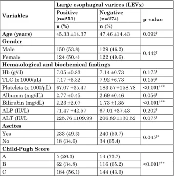
The difference in majority of the variables were insignificant among patients with and without LEVx including age (p-value 0.092), gender (p-value 0.442), Hb (p-value 0.175), TLC (p-value 0.159), ALP (p-value 0.202), and ALT (p-value0.075). While, the platelet count (p-value < 0.001), Bilirubin (p-value < 0.001), pres-ence of ascites (p-value 0.045) and Child-Pugh Score (p-value < 0.001) were the only variables found significantly related with LEVx. (Table II)
The univariate analysis revealed that platelet counts (OR : 0.98, 95% CI : 0.97-0.99),Child score A (OR : 0.28, 95% CI : 0.98-0.79) and Child Score B (OR : 0.42, 95% CI : 0.29-0.61)
Figure 1 : Difference between occurrence of large size esophageal varices versus no large size esophageal varices in thrombocytopenic and non-thrombocytopenic patients (n = 525)
Table I : Diagnostic accuracy of thrombocytopenia in predicting large esophageal varices (n = 525)
Thrombocytopenia Large scale esophageal varices
Yes No Total Yes 221 110 331 No 30 164 194 Total 251 274 525 Sensitivity : 88.05% Specificity : 59.85%
Positive Predicted Value (PPV) : 66.77% Negative Predicted Value (NPV) : 84.54% Test Accuracy : 73.33%
250
K. Mahmood, et al. Non-invasive assessment of esophageal variceswere less likely whereas bilirubin level (OR : 1.18, 95% CI : 1.06-1.31) and presence of ascites (OR : 1.83, 95% CI : 1.01-3.34) were more likely to have LEVx. Similarly, on multivariate analysis (adjusted for platelet counts, bilirubin, ascites and Child-Pugh score), platelet counts (AOR : 0.98, 95% CI : 0.97-0.99),Child score A (AOR : 0.26, 95% CI : 0.09-0.75) and Child Score B (AOR : 0.42, 95% CI : 0.28-0.61) were less likely whereas biliru-bin level (AOR : 1.22, 95% CI : 1.07-1.39) and presence of ascites (AOR : 1.87, 95% CI : 1.02-3.49) were more likely to have LEVx. (Table III)
DISCUSSION
This study has shown a higher proportion of patients with liver cirrhosis having LEVx. Low platelet counts, high bilirubin level, presence of ascites, severity of cirrhosis (A, and CTP-B) were found to have predictive ability on univariate analysis. Earlier studies (2, 4, 15-17) have also shown an independent predictive ability of low platelet count in liver cirrhosis patients to predict LEVx.
A palpable or enlarged spleen is reported to be an important predictor in most of the studies (2, 6, 18-20). Various studies have recommended “platelet count/spleen diameter” (PC/SD) ratio as the most viable non-invasive tool in the prediction of the varices (4, 19-23). However, as this study is retrospective PC/SD ratio of our patients was not calculated. Though mostly earlier studies have shown PC/SD ratio as an effective non-invasive parameter for prediction of LEVx, but one local study in Pakistan has shown contrary findings (24) where PC/SD ratio could not reliably pre-dict the esophageal varices in cirrhotic patients. Furthermore, above referred study (24) have also reported an insignificant association of low platelet count with esophageal varices.
Low platelet count and ascites are widely reported as a nonin-vasive predictor of esophageal varices by earlier published stud-ies (25-31) which is in agreement to this study. Prior studstud-ies (32, 33) have validated both clinical and sonologically determined ascites as an independent predictor for LEVx.
High level of serum bilirubin has shown a significant predic-tor for LEVx in this study which is in accordance to an earlier study (29). Schwarzenberger et al. in their study reported various
parameters like ascites, splenomegaly, serum albumin concen-tration, Child score, and portal vein diameter as predictive vari-ables for LEVx (30). Recently, Chen et al evaluated the combined effect of “albumin bilirubin grade and platelet count” to predict risk of large varices and variceal haemorrhage (5). Due to the diversity of liver diseases and variations in the characteristics among different populations earlier studies have shown different parameters to predict large esophageal varices. In fact, a highly accurate non-invasive predictive model for LEVx is still awaited (4).
Findings of this study could be ascertained in the light of the limitations that due to retrospective nature of the study, spleen diameter values of our patients were not collected. Thus, PC/ SD ratio of our patients was not evaluated which if determined would have a more statistical impact. However, to the best of our knowledge, this is the first study from Pakistan reporting the predicting factor for LEVx from such a larger population.
CONCLUSION
Low platelet count, high bilirubin level and presence of ascites can be used as independent non-invasive predictive factors for LEVx among patients with liver cirrhosis. Further validation of these non invasive parameters through large prospective studies in resource-limited areas like Pakistan where endoscopy is not available everywhere,would definitely provide a non invasive alternative to endoscopy with substantial reduction in financial burden.
REFERENCES
1. Pagliaro L, D’amico G, Pasta L, Tiné F, Aragona E, Politi F, Malizia G, Puleo A, Peri V, D’Antoni A, Simonetti R : Efficacy and efficiency of treatments in portal hypertension. InPortal Hypertension II. Proceedings of the second Baveno Inter-Table II : Comparison of Large esophageal varices with predictor
variables (n = 525) Variables
Large esophageal varices (LEVx) Positive (n=251) Negative(n=274) p-value n (%) n (%) Age (years) 45.33 ±14.37 47.46 ±14.43 0.092† Gender Male 150 (53.8) 129 (46.2) 0.442‡ Female 124 (50.4) 122 (49.6)
Hematological and biochemical findings
Hb (g/dl) 7.05 ±0.83 7.14 ±0.73 0.175† TLC (x 1000/μL) 7.17 ±5.32 7.92 ±6.73 0.159† Platelets (x 1000/μL) 67.07 ±35.47 183.57 ±158.78 <0.001†** Albumin (mg/dL) 2.77 ±0.45 2.69 ±0.46 0.056† Bilirubin (mg/dL) 2.23 ±2.07 1.73 ±1.35 <0.001†** ALP (IU/L) 71.47 ±42.57 67.01 ±37.43 0.202† ALT (IU/L 225.76 ±109.99 206.89 ±130.52 0.075† Ascites Yes 233 (49.3) 240 (50.7) 0.045‡* No 18 (34.6) 34 (65.4) Child-Pugh Score A 5 (26.3) 14 (73.7) <0.001‡** B 62 (34.8) 116 (65.2) C 184 (56.1) 144 (43.9)
†Independent t-test applied, ‡Chi-square test applied, *p-value < 0.05, **p-value < 0.001
ALP : Alkaline Phosphate, ALT : Alanine Transaminase, TLC : Total Leukocyte Count, Hb : Hemoglobin
Table III : Regression analysis of factors associated with large esophageal varices (n = 525)
OR (95% CI) p-value AOR (95% CI) p-value
Age (years) 0.99 (0.97-1.01) 0.092
-Gender
Male 0.874 (0.62-1.23) 0.442 -Female 1
Hematological and biochemical findings
Hb (g/dl) 0.85 (0.69-1.07) 0.175 -TLC (x 1000/μL) 0.98 (0.95-1.01) 0.159 -Platelets (x 1000/μL) 0.98 (0.97-0.99) <0.001 0.98 (0.97-0.99) <0.001 Albumin (mg/dL) 1.44 (0.98-2.09) 0.057 -Bilirubin (mg/dL) 1.18 (1.06-1.31) <0.001 1.22 (1.07-1.39) 0.003 ALP (IU/L) 1.01 (0.99-1.02) 0.202 -ALT (IU/L) 0.99 (0.99-1.01) 0.120 -Ascites Yes 1.83 (1.01-3.34) 0.047 1.87 (1.02-3.49) 0.040 No 1 1 Child-Pugh Score A 0.28 (0.98-0.79) 0.017 0.26 (0.09-0.75) 0.0.12 B 0.42 (0.29-0.61) <0.001 0.42 (0.28-0.61) <0.001 C 1 1
CI : Confidence Interval, OR : Odds ratio, AOR : Adjusted Odds Ratio, ALP : Alkaline Phosphate, ALT : Alanine Transaminase, TLC : Total Leukocyte Count, Hb : Hemoglobin
251
The Journal of Medical Investigation Vol. 66 August 2019
national Consensus Workshop on Definitions, Methodology and Therapeutic Strategies. Oxford, Blackwell Science, 1996, pp.159-179
2. Sharma SK, Aggarwal R : Prediction of large esophageal varices in patients with cirrhosis of the liver using clinical, laboratory and imaging parameters. J Gastroenterol Hepa-tol 22 : 1909-1915, 2007
3. Elalfy H, Elsherbiny W, Rahman AA, Elhammady D, Shaltout SW, Elsamanoudy AZ, El Deek B : Diagnostic non-invasive model of large risky esophageal varices in cirrhotic hepatitis C virus patients. World J Hepatol 8 : 1028-1037, 2016
4. González-Ojeda A, Cervantes-Guevara G, Chávez-Sánchez M, Dávalos-Cobián C, Ornelas-Cázares S, Macías-Amezcua MD, Chávez-Tostado M, Ramírez-Campos KM, del Rocío Ramírez-Arce A, Fuentes-Orozco C : Platelet count/spleen diameter ratio to predict esophageal varices in Mexican patients with hepatic cirrhosis. World Journal of Gastroen-terology : WJG 20 : 2079, 2014
5. Chen PH, Hsieh WY, Su CW, Hou MC, Wang YP, Hsin IF, Yang TC, Liao WC, Lin HC, Lee FY, Wu JC : Combination of Albumin-Bilrubin Grade and Platelet to Predict Compen-sated Patient with Hepatocellular Carcinoma Who Do Not Require Endoscopic Screening for Esophageal Varices. Gas-trointestinal endoscopy 88 : 230-239, 2018
6. Sarangapani A, Shanmugam C, Kalyanasundaram M, Rangachari B, Thangavelu P, Subbarayan JK : Noninvasive prediction of large esophageal varices in chronic liver disease patients. Saudi J Gastroenterol 16 : 38, 2010
7. Cherian JV, Deepak N, Ponnusamy RP, Somasundaram A, Jayanthi V : Non-invasive predictors of esophageal varices. Saudi J Gastroenterol 17 : 64, 2011
8. Cales P, Sacher-Huvelin S, Valla D, Bureau C, Oberti F, Boursier J, GalmicheJP : Screening of large esophageal varices by a simple algorithm of blood markers. J Hepatol 66 : S565-S566, 2017
9. Memon MS, ZakiM : Burden of chronic liver disease and liver transplantation in Sindh. JLUMHS 12 : 01, 2013
10. Garcia-Tsao G, Sanyal AJ, Grace ND, Carey W : for the Practice Guidelines Committee of the American Association for the Study of Liver Disease and the Practice Parameters Committee of the American College of Gastroenterology. Prevention and management of gastroesophageal varices and variceal hemorrhage in cirrhosis. Hepatology 46 : 922-938, 2007
11. Andreani T, Poupon RE, Balkau BJ, Trinchet JC, Grange JD, Peigney N, Beaugrand M, PouponR : Preventive therapy of first gastrointestinal bleeding in patients with cirrhosis : re-sults of a controlled trial comparing propranolol, endoscopic sclerotherapy and placebo. Hepatology12 : 1413-1419, 1990 12. Groszmann RJ, Garcia-Tsao G, Bosch J, Grace ND,
Bur-roughs AK, Planas R, Escorsell A, Garcia-Pagan JC, Patch D, Matloff DS, Gao H : Beta-blockers to prevent gastroesophage-al varices in patients with cirrhosis. N Engl J Med 353 : 2254-2261, 2005
13. Naqvi IH, Mahmood K, Salekeen S, Akhter ST : Determining the frequency and severity of malnutrition and correlating it with the severity of liver cirrhosis. Turk J Gastroenterol 24 : 415-22, 2013
14. Dagradi AE, Stempien SJ, Owens LK : Bleeding esophago-gastric varices. An endoscopic study of 50 cases. Arch Surg 92 : 944-947, 1966
15. Rockey DC, Elliott A, Lyles T : Prediction of esophageal var-ices and variceal hemorrhage in patients with acute upper gastrointestinal bleeding. J Investig Med 64 : 745-751, 2016 16. Amin MA, El-Badry AE, Fawzi MM, Muhammed DA, Moussa
SM : New Non-Invasive Index for Detecting Esophageal Vari-ces in Patients with Liver Cirrhosis. Open Journal of Internal Medicine6 : 101, 2016
17. Marot A, Trépo E, Doerig C, Schoepfer A, Moreno C, Deltenre P : Systematic review with meta-analysis : liver stiffness and platelet count for identifying patients with compensated liver disease at low risk of variceal bleeding. J Hepatol66 : S133, 2017
18. Stefanescu H, ProcopetB : Noninvasive assessment of portal hypertension in cirrhosis : liver stiffness and beyond. World J Gastroenterol 20 : 16811-16819, 2014
19. Ying L, Lin X, Xie ZL, Hu YP, Shi KQ : Performance of plate-let count/spleen diameter ratio for diagnosis of esophageal varices in cirrhosis : a meta-analysis. Dig Dis Sci 57 : 1672-1681, 2012
20. Amin K, Muhammad D, Anjum A, Jamil K, Hassan A : Plate-let count to splenic diameter ratio as a predictor of esophageal varices in patients of liver cirrhosis due to hepatitis C virus. JUMDC 3 : 6-11, 2012
21. Sezer OB, Çelik D, Tutar N, ÖzçayF : Can platelet count/ spleen diameter ratio be used for cirrhotic children to predict esophageal varices? World J Hepatol 8 : 1466, 2016
22. Jamil Z, Malik M, DurraniAA : Platelet count to splenic diameter ratio and other noninvasive markers as predictors of esophageal varices in patients with liver cirrhosis. Turk J Gastroenterol 28 : 347-352, 2017
23. Zimbwa TA, Blanshard C, Subramaniam A : Platelet count/ spleen diameter ratio as a predictor of oesophageal varices in alcoholic cirrhosis. Gut 53 : 1055, 2004
24. Sanvar SS, Alam A, Khan AA, Butt AK, Shafqat F, Shah WH : Platelet count/splenic size ratio : Can it predict the presence of varices in patients of Cirrhosis of liver. Proceeding Shaikh Zayed Postgrad Med Inst 18 : 21-6l, 2004
25. Zein CO, Lindor KD, Angulo P : Prevalence and predictors of esophageal varices in patients with primary sclerosing chol-angitis. Hepatology 39 : 204-210, 2004
26. Zaman A, Becker T, Lapidus J, Benner K : Risk factors for the presence of varices in cirrhotic patients without a history of variceal hemorrhage. Arch Intern Med 161 : 2564-2570, 2001 27. Riggio O, Angeloni S, Nicolini G, Merli M, Merkel C : Endo-scopic screening for esophageal varices in cirrhotic patients. Hepatology 35 : 501-502, 2002
28. Bressler B, Pinto R, El-Ashry D, Heathcote EJ : Which pa-tients with primary biliary cirrhosis or primary sclerosing cholangitis should undergo endoscopic screening for oesopha-geal varices detection? Gut 54 : 407-410, 2005
29. Sanyal AJ, Fontana RJ, Di Bisceglie AM, Everhart JE, Doherty MC, Everson GT, Donovan JA, Malet PF, Mehta S, Sheikh MY, Reid AE : The prevalence and risk factors associ-ated with esophageal varices in subjects with hepatitis C and advanced fibrosis. Gastrointest Endosc 64 : 855-864, 2006 30. Schwarzenberger E, Meyer T, Golla V, Sahdala NP, Min
AD : Utilization of platelet count spleen diameter ratio in predicting the presence of esophageal varices in patients with cirrhosis. J Clin Gastroenterol 44 : 146-150, 2010
31. Thomopoulos KC, Labropoulou-Karatza C, Mimidis KP, Katsakoulis EC, Iconomou G, Nikolopoulou VN : Non-inva-sive predictors of the presence of large oesophageal varices in patients with cirrhosis. Dig Liver Dis 35 : 473-478, 2003 32. Ng FH, Wong SY, Loo CK, Lam KM, Lai CW, Cheng CS :
Prediction of oesophagogastric varices in patients with liver cirrhosis. J Gastroenterol Hepatol 14 : 785-790, 1999 33. Sersté T, Melot C, Francoz C, Durand F, Rautou PE, Valla
D, Moreau R, Lebrec D : Deleterious effects of beta-blockers on survival in patients with cirrhosis and refractory ascites. Hepatology 52 : 1017-1022, 2010