Fukushima Medical University
This document is downloaded at: 2021-11-08T00:15:13Z
Title Potential of laryngeal muscle regeneration using induced pluripotent stem cell-derived skeletal muscle cells( 本文 )
Author(s) Dirja, Bayu Tirta
Citation
Issue Date 2018-03-21
URL http://ir.fmu.ac.jp/dspace/handle/123456789/740
Rights
This is an Accepted Manuscript of an article published by Taylor & Francis in "Acta oto-laryngologica" on 29 Jan 2016, available online:
https://doi.org/10.3109/00016489.2015.1126351.
DOI
Text Version ETD
Potential of laryngeal muscle regeneration using induced pluripotent stem cell-derived skeletal muscle cells
Bayu Tirta Dirja, Susumu Yoshie, Masakazu Ikeda, Mitsuyoshi Imaizumi, Ryosuke Nakamura, Koshi Otsuki, Yukio Nomoto, Ikuo Wada, Akihiro Hazama, Koichi Omori
This is an Accepted Manuscript of an article published by Taylor & Francis in Acta Oto-Laryngologica, 2016 available online at
https://www.tandfonline.com/doi/full/10.3109/00016489.2015.1126351
CONTENTS CONTENTS
ABSTRACT INTRODUCTION
MATERIAL AND METHODS RESULTS
DISCUSSION CONCLUSION REFERENCES FIGURES TABLE
ACKNOWLEDGEMENT
Abstract
Conclusion: induced pluripotent stem (iPS) cells may be a new potential cell source for laryngeal muscle regeneration in the treatment of vocal fold atrophy after recurrent laryngeal nerve paralysis. Objectives: Unilateral vocal fold paralysiscan lead to degeneration, atrophy, and loss of force of the thyroarytenoidmuscle. At present, there are some treatments such as thyroplasty, arytenoid adduction, and vocal fold injection. However, such treatments cannot restore reduced mass of the thyroarytenoid muscle. iPS cells have been recognized as supplying a potential resource for cell transplantation. The aim of this study was to assess the effectiveness of the use of iPS cells for the regeneration of laryngeal muscle through the evaluation of both in vitro and in vivo experiments. Methods: Skeletal muscle cells were generated from tdTomato-labeled iPS cells using embryoid body formation. Differentiation into skeletal muscle cells was analyzed by gene expression and immunocytochemistry. The tdTomato-labeled iPS cell-derived skeletal muscle cells were transplanted into the left atrophied thyroarytenoid muscle.To evaluate the engraftment of these cells after transplantation,immunohistochemistry was performed. Results:
The tdTomato-labeled iPS cells were successfully differentiated into skeletal muscle cells through an in vitro experiment. These cells survived in the atrophied thyroarytenoid muscle after transplantation.
Introduction
Vocal fold paralysis is a result of vocal fold immobility due to the loss of its nerve supply, and is a common cause of voice and swallowing disorders. Recurrent laryngeal nerve injury caused by intubationor inadvertent injury during surgery of the head and neck region or non-laryngeal malignancies are often associated with vocal fold paralysis [1]. Unilateral denervation of the thyroarytenoidmuscle leads to degeneration, atrophy, and loss of the thyroarytenoid muscle force.
In patients with vocal fold paralysis, localized unilateral denervation of thethyroarytenoid muscle leads to an inability to close the glottis [2]. To date, there are several therapies, including thyroplasty, arytenoid adduction, and vocal fold injection, thatcan effectively improve glottic closure during vocal fold adduction, none of which, however, can return the decrement in mass or force of the thyroarytenoid muscle [3,4].Vocal fold paralysis requires an ideal treatment to return the muscle mass and dynamic function.
Skeletal muscle stem cell transplantation has been recognized as one of the most promising therapies for muscle regeneration. The injection of autologous muscle stem cells (myoblasts) into the thyroarytenoid muscle for the treatment of vocal fold paralysis has been reported [1].
However, obtaining autologous muscle stem cell requires an invasive procedure.The mesenchymal stem cells (MSCs) can give rise to differentiated cells such asmyocytes,osteocytes, chondrocytes, and adipocytes. Nevertheless, MSCs gained from bone marrow are slightly limited inpopulation, and their isolation is also invasive and have a risk [5]. Chang et al have generated myogenic lineage cells from embryonic stem (ES) cells [6]. The ES cells are one of the attractive cell sources, since they have the capacities of self-renewal and pluripotency[7]. However, the establishment of ES cellswas required for destruction of pre-implantation embryos at the
blastocyst stage, which is morally unadaptable. Moreover, the transplantation of ES cells for therapeutic purposes can trigger host immune rejection.
In 2006, Yamanaka and co-workers reported the generation of induced pluripotent stem (iPS) cells from mouse somatic fibroblasts by the transfection of Oct3/4, Sox2, Klf4, and c-Myc genes [8]. The iPS cells alsoexhibitproperties of self-renewal and pluripotency.They have a potential to provide an affluent cell source for improvementof damaged tissues in the body. Under the appropriate conditions, these cells can be induced to become various cell types with a specific function, including skeletal muscle cells. [9,10]. In addition, the use of iPS cells derived from the recipients can prevent immune rejection.
The aim of this study was to assess the effectiveness of iPS cells for the regeneration of laryngeal muscle through the evaluation of both in vitro and in vivo experiments.
Materials and Methods
iPS cells culture
The td Tomato-labeled iPS cells (a kind gift from Dr. S. Yoshie) [11] were cultured on a feeder- layer of 10 µg/ml Mitomycin C (Sigma-Aldrich, MO, USA)-inactivated mouse embryonic fibroblasts (MEFs). MEFsina cultivation medium consisting of Dulbecco’s modified Eagle’s medium (DMEM; Gibco, Invitrogen, Grand Island, New York) were supplemented with 15%
fetal bovine serum (FBS: Invitrogen), 2 mmol/L L-glutamine (Wako, Saitama, Japan), 100 µM 2-mercaptoethanol (Sigma-Aldrich), 1000 U/ml mouse LIF (Wako), 100 µM non-essential amino acids (Invitrogen), and penicillin streptomycin (Invitrogen). The iPS cells were passaged every second day by using 0.25% trypsin-EDTA (Invitrogen).
invitro differentiation of tdTomato-labeled iPS cells into skeletal muscle cells
A six well plate containing iPS cells with 80% confluency were rinsed with phosphate buffered saline (PBS) and were dispersed into a single cell suspension with 0.25% trypsin, then transferred onto tissue culture dishes coated with 0.1% gelatin in iPS cell medium and incubated for 30 mins at 37°C for removal of the feeder cells. According to Mizuno’s protocol, the iPS cells were cultured in hanging drops, each of which contained approximately 800 cells in a 20 µl volume of differentiation medium, consisting of DMEM supplemented with 10% fetal bovine serum, 5% horse serum (Sigma-Aldrich), 100 µM 2-mercaptoethanol, 100 µM non-essential amino acids, and penicillin streptomycin. These cells were kept for three days on the lids of dishes filled with PBS. Embryoid bodies (EBs) were then transferred to a suspension culture in the differentiation medium for three additional days. Every EB was plated onto 24-well tissue
culture plates coated with Matrigel Basement Membrane Matrix (Corning Life Sciences, MA, USA). The medium was subsequently replaced every five days [10].
RT-PCR
Total RNA samples were isolated from tdTomato-labeled iPS cells using TRIzol reagent (Invitrogen). Superscript Reverse Transcriptase (TaKaRa Bio, Shiga, Japan) and 1 µg total RNA were used to produce a single first-strand cDNA. The cDNA samples were subjected to PCR amplification with a termal cycler (T100; Bio-Rad,CA, USA). The amplification program was required 30 cycles of 94ºC denaturation for 30 sec. The annealing temperature depended on each primer set and required 30 sec. Elongation at 72ºC for 30 sec and a final incubation of 7 mins at 72ºC, were performed. The PCR products were visualized in 1.5% agarose gel with ethidium bromide. The housekeeping gene β-actin was used for comparison of the gene expression data, and was also used as the internal control. The primer sets and annealing temperatures are shown in Table I.
Quantitative real-time RT-PCR
Quantitative RT-PCR reactions were carried out using an Applied BiosystemsStepOnePlus Real- Time PCR System (Invitrogen).All procedures were performed according to the manufacturer’s instructions. The cycling protocol used was 95ºC for 10 sec, followed by 95ºC for 5 sec, and 60ºC for 30 sec with a total 40 cycles. Quantitative RT-PCR for Nanog, GFP, Pax3, Pax7, MyoD, Myogenin, and β-actin genes was carried out with specific primer pairs. The primer sets are shown in Table II.
Immunocytochemistry and immunohistochemistry
The EBs at day 20 of expansion culture were fixed for 15 mins in 4% paraformaldehyde (PFA) at room temperature. These cells were rinsed three times in PBS for 10 minseach and permeabilized with 0.1% Triton X-100 in PBS for 30 mins. Thesamples were then rinsed three times in PBS for 10 mins each andincubated with 3% Goat serum in PBS for 30 mins. The samples werestained with primary antibodiesto anti-myosin heavy chain (MHC: mouse monoclonal) and td-Tomato (rabbit polyclonal) at 4ºC over-night, and then incubated with secondary antibodiesfor 1 h at room temperature. The secondary antibodiesused in this study were alexa fluor 488 goatanti-mouse IgG and alexa fluor 568 goat anti-rabbit IgG. DAPI was used for nuclear staining. For immunohistochemistry, the engrafted cells at two weeks after transplantation were isolated and fixed in 4% PFA, and embedded in paraffin. After deparaffinization and rehydration, the samples were treated in a microwave oven for 15 mins and rinsed three times in PBS. The samples were stained with above primary antibodies then conjugated with secondary antibodies. Each sample was then examined using a confocal laser scanning microscopy A1R (Nikon Instech, Tokyo, Japan).
Transplantation of tdTomato-labeled iPS cell-derived skeletal muscle cells into thyroarytenoid muscle atrophy
All experiments were performed in accordance with the guidelines of the Animal Care and Use Committee in fulfillment with the Animal Research Center at Fukushima Medical University School of Medicine, Fukushima, Japan. Six-week-old F344/NJcl-rnu/rnu nude rats (CLEA Japan, Tokyo, Japan) were used in this study. The animals were sedated by inhalation of isoflurane and then anesthetized with an intraperitoneal injection of a mixture of midazolam (1.9 mg/kg;
Astellas Pharma, Tokyo, Japan), butorphanol tetrate(2.4 mg/kg; Meiji Seika Pharma, Tokyo,
Japan), and medetomidine hydrochloride(0.14 mg/kg; Nippon Zenyaku Kogyo, Fukushima, Japan). After exposure of the recurrent laryngeal nerve via a vertical midline incision of the cervical skin, left recurrent laryngeal nerve resection was performed. Four weeks after said resection, transplantation of tdTomato-labeled iPS cell-derived skeletal musclecells was carried out. The cells (approximately 1 x 10⁶) derived from EB on day 20 of expansion culture in PBS were directly injected into the atrophiedthyroarytenoid muscle (Figure 1).Four weeks after resection, the nude ratwas humanely sacrificed, and the larynxes were dissected and fixed in 4%
paraformaldehyde. Paraffin-embedded tissues were sliced at a thickness of 4 µm per section for hematoxylin-eosin (H&E) staining.
Results
Confirmation of undifferentiated tdTomato-labeled iPS cells
The tdTomato-labeled iPS cells were used to visualize the iPS cell-derived cells.These cells formed dome-shaped and refractive colonies (Figure 2A). Furthermore, they expressedthe undifferentiated cell markers such as Sox2, Oct3/4, Nanog, GFP, and Rex1 in RT-PCR analysis (Figure 2B).
Generation of skeletal muscle cells from tdTomato-labeled iPS cells
To obtain an efficient differentiation protocol for inducing skeletal muscle cells from iPS cells, an EB-based method was used, which can generate skeletal muscle differentiation from mouseiPS cells [11]. In the expansion culture, the spherical shape of the EB was still maintained on day 0 (Figure 3a,b). The cells in EB outgrowth on day 6 (Figure 3c,d) and 13 (Figure 3e,f). At day 20 after plating, thefibers-like tissue was appeared (Figure 3g,h).
Detection of gene expression by Quantitative RT-PCR analysis and immunocytochemistry
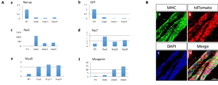
Real time-PCR was used to examine the quantitative gene expression of undifferentiated cell markers and skeletal muscle markers during the induction of differentiation. The expression levels of Nanog and GFP, which are undifferentiated cell markers, were gradually decreased under the differentiation condition of the expansion culture (Figure 4Aa,b). The expression levels of satellite markers Pax3 and Pax7 were transiently up-regulated (Figure 4Ac,d). The expression levels of MyoD, which is a terminal specification marker for the muscle lineage differentiation, was transiently up-regulated (Figure 4Ae). Furthermore, the expression levels of a terminal differentiation marker Myogenin was gradually increased (Figure 4Af).In immunocytochemistry,
the differentiated cells were positive for MHC, a specific marker of mature myofibers (Figure 4Ba-d).
Transplantation of tdTomato-labeled iPS cell-derived skeletal muscle cells
The short-term engraftmentof tdTomato-labeled iPS cell-derived skeletal muscle cells was examined. These cells were transplanted into the atrophiedthyroarytenoid muscleof nude rats(Figure 5Aa-c).The engrafted cells were analyzedby immunohistochemistry at twoweeks after transplantation.ThetdTomato- and MHC-positive cells were detected in the atrophied thyroarytenoid muscle, indicating that these cells survived in the atrophied thyroarytenoid muscle after transplantation (Figure 5Ba-f).
Discussion
The vocal folds have an important role for producing sound and maintaining breathing, as well as swallowing. Unilateral vocal fold paralysis occurs from a dysfunction of the recurrent laryngeal nerve. It causes a characteristic breathy voice often accompanied by swallowing inability, a weak cough, and the sensation of shortness of breath.At present, there are several treatments, including thyroplasty, arytenoid adduction, and vocal fold injection, thatcan effectively improve glottic closure during vocal fold phonation, none of which, however, can restore the decrement in mass or force of the thyroarytenoid muscle [3,4].Vocal fold paralysis requires an ideal treatment to return muscle mass and dynamic function.
To overcome this issue, iPS cells were used for muscle regeneration. These cellshave a potential to provide an affluent cell source for the regenerationof damaged tissues in the body.
Furthermore, the use of iPS cells derived from recipients can also prevent ethical issues and immune system rejection of ES cells. Hence, iPS cell-derived skeletal muscle cells are expected to be a useful cell source for muscle regeneration therapy in future. This study demonstrated the regeneration potential of thyroarytenoid muscle atrophy through the use of tdTomato labeled-iPS cells. This study is the first report for the regeneration of laryngeal muscle using iPS cells.
First, the tdTomato-labeled iPS cells were confirmed in undifferentiated state before beginning in vitro experiments. These cells could maintain the undifferentiated state, assessing from their morphology and the expression of undifferentiated cell markers such as Sox2, Oct3/4, Nanog, and Rex1.The expression of GFP, which is controlled by the Nanog enhancer/promoter [12], was also detected in the tdTomato-labeled iPS cells, indicating that these cells were in undifferentiated state. In expansion culture,when the EBs were plated on a Matrigel-coated plate with DMEM containing horse serum, the EBs were gradually towards the differentiated state.
The matrigel and horse serum have an important role during skeletal muscle cell differentiation.
It is well known that matrigel is obtained from tumor rich in extracellular matrix protein. The major component of matrigel is laminin, a major protein in the basal lamina. Normally, satellite cells are quiescent beneath the myofiber basal lamina [13]. Then, the horse serum contains insulin-like growth factor, an effective anabolic factor which can stimulate the differentiation of skeletal muscle cells [14,15]. Therefore, both the matrigelenvironment and horse serum can induce iPS cells into skeletal muscle lineage cells, and then form myofibers in vitro.In quantitative RT-PCR, the differentiated cells expressed satellite cell markers (Pax3 and Pax7) [13], a terminal specification marker (MyoD), and a terminal differentiation marker (Myogenin) [16]. Furthermore, tdTomato and a specific marker of mature myofibers (MHC) double-positive cells were observed by immunocytochemistry. These results suggest that the cells differentiated from tdTomato-labeled iPS cells have the phenotype of skeletal muscle cells.The in vivo potential of tdTomato-labeled iPS cell-derived skeletal muscle cells was examined. These cells were transplantedinto the atrophied thyroarytenoid muscle in nude rats. The engrafted cells were analyzed by immunohistochemistry and the tdTomato-and MHC-positive cells were successfully detected in the atrophied thyroarytenoid muscle.Thus, it is considered thatiPS cell-derived skeletal muscle cellswill be one of the promising treatment modality for laryngeal muscle regeneration.However, the number of these cells were a few in the atrophied thyroarytenoid muscle. As a next study, the transplantation of iPS cell-derived skeletal muscle progenitor cells may develop enhancement of survival number of iPS cell-derived skeletal muscle cells in the atrophied thyroarytenoid muscle.
Conclusion
This study demonstrated thattdTomato-labeled iPS cells were successfully differentiated into skeletal muscle cells through an in vitro experiment. These cells survived in the atrophiedthyroarytenoidmuscle after transplantation in vivo experiment. With the further study, iPS cells may be a new potential cell source for laryngeal muscle regeneration in the treatment of vocal fold atrophy after recurrent laryngeal nerve paralysis
References
1. Halum SL, Naidu M, Delo DM, Atala A, Hingtgen CM. Injection of autologous muscle stem cells (Myoblasts) for the treatment of vocal fold paralysis: a pilot study.
Laryngoscope 2007;117:917–22
2. Lee WT, Milstein C, Hicks D, Akst LM, Esclamado RM. Results of ansa to recurrent laryngeal nerve reinnervation. J Otolaryngol Head Neck Surg 2007;136:450-4
3. Mat Baki M, Yu R, Rubin JS, Chevretton E, Sandhu G, Birchall MA. Patient perception of a randomised, controlled trial of laryngeal reinnervation versus thyroplasty for unilateral vocal fold paralysis. J LaryngolOtol 2015;129:693–701
4. Miyamaru S, Kumai Y, Ito T, Yumoto E. Effects of long-term denervation on the rat thyroarytenoid muscle. Laryngoscope 2008;118:1318-23
5. Sun S, Guo Z, Xiao X, Liu B, Liu X, Tang PH, et al. Isolation of mouse marrow mesenchymal progenitors by a novel and reliable method. Stem cells 2003; 21:527-35 6. Chang H, Yoshimoto M, Umeda K, Iwasa T, Mizuno Y, Fukada S, et al. Generation of
transplantable, functional satellite-like cells from mouse embryonic stem cells. FASEB J 2009;23:1907-19
7. Evans MJ, Kaufman MH. Establishment in culture of pluripotential cells from mouse embryos. Nature 1981;292:154-6
8. Takahashi K, Yamanaka S. Induction of pluripotent stem cells from mouse embryonic and adult fibroblast cultures by defined factors. Cell 2006;126:663-76
9. Geoghegan E, Byrnes L. Mouse Induced Pluripotent Cells. Int J DevBiol 2008;52:1015- 22
10. Mizuno Y, Chang H, Umeda K,Niwa A, Iwasa T, Awaya T, et al. Generation of skeletal muscle stem/progenitor cells from murine induced pluripotent stem cells. FASEB J 2010;24:2245-53
11. Yoshie S, Ikeda M, Imaizumi M, Otsuki K, Nomoto Y, Wada I, et al. Visualization of mouse induced pluripotent stem cells for evaluation of tracheal regeneration. Acta Otolaryngol 2015;135:395–401
12. Okita K, Ichisaka T, Yamanaka S. Generation of germline-competent induced pluripotent stem cells. Nature 2007;448:313-17
13. Tedesco FS, Dellavalle A, Manera JD, Messina G, Cossu G. Repairing skeletal muscle:
regenerative potential of skeletal muscle stem cells. J Clin Invest 2010;120:11-19
14. Prosser CG, Mclaren RD. Horse serum contains a novel insulin-like growth factor binding protein. Proc New Zeal Soc An 1993;53;433-6
15. Grabiec K, Gajewska M, Milewska, Blaszczyk M, Grzelkowska-Kowalczyk K. The influence of high glucose and high insulin on mechanism controlling cell cycle progression and arrest in mouse C2C12 myoblasts; the comparison with IGF-I effect. J Endocrinol Invest 2014;37;233-45
16. Bentzinger CF, Wang YX, Rudnicki MA. Building muscle; molecular regulation of myogenesis. Cold Spring HarbPerspectBiol 2012;4:a008342
Figure legends
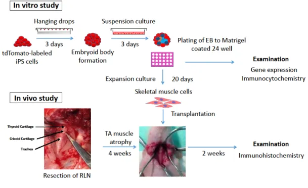
Figure 1. Experimental design forlaryngeal muscle regeneration from tdTomato-labeled iPScellsin vitro and in vivo. Skeletal muscle cells were generated from tdTomato-labeled iPS cells using the embryoid body method. The tdTomato-labeled iPS cell-derived skeletal muscle cells were analyzed by gene expression and immunocytochemistry.After confirming skeletal muscle celldifferentiation, these cells were transplanted into the atrophiedthyroarytenoidmuscle in nude rats.At 2 weeks post-transplantation, the immunohistochemistry was examined.
Figure 2. Undifferentiated state oftdTomato-labeled iPS cells. (A) Phase-contrast (left panel) and tdTomato (right panel). Scale bars = 100µm. (B)RT-PCR analysis of gene expression intdTomato-labeled iPS cells and MEF, using undifferentiatedcell markers. β-actin was used as housekeeping gene.
Figure 3. Expansion culture of EBs from tdTomato-labeled iPS cells. The samples were obtained from EB on day 0 (a,b), day 6 (c,d), day 13 (e,f), and day 20 (g,h). Top row: phase contrast (a,c,e,g). Bottom row: tdTomato (b,d,f,h). Scale bars = 200 µm
Figure 4. Time course of skeletal musclecell differentiation from tdTomato-labelediPS cells. (A) Quantitative gene expression of undifferentiated markers (a,b),satellite markers (c,d), terminal specification marker (e), and terminal differentiation marker (f). The samples were obtained fromundifferentiatedtdTomato-labelediPS cells, EBs on day 6, day 13, and day 20. (B) Immunocytochemistry of tdTomato-labeled iPS cell-derived embryoid body (EB) using MHC antibody (a-d). TheSample was obtained from EB on day 20 of expansion culture. DAPI (blue) was used for nuclear staining. Scale bars = 50 µm.
Figure 5. Survival of tdTomato-labeled iPS cell-derived skeletal muscle cells after transplantation. (A) H&E staining,the boxed region in (aR1,R2) is magnified and shown in normal thyroarytenoid muscle(bR1)and atrophy model (cR2).The sample was obtained from the thyroarytenoid muscle afterresection of left recurrent laryngeal nerve. (B)The tdTomato-and MHC-positive cells were found in atrophied thyroarytenoid muscle (a,b,c,d,e,f). The boxed region in (a,b,c) is magnified and shown in region (d,e,f). The sample was obtained from the atrophied thyroarytenoid muscle at two weeks after transplantation. DAPI was used for nuclear staining. Scale bars = 2.0 mm (A), and 100 µm (B).
TABLE
Table I. Primer sets used for PCR amplification of cDNA
Gene Sense primer Antisense primer Anealing tempeature (ºC)
Sox2 TAGAGCTAGACTCCGGGCGATGA TTGCCTTAAACAAGACCACGAAA 65
Oct3/4 TCTTTCCACCAGGCCCCCGGCTC TGCGGGCGGACATGGGGAGATCC 65
Nanog AGGGTCTGCTACTGAGATGCTCTG CAACCACTGGTTTTTCTGCCACCG 65
Rex1 ACGAGTGGCAGTTTCTTCTTGGGA TATGACTCACTTCCAGGGGGCACT 65
GFP ACGTAAACGGCCACAAGTTC CACCTTGATGCCGTTCTTCT 65
β-actin TTCCTTCTTGGGTATGGAAT GAGCAATGATCTTGATCTTC 60
Table II. Primer sets used for quantitative real-time RT-PCR Gene Sense primer Antisense primer Product Nanog GCTTACAAGGGTCTGCTACT CCTCAGGACTTGAGAGCTTT 131
GFP CTGAAGGGCATCGACTTCAA CACCTTGATGCCGTTCTTCT 117
Pax3 AACACTGGCCCTCAGTGAGTTCTAT ACTCAGGATGCCATCGATGCTGTG 142
Pax7 GTACAGCCAGTATGGCCAAA CCGTTTCCACAGGAAGAAGT 121
MyoD ACTACAGTGGCGACTCAGAT TAGGCGGTGTCGTAGCCATTCT 114
Myogenin AGCGCCATCCAGTACATTGA CTGTGGGAGTTGCATTCACT 127
β-actin CATCCGTAAAGACCTCTATGCCAAC ATGGAGCCACCGATCCACA 170
Acknowledgments
We wish to thank Ms Etsuko Sato for her skillful technical assistance. This studywas supported by the Grant-in-Aid for Scientific Research (B) 26293371, SPIO grant, and Fukushima Medical University.