単発肝細胞癌における
切除断端陰性かつ切除断端距離
1mm
未満症例の検討日本大学大学院医学研究科博士課程 外科系消化器外科学専攻
小林 直哉 修了年 2021 年 指導教員 高山 忠利
目次
Ⅰ. 本文
1. 要旨・・・・・・・・・・・・・・・・・・・・・・・・・・・・・・・・・・・・・・・・1 2. 緒言・・・・・・・・・・・・・・・・・・・・・・・・・・・・・・・・・・・・・・・・3 3. 対象と方法
3.1 患者背景・・・・・・・・・・・・・・・・・・・・・・・・・・・・・・・・・・・・4 3.2 肝切除・・・・・・・・・・・・・・・・・・・・・・・・・・・・・・・・・・・・・5 3.3 術後評価・・・・・・・・・・・・・・・・・・・・・・・・・・・・・・・・・・・・6 3.4 統計学的解析・・・・・・・・・・・・・・・・・・・・・・・・・・・・・・・・・・6 4. 結果
4.1 患者背景・・・・・・・・・・・・・・・・・・・・・・・・・・・・・・・・・・・・7 4.2 長期生存・・・・・・・・・・・・・・・・・・・・・・・・・・・・・・・・・・・・7 4.3 再発形式・・・・・・・・・・・・・・・・・・・・・・・・・・・・・・・・・・・・7 4.4 無再発生存における予後規定因子・・・・・・・・・・・・・・・・・・・・・・・・・8 4.5 全生存における予後規定因子・・・・・・・・・・・・・・・・・・・・・・・・・・・8 4.6 傾向スコア後の長期生存と再発形式・・・・・・・・・・・・・・・・・・・・・・・・8 5. 考察・・・・・・・・・・・・・・・・・・・・・・・・・・・・・・・・・・・・・・・10 6. まとめ・・・・・・・・・・・・・・・・・・・・・・・・・・・・・・・・・・・・・・12
Ⅱ. 謝辞・・・・・・・・・・・・・・・・・・・・・・・・・・・・・・・・・・・・・・・・13
Ⅲ. 表・・・・・・・・・・・・・・・・・・・・・・・・・・・・・・・・・・・・・・・・・14
Ⅳ. 図・・・・・・・・・・・・・・・・・・・・・・・・・・・・・・・・・・・・・・・・・20
Ⅴ. 図説・・・・・・・・・・・・・・・・・・・・・・・・・・・・・・・・・・・・・・・・26
Ⅵ. 引用文献・・・・・・・・・・・・・・・・・・・・・・・・・・・・・・・・・・・・・・28
Ⅶ. 研究業績・・・・・・・・・・・・・・・・・・・・・・・・・・・・・・・・・・・・・・32
1
Ⅰ 本文 1. 要旨
【目的】: 肝細胞癌肝切除において適切な切除断端距離は明らかではない. 肝細胞癌肝切除 において切除断端距離が予後に寄与するかを明らかにするために, 最低限度である切除断 端陰性かつ切除断端距離 1mm 未満症例を検討した.
【方法】: 2001 年から 2012 年までに施行した単発単結節型肝細胞癌の初回治癒切除症例
454 例を対象とし, 術後病理にて切除断端陰性かつ切除断端距離が 1 mm 未満である 90 例
[Marginal Resection 群 (MR 群)] と切除断端距離が 1 mm 以上である 364 例 [Non-
Marginal Resection 群 (non-MR 群)] の 2 群に分けた. さらに傾向スコアを用いて背景因子
を調整し MR 群 90 例, non-MR 群 90 例として全生存及び無再発生存, また再発形式に有
意差があるか比較検討した.
【結果】: 手術因子では腫瘍径 [40mm (10-205) vs 30mm (7-205), P = 0.002]は MR 群の
方が non-MR 群よりも有意に大きく, 術中出血量 [420ml (0-2950) vs. 238ml (0-2988), P =
0.001]は MR 群の方が有意に多かった. 系統的切除の割合は MR 群が有意に低頻度であっ
た (33.3% vs 45.3%, P = 0.04). 長期生存においては無再発生存では MR 群の方が non-MR
群よりも有意に不良であった (P = 0.01). 全生存では両群に有意差を認めなかった (P =
0.051). Cox 比例ハザードモデルにて切除断端 1mm 未満は全生存, 無再発生存ともに独立
した予後規定因子ではなかった. 傾向スコアで背景因子を調整後, 全生存, 無再発生存とも
両群に有意差を認めなかった (P = 0.50, 0.38). 再発形式については肝外, 肝内ともに有意
2
差を認めなかった (P = 0.16, 0.65).
【結語】: 肝細胞癌肝切除において最低限度である切除断端陰性かつ切除断端距離 1mm 未
満の切除症例は, 傾向スコア後, 無再発生存, 全生存, 再発形式に差を認めず, 切除断端陰
性かつ 1mm 未満となる切除は許容される.
3
2. 緒言
悪性腫瘍に対する手術における至適な切除断端は, 根治性の面から距離を十分に 確保するほうが望ましく, 安全性および臓器の機能温存の面からは切除断端距離を最小限 にするべきと考えられ, 悪性腫瘍の発生した臓器, 性質, 残存臓器の機能の重要性によって 各々の至適な切除断端距離の確保は決定される (1). 肝細胞癌ではそのほとんどは背景に
慢性肝炎, 肝硬変を有しており, 切除断端距離の確保は肝予備能, また腫瘍の位置や大きさ によっても限定される (2, 3).
肝細胞癌は被膜におおわれ, 膨張性の発育をする特徴があり, 切除断端距離の確保 の必要性は他臓器の癌よりも少ないとされている (4-6). 切除断端距離については 5mm 未
満の群と以上の群, また 10mm 未満の群と以上の群に対する比較検討で, 全生存が有意に
不良であるため少なくとも 5mm また 10mm の距離の確保が必要であると報告がみられた
(7-9). しかしながら他の研究では, 5mm から 10mm の切除断端距離は無再発生存に有意差
がなかったと報告もあり (2, 10-14), 肝細胞癌肝切除における至適な切除断端距離は明ら かでない.
本研究では肝細胞癌肝切除において切除断端距離が予後に寄与するかを明らかに するために, 最低限度である切除断端陰性かつ切除断端距離 1mm 未満症例の長期予後, 局
所再発について検討した.
4
3. 対象と方法 3-1. 患者背景
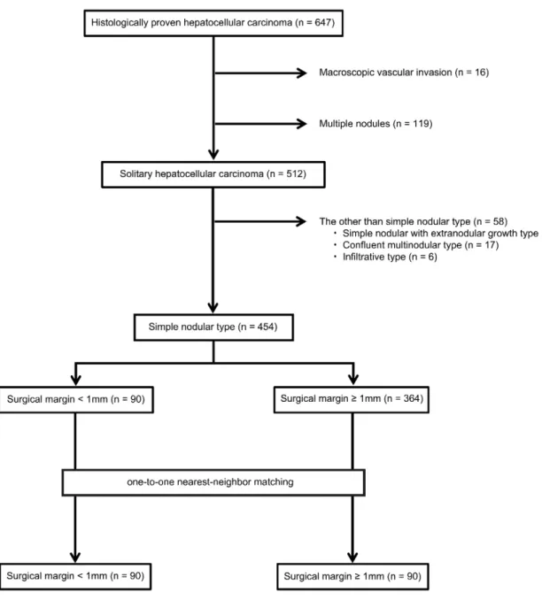
研究期間は 2001 年から 2012 年で, 患者は肝細胞癌初回治癒切除例 647 例を対象
とした. 検体の肉眼所見については日本肝癌研究会の分類によって腫瘍の形態を分類した
(15, Figure 1). また肝細胞癌の肉眼型について, 単純結節周囲増殖型や多結節癒合型の場合,
多発肝細胞癌との鑑別が困難であることがあり, 浸潤型の場合, 境界が不明瞭で切除断端距 離を正確に計測することが難しいため, 今回の検討ではほとんどが繊維性被膜で包まれて いる単結節型を対象とした (16-18). 腫瘍の個数については肝細胞癌術後の予後規定因子 であり, また多発肝細胞癌の場合, 切除断端距離が混在している可能性が高い. そのため今 回の検討では多発の肝細胞癌症例は除外した (2, 19). 647 例のうち, 以下の 193 例が除外さ
れた. (1)16 例は肉眼的に脈管侵襲が認められた. (2)119 例は多発肝細胞癌であった. (3)58 例は単純結節型以外のものであった (35 例は単純結節周囲増殖型, 17 例は多結節癒合型, 6 例は浸潤型). 最終的に単発で単純結節型の 454 例を今回の研究の対象とした. 本研究で
Marginal Resection の定義は, 組織学的に切除断端陰性かつ切除断端距離が 1mm 未満とな
った切除とした. 切除断端陰性かつ切除断距離が 1mm 未満であった症例群を Marginal
Resection 群 (MR 群)とした. 切除断端距離は組織学的に測定し, 組織学的評価方法は病理
報告書に基づいた. 今回の研究対象となった 454 例を MR 群 90 例と, 切除断端距離が 1mm
以上である Non-Marginal Resection 群 (non-MR 群) 364 例の 2 群に分けた. さらに傾向ス
コアを用いて背景因子を調整し MR 群 90 例, non-MR 群 90 例として全生存及び無再発生
5
存, また再発形式に有意差があるか比較検討した (Figure 2).
本研究はヘルシンキ宣言の倫理的なガイドラインに準拠した. 本研究は日本大学 医学部附属板橋病院の臨床研究倫理審査委員会の承認を得た (承認番号: RK-200114-7).
3-2. 肝切除
肝切除の術式と適応は, 本邦で最も肝切除術に頻用されている幕内基準に準じて いる (20). この基準は C 型肝炎を背景とした肝硬変併存の病態を想定して作成され安全性
を担保したもので, 肝機能条件毎に可能な最大術式が決定される (Figure 3). まずコントロ ールの悪い腹水併存患者は外科切除の適応外とし, 肝機能条件は血清ビリルビン値, および indocyanine green 15-minute retention rate (ICG-R
15)によって評価される. 標準的な肝切除
術式は小型の肝細胞癌(5cm 以下)に対しては, 系統的切除, あるいは縮小手術としての部分 切除(特に肝機能不良例)が選択され, 大型の肝細胞癌に対しては 2 区域以上の拡大切除(片
肝切除を含む)が選択される(20). 系統的切除とは Couinaud 分類で肝 S1 から S8 に分けら
れる亜区域の切除であり, 超音波ガイド下に担癌領域の門脈枝を穿刺し色素を注入して肝 表における当該の肝領域を同定して切除する術式である(20). 肝離断は間欠的肝流入血流 遮断法を施行し, 鉗子圧座法で行った. 肝動脈, 門脈血流を遮断し, 圧挫は 15 分間で施行し
血流遮断解除は 5 分間とした (21).
6
3-3. 術後評価
術後は 3-4 か月毎に血清腫瘍マーカー値の測定と腹部単純相と肝臓 3 相造影 CT(肝
動脈相, 門脈相, 平衡相)を施行した. 再発は CT で典型的な肝細胞癌と診断されたものと定
義した (22). 無再発生存は手術日から最初に再発が認められた日までか最後に観察された 日までの期間とした. 再発部位は肝内再発と肝外再発で分け, 肝内再発は断端再発, 同一区 域内再発, その他の区域の再発の 3 つに分類した.
3-4. 統計学的解析
統計手法は連続変数は Mann-WhitneyU 検定, 質的変数の比較はχ
2検定で行った.
生存率をログランク検定で, Cox 比例ハザードモデルで多変量解析を行った. アルブミン値、
腫瘍径、血小板数がロジスティック回帰で選択され、傾向スコアを作成した. 有意差は P 値
< 0.05 とし, 統計処理は IBM SPSS 19 を用いた.
7
4. 結果 4-1. 患者背景
患者背景は男性の比率が MR 群の方が non-MR 群よりも有意に高頻度であった
(83.3% vs. 72.5%, P = 0.03). 肝機能などその他の因子では両群で有意差を認めなかった.
手術因子では腫瘍径は MR 群の方が non-MR 群より有意に大きかった [40mm (10-205) vs
30mm (7-205), P = 0.002]. 術中出血量は MR 群の方が non-MR 群より有意に多かった
[420ml (7-2988) vs 238ml (7-2895), P = 0.001]. 系統的切除の割合は MR 群の方が non-MR
群より有意に低頻度であった (33.3% vs 45.3%, P = 0.04; Table 1). また non-MR 群の切除
断端距離の中央値は 3.0mm (1.0-40.0)であった.
4-2 長期生存
無再発生存は MR 群が non-MR 群よりも有意に不良であった (P = 0.012; Figure
4a). 全生存は両群で有意差を認めなかった (P = 0.051; Figure 4b).
4-3 再発形式
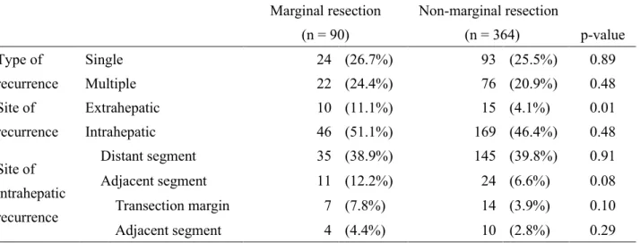
肝外再発率は MR 群が有意に不良であった (P = 0.01; Table 2). 肝内再発につい
ては断端再発、同一区域内再発、その他の区域の再発に分けて検討し, それぞれ両群で有意
差を認めなかった (P = 0.10, 0.29, 0.91; Table 2).
8
4-4 無再発生存率における予後規定因子
Cox 比例ハザードモデルにより多変量解析を行い予後規定因子の抽出を行った.
ICGR
15> 15% [hazard ratio (HR) 1.510, 95% Confidence interval (CI) 1.151-1.981, P =
0.003], 腫 瘍 径 ≧ 30mm (HR 1.491, 95% CI 1.150-1.935, P = 0.003), Aspartate
aminotransferase (AST) ≧ 35U/L (HR 1.376, 95% CI 1.065-1.776, P = 0.014)が独立した
予後規定因子であり, 切除断端 1mm 以下は独立した予後規定因子ではなかった (Table 3).
4-5 全生存における予後規定因子
Cox 比例ハザードモデルにより多変量解析を行い予後規定因子の抽出を行った.
アルブミン < 3 .0 g/dL (HR 2.252, 95% CI 1.221-4.153, P = 0.009), 腫瘍径 ≧ 30mm(HR 1.966, 95% CI 1.375-2.812, P = 0.0002), AST ≧ 35U/L (HR 1.648, 95% CI 1.160-2.341, P
= 0.005), 肝硬変の合併(HR 1.568, 95% CI 1.103-20.229, P = 0.012)が全生存における独立
した予後規定因子であり, 切除断端 1mm 未満は独立した予後規定因子ではなかった
(Table 4).
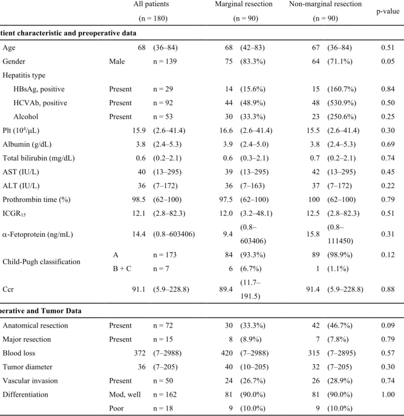
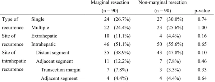
4-6 傾向スコア後の長期生存と再発形式
傾向スコア後, 対象患者は MR 群 90 例, non-MR 群 90 例に調整された (Figure
2). 傾向スコア後の患者背景に有意差は認めなかった (Table 5). 傾向スコア後の無再発生
存と全生存に有意差を認めなかった (P = 0.375, 0.496; Figure 5a and 5b). 再発形式につい
9
ては両群間で肝外再発, 肝内再発とも有意差を認めなかった(P = 0.16, 0.65; Table 6). 肝
内再発については断端再発, 同一区域内再発, その他の区域の再発それぞれで有意差を認
めなかった(P = 0.33, 0.64, 0.10; Table 6).
10
5. 考察
本検討では単発単結節型肝細胞癌において, 傾向スコア後 MR 群と non-MR 群は
無再発生存, 全生存, 再発形式において有意差を示さなかった.
今回, MR 群と non-MR 群の間で肝機能は有意差を認めなかったが, 術中出血量に
ついては MR 群の方が non-MR 群よりも有意に多かった. MR 群で出血量が多くなる原因と
して腫瘍が non-MR 群よりも有意に大きいことや, 腫瘍が肝臓の中心部に位置し主要な脈
管と接する症例など non-MR 群と比較して切除がより困難な症例が多かったことが考えら
れる. 主要な脈管に接しており肝機能が十分でないため脈管を合併切除できないような症 例は, 肝細胞癌の被膜に沿うような切除を行わざるをえない場合があり, 切除断端は最低限 度となる (23). 切除断端が最低限度である MR 群は今回の検討では無再発生存が有意に不
良であったが, 傾向スコアで予後不良因子である腫瘍径を含めた背景因子が調整されると, 無再発生存, 全生存ともに有意差は認めなかった (P = 0.375, 0.496).
これまで肝切除における切除断端陰性かつ切除断端距離 1mm 未満あるいは以下 であった症例について 3 編の報告があった (12-14). Lee et al. は 3 編の検討のなかで唯一
傾向スコアを用いており, 切除断端距離が 1mm 以下の群と、 1mm より大きい群で傾向スコ
ア後の全生存と無再発生存, また再発形式に差はないと我々と同様の結果を示しているが 対象症例の約 4 分の 1 が多発の肝細胞癌である (12). Matsui et al. は切除断端距離が 1mm
未満の群と 1mm 以上の群を比較し, 1mm 未満の群対象症例の約 4 分の 1 に多発の肝細胞癌
を含んでいるが MR 群と non-MR 群で比較し無再発生存と全生存に差はないと報告してい
11
る (13). Nara et al. は切除断端距離が 1mm 以下の群と 1mm より大きい群で比較し我々の
検討と同様に単発で単結節型の肝細胞癌を対象とし, MR 群と non-MR 群の間で再発率, 再
発形式に差がないため切除断端距離 1mm 以下は許容されると報告している (14).
肝内再発は腫瘍からの門脈浸潤によって起きる肝内転移と, 多中心性の発がんに よる 2 つの機序とされており, これらは切除断端距離の大きさとは無関係のものである (2,
3). これまでの肝細胞癌に対する切除断端距離が 1mm 未満あるいは以下についての 3 編の
検討では切除断端陰性で、かつ切除断端距離が 1mm 未満あるいは以下であった場合でも断
端再発率は低く (3-8%), non-MR 群と比較して有意差は認めなかった (12-14). 本検討で は同様の結果となり, MR 群の断端再発率は 7.78%, non-MR 群は 3.85%であり, 傾向スコア
後の non-MR 群は 3.33%で, いずれも 2 群間で有意差は認めなかった (P = 0.10, 0.33). こ
の結果は肝内再発のほとんどが腫瘍からの門脈浸潤による肝内転移か多中心性発がんのい ずれかの機序で起きていることと矛盾しない.
本研究の課題は単一施設での研究であり, 結果にいくつかの偏りがある可能性が
あること. また後方視的な研究であり無作為化比較試験ではないため, 傾向スコアを用いて
いるにも関わらず選択バイアスがある可能性があることである.
12
6. まとめ
肝細胞癌肝切除において最低限度である切除断端陰性かつ切除断端距離 1mm 未満
の切除症例は, 傾向スコア後, 無再発生存, 全生存, 再発形式に差を認めず, 切除断端陰性
かつ 1mm 未満となる切除は許容される.
13
Ⅱ 謝辞
本研究に関して, 研究の御指導, ならびに学位論文の御指導, ご校閲を賜りました日本大
学医学部外科学系消化器外科分野 高山忠利教授に深謝いたします.
14
Ⅲ 表
Table 1. Clinical history of patients
All patients Marginal resection Non-marginal
resection p-value
(n = 454) (n = 90) (n = 364)
Patient characteristic and preoperative data
Age 68 (36–84) 68 (42–83) 69 (36–84) 0.92
Gender Male n = 339 75 (83.3%) 264 (72.5%) 0.03
Hepatitis type
HBsAg, positive Present n = 66 14 (15.6%) 52 (140.3%) 0.76
HCVAb, positive Present n = 254 44 (48.9%) 210 (570.9%) 0.13
Alcohol Present n = 126 30 (33.3%) 96 (260.4%) 0.19
Plt (104/μl) 15.9 (2.6–41.4) 16.6 (2.6–41.4) 15.5 (2.6–41.4) 0.30
Albumin (g/dL) 3.9 (2.3–5.3) 3.9 (2.4–5.0) 3.8 (2.3–5.3) 0.87
Total bilirubin (mg/dL) 0.6 (0.2–3.4) 0.6 (0.3–2.1) 0.7 (0.2–3.4) 0.67
AST (IU/L) 40 (12–295) 39 (13–295) 40 (12–295) 0.86
ALT (IU/L) 35 (5–253) 36 (7–163) 35 (5–253) 0.57
Prothrombin time (%) 98.0 (53–100) 97.5 (62–100) 98 (53–100) 0.72
ICGR15 12.0 (1.3–82.3) 12.0 (3.2–48.1) 12.0 (1.3–82.3) 0.90
α-Fetoprotein (ng/mL) 13.7 (0.6–603406) 9.4 (0.8–603406) 14.0 (0.6–143200) .54
Child-Pugh classification A n = 439 84 (93.3%) 355 (97.5%) 0.09
B+C n = 15 6 (6.7%) 9 (20.5%)
Ccr 89.4 (5.9–281.5) 89.4 (11.7–191.5) 89.4 (5.9–281.5) 0.79
Operative and tumor data
Anatomical resection Present n = 195 30 (33.3%) 165 (45.3%) 0.04
Major resection Present n = 27 8 (8.9%) 19 (5.2%) 0.19
Blood loss 257 (7–2988) 420 (7–2988) 238 (7–2895) 0.0008
Tumor diameter 32 (7–205) 40 (10–205) 30 (7–205) 0.002
Microvascular invasion Present n = 130 24 (26.7%) 106 (29.1%) 0.64
Differentiation Mod,well n = 408 81 (90%) 327 (89.8%) 0.96
Poor n = 46 9 (10%) 37 (100.2%) 0.96
Values are expressed as n (%) or median (range).
AST, aspartate aminotransferase; ALT, alanine aminotransferase; HBsAg, Hepatitis B virus surface antigen; HCVAb, hepatitis C virus antibody; ICGR15, indocyanine green retention rate at 15 minutes
15
Table 2. Recurrence site after liver resection
Marginal resection (n = 90)
Non-marginal resection
(n = 364) p-value Type of
recurrence
Single 24 (26.7%) 93 (25.5%) 0.89
Multiple 22 (24.4%) 76 (20.9%) 0.48
Site of recurrence
Extrahepatic 10 (11.1%) 15 (4.1%) 0.01
Intrahepatic 46 (51.1%) 169 (46.4%) 0.48
Site of intrahepatic recurrence
Distant segment 35 (38.9%) 145 (39.8%) 0.91
Adjacent segment 11 (12.2%) 24 (6.6%) 0.08
Transection margin 7 (7.8%) 14 (3.9%) 0.10
Adjacent segment 4 (4.4%) 10 (2.8%) 0.29
16
Table 3. Prognostic factors for recurrence-free survival in patients with hepatocellular carcinoma by uni- and multivariate analysis
Values are expressed as n (%) or median (range).
AST, aspartate aminotransferase; ALT, alanine aminotransferase; HBsAg, Hepatitis B virus surface antigen; HCVAb, hepatitis C virus antibody; ICGR15, indocyanine green retention rate at 15 minutes
Univariate Multivariate
Hazard Ratio (95% CI) P-value Hazard Ratio (95% CI) p-value Age
≥ 651.005 (0.778–1.298) 0.97 -
Gender Male 0.972 (0.727–1.299) 0.85 -
Alcohol Present 1.000 (0.757–1.320) 1.00 -
HBsAg Positive 0.995 (0.682–1.451) 0.98 - HCVAb Positive 1.284 (0.990–1.665) 0.06 - Liver cirrhosis Present 0.860 (0.501–1.475) 0.58 - Albumin (g/dL) < 3.0 1.367 (0.797–2.346) 0.26 - Total bilirubin (mg/dL)
≥ 1.01.360 (0.965–1.917) 0.08 -
AST
≥ 351.370 (1.057–1.776) 0.02 1.394 (1.074–1.810) 0.013 ALT
≥ 351.484 (1.152–1.913) 0.002 -
Prothrombin time (%)
≥ 800.719 (0.493–1.048) 0.09 -
α-Fetoprotein (ng/mL) ≥ 20
1.506 (1.171–1.937) 0.001 1.376 (1.065–1.776) 0.014 ICGR
15 ≥ 151.554 (1.202–2.009) 0.001 1.510 (1.151–1.981) 0.003 Child-Pugh A vs. B 1.547 (0.865–2.765) 0.14 -
Tumor size (mm) < 30 1.378 (1.071–1.773) 0.01 1.491 (1.150–1.935) 0.003 Vascular invasion Present 1.190 (0.905–1.565) 0.21 -
Anatomical resection Present 1.396 (1.078–1.809) 0.01 1.267 (0.939–1.656) 0.084 Major resection Absent 0.860 (0.501–1.475) 0.58 -
Blood loss (mL)
≥ 5001.532 (1.020–2.303) 0.04 - Blood transfusion Absent 0.997 (0.529–1.877) 0.99 -
Surgical margin (mm) < 1.0 1.459 (1.081–1.970) 0.01 1.350 (0.993–1.836) 0.056
17
Table 4. Prognostic factors for overall survival in patients with hepatocellular carcinoma by uni- and multivariate analysis
Univariate Multivariate
Hazard Ratio (95% CI) p-value Hazard Ratio (95% CI) p-value Age
≥ 651.143 (0.806–1.622) 0.453 -
Gender male 0.980 (0.658–1.459) 0.919 -
Alcohol Present 1.038 (0.7141–1.507) 0.847 -
HBsAg Positive 0.687 (0.387–1.219) 0.199 -
HCVAb Positive 1.140 (0.799–1.627) 0.469 -
Liver cirrhosis Present 1.550 (1.102–2.18) 0.012 1.568 (1.103–2.229) 0.012 Albumin (g/dL) < 3.0 2.965 (1.631–5.393) 0.0004 2.252 (1.221–4.153) 0.009 Total bilirubin (mg/dL)
≥ 1.01.225 (0.768–1.952) 0.395 -
AST
≥ 351.593 (1.110–2.286) 0.012 1.648 (1.160–2.341) 0.005 ALT
≥ 351.553 (1.098–2.197) 0.013 -
Prothrombin time (%)
≥ 800.656 (0.407–1.057) 0.083 -
α-Fetoprotein (ng/mL) ≥ 201.549 (1.101–2.178) 0.012 - ICGR
15 ≥ 151.444 (1.018–2.048) 0.039 -
Child-Pugh A 1.676 (0.850–3.305) 0.136 -
Tumor size (mm) < 30 1.756 (1.241–2.486) 0.001 1.966 (1.375–2.812) 0.0002 Vascular invasion Present 1.563 (1.097–2.227) 0.013 -
Anatomical resection Present 1.223 (0.863–1.734) 0.259 - Major resection Absent 0.734 (0.300–1.796) 0.498 -
UICC 1, 2 vs. 3 4.173 (2.109–8.257) 0.00004 -
Blood loss (mL)
≥ 5001.546 (1.086–2.202) 0.016 - Blood transfusion Absent 1.115 (0.519–2.397) 0.780 - Surgical margin (mm) < 1.0 0.672 (0.449–1.006) 0.053 -
AST, aspartate aminotransferase; ALT, alanine aminotransferase; HBsAg, Hepatitis B virus surface antigen; HCVAb, hepatitis C virus antibody; ICGR15, indocyanine green retention rate at 15 minutes
18
Table 5. Clinical history of patients after propensity match
All patients Marginal resection Non-marginal resection
p-value
(n = 180) (n = 90) (n = 90)
Patient characteristic and preoperative data
Age 68 (36–84) 68 (42–83) 67 (36–84) 0.51
Gender Male n = 139 75 (83.3%) 64 (71.1%) 0.05
Hepatitis type
HBsAg, positive Present n = 29 14 (15.6%) 15 (160.7%) 0.84
HCVAb, positive Present n = 92 44 (48.9%) 48 (530.9%) 0.50
Alcohol Present n = 53 30 (33.3%) 23 (250.6%) 0.25
Plt (104/μL) 15.9 (2.6–41.4) 16.6 (2.6–41.4) 15.5 (2.6–41.4) 0.30
Albumin (g/dL) 3.8 (2.4–5.3) 3.9 (2.4–5.0) 3.8 (2.4–5.3) 0.69
Total bilirubin (mg/dL) 0.6 (0.2–2.1) 0.6 (0.3–2.1) 0.7 (0.2–2.1) 0.74
AST (IU/L) 40 (13–295) 39 (13–295) 42 (13–295) 0.45
ALT (IU/L) 36 (7–172) 36 (7–163) 37 (7–172) 0.22
Prothrombin time (%) 98.5 (62–100) 97.5 (62–100) 100 (62–100) 0.79
ICGR15 12.1 (2.8–82.3) 12.0 (3.2–48.1) 12.5 (2.8–82.3) 0.51
α-Fetoprotein (ng/mL) 14.4 (0.8–603406) 9.4 (0.8–
603406) 15.8 (0.8–
111450) 0.31
Child-Pugh classification A n = 173 84 (93.3%) 89 (98.9%) 0.12
B + C n = 7 6 (6.7%) 1 (1.1%)
Ccr 91.1 (5.9–228.8) 89.4 (11.7–
191.5) 91.4 (5.9–228.8) 0.88
Operative and Tumor Data
Anatomical resection Present n = 72 30 (33.3%) 42 (46.7%) 0.09
Major resection Present n = 15 8 (8.9%) 7 (7.8%) 0.79
Blood loss 372 (7–2988) 420 (7–2988) 315 (7–2895) 0.57
Tumor diameter 36 (7–205) 40 (10–205) 32 (7–205) 0.30
Vascular invasion Present n = 50 24 (26.7%) 26 (28.9%) 0.74
Differentiation Mod, well n = 162 81 (90.0%) 81 (90.0%) 1.00
Poor n = 18 9 (10.0%) 9 (10.0%)
Values are expressed as n (%) or median (range).
AST, aspartate aminotransferase; ALT, alanine aminotransferase; HBsAg, Hepatitis B virus surface antigen; HCVAb, hepatitis C virus antibody; ICGR15, indocyanine green retention rate at 15 minutes
19
Table 6. Recurrence site after propensity match.
Marginal resection (n = 90)
Non-marginal resection
(n = 90) p-value Type of
recurrence
Single 24 (26.7%) 27 (30.0%) 0.74
Multiple 22 (24.4%) 23 (25.6%) 1.00
Site of recurrence
Extrahepatic 10 (11.1%) 4 (4.4%) 0.16
Intrahepatic 46 (51.1%) 50 (55.6%) 0.65
Site of intrahepatic recurrence
Distant segment 35 (38.9%) 43 (47.8%) 0.10
Adjacent segment 11 (12.2%) 7 (7.8%) 0.46
Transection margin 7 (7.8%) 3 (3.3%) 0.33
Adjacent segment 4 (4.4%) 4 (4.4%) 0.64
20
Ⅳ. 図
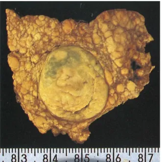
Figure 1 The macroscopic classification of the Liver Cancer Study Group of Japan
Simple nodular type: The nodule is clearly divided from around liver parenchyma and have a thick and firm fibrous capsule.
Simple nodular type with extranodular growth: There is the extra
growth nodule continuous to the main nodule.
21
Confluent multinodular type: The nodules take the form of multiple fusing.
Infiltrative type:A boundary between cancerous and non-cancerous
part is irregularity and inflating growth of the nodule is conspicuous.
22
Figure 2 Flow chart of the patients with hepatocellular carcinoma
23
Figure 3 Makuuchi’s criteria
No or controllable Incontrollable
Total bilirubin
Normal
Limited resection 1.1-1.9 mg/dL
ICG 15’
2.0 mg/dL≤
No hepatectomy
Normal 10%-19% 20%-29% 30%≤
Trisegmentectomy Rt hepatectomy
Lt hepatectomy Rt segmentectomy
Subsegmentectomy
Limitedresection
Algorithm for treatment in Japanese hepatocellular carcinoma guideline.
Acites
24
Figure 4
a. Recurrence-free survival of the surgical margin < 1 mm and surgical margin ≥ 1 mm groups in the entire study population
b. Overall survival of surgical margin < 1 mm and surgical margin ≥ 1
mm groups in the entire study population
25
Figure 5
a. Recurrence-free survival of the surgical margin < 1 mm and surgical margin ≥ 1 mm groups after propensity score matching
b. Overall survival of surgical margin < 1 mm group and surgical
margin ≥ 1 mm group after propensity score matching
26
Ⅴ 図説
Figure 1. The macroscopic classification of the Liver Cancer Study Group of
Japan; Simple nodular type, simple nodular type with extranodular growth,
confluent multinodular type and Infiltrative type.
Figure 2. Flow chart of the patients with hepatocellular carcinoma
Figure 3.
Algorithm before proceeding to safety hepatectomy for hepatocellular carcinoma
with cirrhotic liver. Mukuuchi’s criteria include three factors: ascites, total serum bilirubin, and the ICG-R
15. This algorithm shows the maximal area for which anoperation can be performed safely.
Figure 4.
a. Recurrence-free survival of the surgical margin < 1 mm [marginal resection
(MR)] and non-MR groups in the entire study population. The recurrence-free
survival was significantly shorter in the MR group than in the non-MR group (p
= 0.012).
27
b. Overall survival in the MR and non-MR groups in the entire study population.
There were marginally significant differences between the two groups in overall
survival (p = 0.051).
Thick line, MR group; thin line, non-MR group.
Figure 5.
a. Recurrence-free survival of the surgical margin < 1 mm [marginal resection
(MR)] and non-MR groups after propensity score matching. There were no
significant differences between the two groups in recurrence-free survival (p =
0.375).
b. Overall survival in the MR and non-MR groups after propensity score matching.
There were no significant differences between the two groups in overall survival
(p = 0.496).
Thick line, MR group; thin line, non-MR group.
28
Ⅵ 引用文献
1. Georgios M, Theodoros S, Ioannis NS, Nikolaos A. Impact of surgical margin
width on recurrence and overall survival following R0 hepatic resection of
colorectal metastases: A systematic review and meta-analysis: Ann Surg. 2018
June; 267(6):1047–1055.
2. Poon RT, Fan ST, Ng IO, Wong J. Significance of resection margin in
hepatectomy for hepatocellular carcinoma: A critical reappraisal. Ann Surg. 2000;
231:544–51.
3. Sakon M, Nagano H, Nakamori S, Dono K, Umeshita K, Murakami T, et al.
Intrahepatic recurrences of hepatocellular carcinoma after hepatectomy: analysis
based on tumor hemodynamics. Arch Surg. 2002; 137:94–9.
4. Shi M, Zhang CQ, Zhang YQ, Liang XM, Li JQ. Micrometastases of solitary
hepatocellular carcinoma and appropriate resection margin. World J Surg. 2004;
28:376–81.
5. Lee KT, Wang SN, Su RW, Chen HY, Shi HY, Ker CG, et al. Is wider surgical
margin justified for better clinical outcomes in patients with resectable
hepatocellular carcinoma? J Formos Med Assoc. 2012; 111:160–70.
6. Zhou XP, Quan ZW, Cong WM, Yang N, Zhang HB, Zhang SH, et al.
29
Micrometastasis in surrounding liver and the minimal length of resection margin
of primary liver cancer. World J Gastroenterol. 2007; 13:4498–503.
7. Zhong FP, Zhang YJ, Liu Y, Zou SB. Prognostic impact of surgical margin in
patients with hepatocellular carcinoma: A meta-analysis. Medicine (Baltimore).
2017; 96(37): e8043.
8. Nonami T, Harada A, Kurokawa T, Nakao A, Takagi H. Hepatic resection for
hepatocellular carcinoma. Am J Surg. 1997; 173:288–91.
9. Regimbeau JM, Kianmanesh R, Farges O, Dondero F, Sauvanet A, Belghiti J.
Extent of liver resection influences the outcome in patients with cirrhosis and
small hepatocellular carcinoma. Surgery. 2002; 131:311–7.
10. Ko S, Kanehiro H, Hisanaga M, Nagao M, Ikeda N, Nakajima Y. Liver
fibrosis increases the risk of intrahepatic recurrence after hepatectomy for
hepatocellular carcinoma. Br J Surg. 2002; 89:57–62.
11. Torzilli G, Montorsi M, Del Fabbro D, Palmisano A, Donadon M, Makuuchi
M. Ultrasonographically guided surgical approach to liver tumours involving the
hepatic veins close to the caval confluence. Br J Surg. 2006; 93:1238–46.
12. Lee JW, Lee YJ, Park KM, Hwang DW, Lee JH, Song KB. Anatomical
resection but not surgical margin width influence survival following resection for
30
HCC, a propensity score analysis. World J Surg. 2016; 40:1429–39.
13. Matsui Y, Terakawa N, Satoi S, Kaibori M, Kitade H, Takai S, et al.
Postoperative outcomes in patients with hepatocellular carcinomas resected with
exposure of the tumor surface: clinical role of the no-margin resection. Arch Surg.
2007; 142:596–602.
14. Nara S, Shimada K, Sakamoto Y, Esaki M, Kishi Y, Kosuge T, et al.
Prognostic impact of marginal resection for patients with solitary hepatocellular
carcinoma: evidence from 570 hepatectomies. Surgery. 2012; 151:526–36.
15. Liver Cancer Study Group of Japan. Classification of Primary Liver Cancer,
The 6th Edition. Tokyo: Kanehara; 2015
16. Hui AM, Takayama T, Sano K, Kubota K, Akahane M, Ohtomo K, Makuuchi
M. Predictive value of gross classification of hepatocellular carcinoma on
recurrence and survival after hepatectomy. J Hepatol. 2000 Dec; 33(6):975–9.
17. Shimada M, Rikimaru T, Hamatsu T, Yamashita Y, Terashi T, Taguchi K,
et al. The role of macroscopic classification in nodular-type hepatocellular
carcinoma. Am J Surg. 2001; 182:177–82.
18. Kondo K, Chijiiwa K, Makino I, Kai M, Maehara N, Ohuchida J, Naganuma
S. Risk factors for early death after liver resection in patients with solitary
31
hepatocellular carcinoma. J Hepatobiliary Pancreat Surg. 2005; 12(5):399–404.
19. Forner A, Llovet JM, Bruix J. Hepatocellular carcinoma. Lancet. 2012;
379:1245–55.
20. Makuuchi M, Kosuge T, Takayama T, Yamazaki S, Kakazu T, Miyagawa S,
et al. Surgery for small liver cancers. Semin Surg Oncol. 1993; 9:298–304.
21. Takayama T, Makuuchi M, Kubota K, Harihara Y, Hui AM, Sano K, Ijichi
M, Hasegawa K. Randomized comparison of ultrasonic vs clamp transection of the
liver. Arch Surg. 2001 Aug; 136(8):922–8.
22. Takayama T, Makuuchi M, Hirohashi S, Sakamoto M, Yamamoto J,
Shimada K, Kosuge T, Okada S, Takayasu K, Yamasaki S. Early hepatocellular
carcinoma as an entity with a high rate of surgical cure. Hepatology. 1998 Nov;
28(5):1241–6.
23. Sasaki K, Shindoh J, Margonis GA, Nishioka Y, Andreatos N, Sekine A, et
al. Effect of background liver cirrhosis on outcomes of hepatectomy for
hepatocellular carcinoma. JAMA Surg. 2017; 152:e165059.
32
Ⅶ 研究業績
小林 直哉
1. 発表 ① 一般発表 5 ② 特別発表 0 2. 論文 ① 原著論文 0 ② 症例報告 0 ③ 総説 なし 3. 著書 なし
以上
33