秋 田 医 学
Akita J Med
39: 73
-79, 2012
INTRAOPERATIVE DETECTION OF LYMPH NODE
MICROMETASTASIS USING A RAPID IMMUNOHISTOCHEMICAL STAINING METHOD IN NON
-SMALL CELL LUNG CANCER
Yasushi Kawaharada
1), Yoshihiro Minamiya
1), Hirosi Nanjo
2), Hajime Saito
1), Hayato Konno
1), Masami Kagaya
3), Yo
-ichi Akagami
3),
Satoru Motoyama
1)and Jun
-ichi Ogawa
1)(received 18 December 2012, accepted 28 December 2012)
1)
Department of Chest Surgery, Akita University Hospital and Akita University Graduate School of Medicine, 1
-1
-1 Hondo, Akita City 010
-8543, Japan
2)
Division of Clinical Pathology, Akita University Hospital and Akita University Graduate School of Medicine, 1
-1
-1 Hondo, Akita City 010
-8543, Japan
3)
Akita Prefectural Research and Development Center, 11
-4 Sanuki Araya, Akita City 010
-1623, Japan
Abstract
Nodal micrometastasis in non
-small cell lung cancer (NSCLC) is associated with a poorer survival rate than node
-negative disease. Furthermore, lymph node micrometastasis often cannot be detected using conventional hematoxylin and eosin staining of frozen sections ; detection requires additional time
-consuming immunohistochemical (IHC) analysis of paraffin
-embedded tis- sue. We previously developed and reported a device that enables us to complete IHC analyses within 11 minutes. In the present study, we used this rapid
-IHC protocol with an anti
-cytokera- tin antibody and analyzed 205 mediastinal lymph nodes dissected during surgery for NSCLC.
Although we modified the original rapid
-IHC procedure to block endogenous peroxidase activity, which could potentially cause misdiagnosis, the staining was still completed within 19 min. On the basis of conventional histological examination, 7 lymph nodes from 3 patients were deemed positive for micrometastasis. By contrast using hematoxylin and eosin staining, 13 lymph nodes from 7 patients were diagnosed positive on the basis of cytokeratin
-detection using routine
-IHC, and the same 13 nodes were diagnosed positive on the basis of rapid
-IHC. That is, all nodes deemed positive with the routine
-IHC procedure were also positive with the rapid
-IHC proce- dure. Assuming the results of the routine
-IHC are correct, the sensitivity, specificity and accu- racy of rapid
-IHC are 100%, 100% and 100%, respectively. These findings demonstrate the utility of our rapid
-IHC analysis for intraoperative diagnosis of micrometastasis. However, our findings are limited by the fact that we tested the method using a single antibody at a single insti- tute. Further investigation in multicenter studies will be needed to confirm the utility of this method.
Key words : non
-small cell lung cancer, lymph node metastasis, lymph node micrometasta- sis, immunohistochemical staining
Correspondence : Yasushi Kawaharada, M.D.
Department of Chest Surgery, Akita University Hospital and Akita University Graduate School of Medicine, 1
-1
-1 Hondo, Akita 010
-8543, Japan
Tel : 81
-18
-884
-6132 Fax : 81
-18
-836
-2615
E
-mail : [email protected]
-u.ac.jp
Introduction
Surgeons performing lung resections to treat non
-small cell lung cancer (NSCLC) rely on timely intraopera- tive assessment of lymph node metastasis when deciding whether to increase the extent of the surgical treatment
(41)
(e.g., with mediastinoscopy or extended segmentecto- my). Lymph node metastasis is commonly diagnosed intraoperatively based on an examination of frozen sec- tions. However, micrometastases within lymph nodes sometimes cannot be detected in hematoxylin and eosin (HE)
-stained frozen sections ; detection must await ad- ditional time
-consuming immunohistochemical (IHC) in- vestigation of paraffin
-embedded tissue. This is note- worthy, as nodal micrometastasis in NSCLC is reportedly associated with a poorer survival rate than node
-negative disease
1,2). Unfortunately, although the sensitivity of IHC analysis using an anti
-cytokeratin (CK) antibody is sufficient to detect micrometastases, the protocol rou- tinely requires 2
-4 hours to complete. To solve this problem, several investigators have proposed rapid meth- ods that enable IHC protocols to be completed within only 12
-30 min
3-11). None of these methods is problem free, however. For example, our use of flow cytometry was limited by the frequency of false
-positives caused by the lack of morphological information. To overcome that limitation, we developed and reported on a device that enables us to complete IHC analyses within 11 min using an alternating current (AC) electric field
12). With this device, we apply a high
-voltage, low
-frequency AC electric field to the sections while they are incubating with the primary antibody. The effect of the device is to reduce the time required for the antigen
-antibody reac- tion to occur. In our earlier report, we focused on the novel method developed and provided no group data or statistical analysis. The aim of the present study, there- fore, was to show the statistical details of the rapid detec- tion of lymph node micrometastases using our rapid
-IHC protocol.
Patients and Methods Patients
Twenty
-five patients with NSCLC were enrolled in the study between May 2011 and July 2012 after obtaining signed informed consent. Surgically resected speci- mens were used under approval of the Institutional Re- view Boards at Akita University School of Medicine and University Hospital. After preoperative evaluation, the patients were taken to an operating room, and the stan-
dard preparations were made for a thoracotomy, lung re- section, and mediastinal lymph node dissection. Lymph nodes from each patient were used for this study.
Tissue preparation
Immediately after surgically resecting the mediastinal lymph nodes, the nodes were embedded in O.C.T. com- pound (Sakura Finetek Japan Co., Ltd., Tokyo, Japan), fro- zen for 30 s in liquid acetone at −80°C using a Histo
-Tek Pino System (Sakura Finetek Japan Co., Ltd., Tokyo, Ja- pan), and transferred to a cryostat (CM1900 Leica, Wetz- lar, Germany).
Routine
-IHC procedure
Frozen sections were cut at 5 μm and placed on slides, then air
-dried for 30 s, fixed in acetone for 10 min at 4°C, air
-dried for 15 s at room temperature, and subjected to the staining procedure. Details of the staining method are listed in Table 1. Briefly, the sections were first in- cubated for 60 min with an anti
-pancytokeratin (CK) anti- body cocktail (AE1/AE3 ; PROGEN Biotechnik GmbH, Heidelberg, Germany) at 1 : 200 dilution (5 μg/mL).
They were then incubated for 30 min with EnVision
TM+ System/HRP Mouse (DAB+), developed using 3,3´di- aminobenzidine substrate, counterstained with hematox- ylin, dehydrated, and mounted on coverslips.
Rapid
-IHC device and procedure
We have developed a device that reduces the time re- quired for IHC analysis (Fig. 1). The device and its mechanism were described in detail previously
12). With this device, we apply a high
-voltage, low
-frequency AC electric field to lymph node sections while they are incu- bating with the primary antibody. This markedly reduc- es the time required for the antigen
-antibody reaction.
For each specimen, frozen sections were cut at 5 μm and placed on slides, then air
-dried for 30 s, fixed in acetone for 2 min at room temperature, air
-dried for 15 s at room temperature, and subjected to the staining procedure.
Details of the staining method are listed in Table 1.
Briefly, the sections were incubated first for 5 min with AE1/AE3 at 1 : 200 dilution (5 μg/mL) and then for 5 min with EnVisionTM+ System/HRP Mouse (DAB+).
Thereafter, the slides were developed with 3,3´diamino-
秋 田 医 学
(43)
benzidine substrate, counterstained with hematoxylin, dehydrated, and mounted on coverslips. Using this method we can complete IHC analyses within 20 min.
Histopathological evaluation
Samples from all dissected lymph nodes were sec- tioned, conventionally stained with HE, and examined by a pathologist. Samples immunohistochemically labeled with anti
-CK (AE1/AE3) antibody using the routine
-IHC and rapid
-IHC procedures were also examined by a pa- thologist. A result was considered positive if positive cell clusters or individual cells with the appropriate tu-
mor cell morphology were recognized. As proposed by the new American Joint Committee on Cancer Staging Manual
11), isolated tumor cells (ITCs) were also consid- ered positive in this study.
Results
The clinical characteristics of the study participants are summarized in Table 2. Using the rapid
-IHC proce- dure outlined in Table 1, IHC analyses were completed within 20 min, and CK
-positive nodes were detected by the pathologist within about 30 min. Using the routine
-Table 1. Procedures used for routine
-IHC and rapid
-IHC
Routine
-IHC procedure Rapid
-IHC procedure
Acetone fixation
10 minutes 4°C15 seconds
Blocking endogenous P.O. activity 5 minutes 15 seconds
Washing with PBS 5 minutes 3 times 30 seconds
Blocking nonspecific antibody binding with 5% BSA 15 minutes 5 minutes
Primary antibody 60 minutes 5 minutes
Washing with PBS 5 minutes 3 times 30 seconds
EnVision
TM+ System/HRP Mouse 30 minutes 5 minutes
Washing with PBS 5 minutes 3 times 30 seconds
3,3´ diaminobenzidine 2 minutes 2 minutes
Approximate time required 167 minutes 19 minutes
Fig. 1. Rapid
-IHC device (prototype No. 1).
IHC procedure, IHC analyses were completed within 167 min, and detection of CK
-positive nodes required an ad- ditional 10 min, on average.
The purpose of intraoperative IHC analysis during sur- gery for NSCLC is detection of lymph node micrometas- tasis without misdiagnosis. This means that to apply the rapid
-IHC procedure in practice, its ability to detect micrometastasis must be at least equal to that of the rou- tine
-IHC procedure. We therefore compared the abili- ties of the routine
-and rapid
-IHC procedures. Typical images of lymph node micrometastasis obtained using the routine
-and rapid
-IHC procedures are shown in Fig.
2. Note that the micrometastases detected using the routine
-IHC procedure were always detectable using the rapid
-IHC procedure.
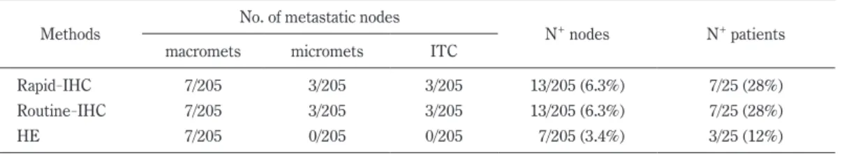
Table 3 summarizes hisotological diagnosis of metasta- sis. We stained 205 mediastinal lymph nodes dissected as part of the surgical treatment for NSCLC. Among those, 7 lymph nodes from 3 patients were deemed posi- tive for micrometastasis on the basis of conventional his-
tological examination. By contrast, 13 lymph nodes from 7 patients were deemed positive on the basis of CK
-detection using routine
-IHC, and the same 13 nodes were deemed positive using rapid
-IHC. In other words, all nodes deemed positive using the routine
-IHC proce- dure were also positive using the rapid
-IHC proce- dure. Assuming the results obtained using routine
-IHC are correct, the sensitivity, specificity and accuracy of the rapid
-IHC would be 100%, 100%, and 100%, respectively.
Discussion
In the present study we demonstrated and confirmed Table 2
Patients 25
Age (y) 68.5 ± 10.5
Sex Male 13
Female 12
Pathological stage Stage IA 15
Stage IB 6
Stage IIA 0
Stage IIB 1
Stage IIIA 3
Histological type Ad 21
Sq 4
Tumor Location RUL 7
RML 0
RLL 7
LUL 7
LLL 4
No. of tested LN Total 205
/patients 8.2 ± 4.8 Ad : adenocarcinoma, Sq : squamous cell carcinoma, RUL : right upper lobectomy, RML : right middle lobec- tomy, RLL : right lower lobectomy, LUL : left upper lobectomy, LLL : left lower lobectomy
Fig. 2. Detection of micrometastasis using rapid
-IHC
After labeling lymph nodes with an anti
-CK antibody
cocktail (AE1/AE3), the result was considered positive
if individual cells or clusters of cells with the appropri-
ate tumor cell morphology were recognized. As pro-
posed by the new American Joint Committee on
Cancer Staging Manual, isolated tumor cells were also
diagnosed as metastasis. The photomicrographs in
panels a, c and e show lymph node metastases stained
using routine
-IHC. Panels b, d and f show lymph
node metastases stained using rapid
-IHC. Panels a
and b, c and d, and e and f show macrometastases,
micro metastases, and isolated tumor cells, respec-
tively.
秋 田 医 学
that lymph node micrometastasis can be diagnosed within 30 min using the rapid
-IHC protocol developed at our in- stitute. Conventional analysis of frozen tissue sections using HE staining enables diagnosis within 20 min at most institutions
10), but the ability of this method to de- tect micrometastasis is comparatively low. The sensi- tivity of IHC analysis using an anti
-CK antibody is suffi- cient to detect micrometastasis, but the standard IHC procedure commonly requires 2
-4 hours to complete.
To address this issue, several investigators have pro- posed rapid methods that enable the IHC procedure to be accomplished within as little as 12
-30 min
5-10,13-15). These methods are not problem
-free, however. For ex- ample, the new EnVision
TMsystem has been described as a very sensitive and rapid IHC
-based detection sys- tem
9,10), which enabled Kämmerer et al.
7)and Mönig et al.
8)to reduce the time needed to immunostain frozen sections to less than 13 min. To do this, however, they applied a higher concentration of primary antibody than is routinely used for IHC. Notably, our rapid
-IHC proce- dure required about the same amount of time as the En- Vision system, and we were able to use the same concen- tration of primary antibody used in the routine IHC procedure. Given the expense of primary antibodies, we believe this represents a significant advantage over the EnVision system. It is also known that microwave irradiation shortens IHC times. For example, Hatta et al.
16)reported that IHC analyses could be completed within 15 min using intermittent microwave irradiation in combination with a prepared immune complex consisting of the primary antibody, a secondary antibody and EnVi- sion
TM-solution. The utility of our rapid
-IHC method is comparable to this microwave approach.
Although the main purpose of the rapid
-IHC is the di- agnosis of sentinel node micrometastasis during surgery,
avoiding axillary lymph node dissection based on the re- sults of sentinel node biopsy is now controversial in breast cancer. Recently, the American College of Sur- geons Oncology Group Z0011 trial demonstrated that, among patients with limited sentinel node metastatic breast cancer, the use of sentinel node dissection alone did not result in inferior survival, as compared to axillary lymph node dissection
17). This result indicates that in- traoperative diagnosis of sentinel node micrometastasis may not be required, at least in breast cancer, and we speculate that it reflects the efficacy of radiation, chemo- therapy, and endocrine therapy in breast cancer. Unfor- tunately, radiation and chemotherapy are not particularly effective in NSCLC. In addition, Ou and Zell demon- strated that the number of lymph nodes removed at sur- gery in patients without apparent lymph node metastasis is a significant prognostic factor in NSCLC
18), and medias- tinal lymph node dissection is still the standard surgical procedure for patients with NSCLC. Therefore, lymph node micrometastasis should not be ignored when apply- ing sentinel node mapping to NSCLC.
The mechanism by which our method promotes the antigen
-antibody reaction is not fully understood. In this study we applied a high
-voltage, low frequency AC electric field to specimens while they were incubating with the primary antibody
12). We previously showed that an interaction force affecting a droplet can be gener- ated as a coulomb force applied from an AC electric field
19). The generated force reflects the difference be- tween the dielectric constants and the electric conductiv- ity. In particular, when a droplet is attracted to the up- per electrode by applying an electric field with a positive polarity, a wave is generated and a stirring phenomenon is caused by changes in the surface of this wave. We speculate that this stirring promotes the antigen
-anti-
(45)
Table 3. Diagnosis of micrometastasis using rapid
-IHC, routine
-IHC and HE
Methods No. of metastatic nodes
N
+nodes N
+patients
macromets micromets ITC
Rapid
-IHC 7/205 3/205 3/205 13/205 (6.3%) 7/25 (28%)
Routine
-IHC 7/205 3/205 3/205 13/205 (6.3%) 7/25 (28%)
HE 7/205 0/205 0/205 7/205 (3.4%) 3/25 (12%)
Macromets : macrometastasis, micromets : micrometastasis, ITC : isolated tumor cells
body reaction.
In our earlier report, we did not block endogenous per- oxidase activity, which enabled us to complete the stain- ing protocol within only 11 min. We were then able to discern CK
-positive cancer cell nests based on their morphology. However, staining due to endogenous per- oxidase could potentially lead to misdiagnosis. We therefore added blocking of endogenous peroxidase to the rapid
-IHC protocol used in the present study (Table 1). This lengthened the staining protocol to 19 min, but we were still able to make a diagnosis of micrometastasis within 30 min, and the quality of the staining was much better.
Our findings demonstrate the utility of our rapid
-IHC analysis for intraoperative diagnosis of micrometasta- sis. However, our findings are limited by the fact that we tested the method using a single antibody at a single institute. Further investigation in multicenter studies will be needed to confirm the utility of this method.
References
1) Gu, C.
-D., Osaki, T., Oyama, T., Inoue, M., Kodate, M., Dobashi, K., Oka, T. and Yasumoto, K. (2002) Detection of micrometastatic tumor cells in pN0 lymph nodes of patients with completely resected nonsmall cell lung cancer : impact on recurrence and Survival. Ann. Surg., 235, 133
-139.
2) Turner, R.R., Ollila, D.W., Krasne, D.L. and Giuliano, A.E. (1997) Histopathologic validation of the senti- nel lymph node hypothesis for breast carcinoma.
Ann. Surg., 226, 271
-278.
3) Mabry, H. and Giuliano, A.E. (2007) Sentinel node mapping for breast cancer : progress to date and prospects for the future. Surg. Oncol. Clin. N. Am.,
16, 55-70.
4) Amersi, F. and Morton, D.L. (2007) The role of sentinel lymph node biopsy in the management of melanoma. Adv. Surg., 41, 241
-256.
5) Richter, T., Nährig, J., Komminoth, P., Kowolik, J. and Werner, M. (1999) Protocol for ultrarapid immu- nostaining of frozen sections. J. Clin. Pathol., 52, 461
-463.
6) Eudy, G.E., Carlson, G.W., Murray, D.R., Waldrop, S.M., Lawson, D. and Cohen, C. (2003) Rapid
immunohistochemistry of sentinel lymph nodes for metastatic melanoma. Hum. Pathol., 34, 797
-802.
7) Kämmerer, U., Kapp, M., Gassel, A.M., Richter, T., Tank, C., Dietl, J. and Ruck, P. (2001) A new rapid immunohistochemical staining technique using the EnVision antibody complex. J. Histochem. Cyto- chem., 49, 623
-630.
8) Mönig, S.P., Luebke, T., Soheili, A., Landsberg, S., Dienes, H.P., Hölscher, A.H. and Baldus, S.E. (2006) Rapid immunohistochemical detection of tumor cells in gastric carcinoma. Oncol. Rep., 16, 1143
-1147.
9) Sabattini, E., Bisgaard, K., Ascani, S., Poggi, S., Pic- cioli, M., Ceccarelli, C., Pieri, F., Fraternali
-Orcioni, G. and Pileri, S.A. (1998) The EnVision++
system : a new immunohistochemical method for diagnostics and research. Critical comparison with the APAAP, ChemMate, CSA, LABC, and SABC techniques. J. Clin. Pathol., 51, 506
-511.
10) Vyberg, M., Horn, T., Francis, D. and Askaa, J. (1990) Immunohistochemical identification of neuron
-spe- cific enolase, synaptophysin, chromogranin and endocrine granule constituent in neuroendocrine tumours. Acta Histochem. Suppl., 38, 179
-181.
11) Greene, F.L., Page, D.L., Fleming, I.D., eds, for the American Joint Committee on Cancer. (2002) AJCC Cancer Staging Manual. 6th ed. Springer
-Ver- lag, New York, NY.
12) Toda, H., Minamiya, Y., Kagaya, M., Nanjo, H., Aka- gami, Y., Saito, H., Ito, M., Konno, H., Motoyama, S.
and Ogawa, J. (2011) A novel immunohistochemical staining method allows ultrarapid detection of lymph node micrometastases while conserving antibody.
Acta Histochem. Cytochem., 44, 133
-139.
13) Matsumoto, M., Natsugoe, S., Ishigami, S., Ueno- sono, Y., Takao, S. and Aikou, T. (2003) Rapid immunohistochemical detection of lymph node micrometastasis during operation for upper gastroin- testinal carcinoma. Br. J. Surg., 90, 563
-566.
14) Nährig, J.M., Richter, T., Kuhn, W., Avril, N., Flatau, B., Kowolik, J., Höfler, H. and Werner, M. (2003) Intraoperative examination of sentinel lymph nodes by ultrarapid immunohistochemistry. Breast J.,
9,277
-281.
15) Dabbs, D.J., Hafer, L. and Abendroth, C.S. (1995)
Intraoperative immunocytochemistry of cytologic
scrape specimens. A rapid immunoperoxidase
秋 田 医 学