Introduction
Pancreatic resection is the only curable treatment for pan-
creatic tumors (1, 2). In the surgical procedure, post-treat- ment morbidity can be a problem and mortality is typically a result of pancreatic fistula and related complications such as intra-abdominal infections or bleeding (1, 3). It is important to predict such morbidity. Pancreatic elasticity or fibrosis is closely related to this complication (4-6). It is difficult to ac- curately evaluate this elasticity by conventional blood test or imaging diagnostic tools such as computed tomography or ultrasonography. A few investigators have examined pancre- atic stiffness using magnetic resonance imaging but this technique has not been clinically applied because of the high
cost and special apparatus requirements (7). Pancreatic stiff- ness is usually evaluated using palpation during operation or it can be histologically diagnosed in specimens. Using these classical diagnostic procedures, an objective evaluation of hardness or elasticity of the pancreas is difficult and can be influenced by the surgeonʼs experience.
Preoperative ultrasonography (US) is an essential tool to
identify tumor location in the pancreas (8). Because of re- cent advances in US function in the field of hepatic diagno- sis, hepatic elasticity has been extensively evaluated using FibroScan® (Echosens Co., Paris, France), which was de- veloped recently (9, 10). Hepatic elasticity detected in the procedure is well correlated with liver fibrosis, which is non- invasive and does not require liver biopsy. While, the acous-
MS#AMN 07188
Evaluation of malignant pancreatic tumor using artificial acoustic radiation force ultrasonography in patients undergoing pancreatectomy
Atsushi N
aNashima, mD,a, b Ayami sakamoto, mD,c Ichiro sakamoto, mD,c Hideyuki hayashi, mD,cYorihisa s
umiDa, mD,b Goushi murakami, mD,b Junichi arai, mD,b Hideo WaDa, mD,b and Takeshi Nagayasu mD, ba Division of Hepato-biliary-pancreas Surgery, Department of Surgery, University of Miyazaki Faculty of Medicine, Miyazaki, Japan
b Division of Surgical Oncology and c Department of Radiology, Nagasaki University Graduate School of Biomedical Sciences, Nagasaki, Japan
To evaluate pancreatic elasticity and tumor diagnosis, we preoperatively investigated the elasticity calculated by a new ultra- sonography (US) technique, acoustic radiation force impulse (ARFI). Materials and Methods: We examined ARFI in non-tumor- ous and tumor regions by push pulse of probe in 30 patients with pancreatic malignancies undergoing pancreatectomy. Measure- ment of stiffness was indicated as the Vs (mm/sec). Results: The Vs in the non-tumor region was measured in the pancreas head in 17 and in the body in 13 patients. The Vs of pancreatic tumors was measured in 14 of 22 patients (64%). The Vs in pancreatic tumors (2.17±0.95 m/sec.) was significantly higher than that in the non-tumorous pancreas (1.41±0.47 m/sec.) (p<0.01). The Vs of the non-pancreatic regions in the pancreas head and body were not significantly different. The Vs of the non-tumorous pan- creas was not associated with patient demographics, laboratory data, the hardness of the pancreatic tissue, or postoperative morbidity. The Vs of the resected pancreatic tumors was not associated with any tumor-related parameters. Conclusions: ARFI imaging elastography can be used to evaluate pancreatic malignant tumor lesion in comparison with the non-tumor lesion.
ACTA MEDICA NAGASAKIENSIA 60: 135−140, 2016 Key words: Pancreas tumor; ultrasonography; acoustic radiation force impulse; pancreatectomy; malignancy
Address correspondence: Atsushi Nanashima, MD., Division of Hepato-biliary-pancreas Surgery, Department of Surgery, University of Miyazaki Faculty of Medicine, 5200 Kihara Kiyotake, Miyazaki, 889-1692 Japan.
Tel: 81-985-85-2905, Fax: 81-985-85-3780, E-mail: [email protected] Received November 18, 2015; Accepted January 25, 2016
tic radiation force impulse (ARFI) imaging US (Siemens AG, Munich, Germany) is a new ultrasound-based modality that has been integrated into conventional US systems and is able to non-invasively evaluate the stiffness of deep tissues (11). Virtual Touch Tissue Quantification applications are not greatly influenced by the examinerʼs ability. The con- verged ultrasonic pulse sound without a press US probe forces pressure to the liver parenchyma and the shear wave velocity in the region of interest (ROI) (12). If stiffness of pancreatic parenchyma is correlated with pathology, it is useful for diagnosing pancreatic malignancies because pan- creatic solid tumors are harder than surrounding tissue. To our knowledge, investigations regarding ARFI US has been gradually reported in the
field of diagnosis for pancreaticdiseases (13-15).
We hypothesized that ARFI US would be applied for eval-
uating pancreatic stiffness or diagnosing pancreas tumor, and applied for predicting post-operative morbidity. Our aim in this preliminary study was to clarify this aspect of the new functional US, and we examined the findings of ARFI US in 30 patients with peri-pancreatic diseases who underwent pancreatectomy. The feasibility or limitations of this method were considered as a supportive diagnostic modality in pan- creatic surgery.
Patients and Methods
A total of 30 patients with peri-pancreatic tumorous dis-
eases admitted to the Division of Surgical Oncology at Na- gasaki University Graduate School of Biomedical Sciences (NUGSBS) between September 2009 and August 2013 were consecutively examined. These pancreatic tumors included invasive ductal carcinoma of the pancreas (PC) in 16 pa- tients, non-invasive intraductal papillary mucin-producing carcinoma (IPMC) in four, pancreatic neuroendocrine tumor in one, pancreatic metastasis originated from the renal cell carcinoma in one, lower bile duct carcinoma in five, and am- pullar carcinoma, duodenal carcinoma and tumor-forming pancreatitis in one patient each. The mean age for the pa- tients at the time of surgery was 54 ± 13 years (range, 40 - 80 years). Fourteen patients were male, and 16 were female.
The operative procedures included standard pancreaticoduo- denectomy (PD) in eight patients, subtotal stomach-preserv- ing PD in eight, distal pancreatectomy in 12, and probe lapa- rotomy in two.
The study protocols were approved by the Human Ethics
Review Board of our institution. Informed consent for the ultrasonic examination and data collection was obtained
from each patient. The examination with ARFI US was pre- operatively performed by radiologists (Dr. A.S., I.S. and H.H.) using an ACUSON S2000
TMUltrasound System (Sie- mens AG, Munich, Germany) and a convex probe (4C1, 1-4.5 MHz, Siemens). The ultrasound probe automatically produces an acoustic
“push” pulse that generates shearwaves, which propels into the pancreas. The speed, mea- sured in meters/second (m/s), is displayed on the screen (16).
The eSie Touch™ elasticity imaging uses gentle compres- sion to provide a high resolution elastogram, and the Virtual Touch™ tissue imaging is an application for ARFI technol- ogy for evaluation of deep tissues. Virtual Touch quantifica- tion uses an acoustic push pulse followed by detection pulses to calculate shear wave speed. The acoustic push pulse ap- plied adjacent to the ROI and, however, size or range of ROI was not clearly established in this study. The tracking beams (sensitive to greater than 1/00 the wavelength of sound) are applied adjacent to the acoustic push pulse. The time be- tween the generation of the shear wave and the passing of the shear wave peak at an adjacent location is utilized to compute the shear wave velocity. The force impulse (F) is calculated by the formula below:
F = 2 * α (attenuation constant) * I (time mean inten sity) / C (sonic speed)
We used this apparatus to examine the measurement of
ARFI as mean stiffness (Vs [millimeter /second]) in three times per lesion of pancreas, and the mean value was deter- mined because of reproducibility could not be always main- tained. The value of the Vs was compared with the patient demographics, laboratory data, histological findings, surgi- cal records, and postoperative complications. The hardness of the resected pancreatic specimens was measured using a muscle durometer (Neutone) (TDM-Z1(RB); TRY-ALL Co., Chiba, Japan). The tumor-node-metastases (TNM) classifi- cations for biliary and pancreas carcinoma were according to
Classification of Pancreatic Carcinoma (3rd English Edi- tion) (17).All continuous data were expressed as mean±SD. The
data for the different groups were compared using one-way
analysis of variance (ANOVA), which was examined by Stu-
dentʼs t-test and the Scheffeʼs multiple comparison test. The
correlation of the continuous data was tested by Spearmanʼs
rank correlation test, and its correlation coefficient (r) was
determined. A two-tailed P value < 0.05 was considered sig-
nificant. All the statistical analyses were performed using the
Statistical Package for the Social Sciences (SPSS) software,
version 18.0 (IBM, Chicago, IL, USA).
Results
The Vs in the non-tumor region was measured in the pan-
creas head in 17 of 30 cases (57%) and in the body in 13 (43%) cases. The Vs of the intra-pancreatic tumor was mea- sured in 14 of 22 pancreatic tumors (64%). The Vs in the pancreatic tumors was 2.17±0.95 m/sec., which was sig-
nificantly higher than that in the pancreas (1.41±0.47 m/
sec.) (p=0.002). The Vs of the non-pancreatic regions in the pancreas head (1.47±0.49 m/sec.) and body (1.30±0.26 m/
sec.) were not significantly different (p=0.25).
The relationship between the Vs in each region and the
clinicopathological parameters were examined (Table 1).
Age or body surface area (m
2), calculated by the patientʼs
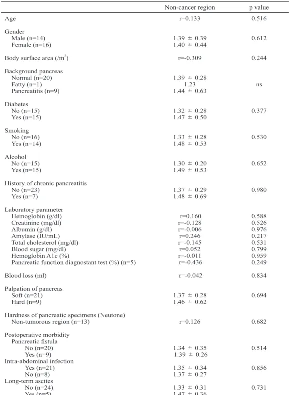
Table 1. Relationship between ARFI-Vs in the pancreas and clinical or surgical parameters.
Non-cancer region p value Age
Gender Male (n=14) Female (n=16) Body surface area (/m2) Background pancreas Normal (n=20) Fatty (n=1) Pancreatitis (n=9) Diabetes
No (n=15) Yes (n=15) Smoking No (n=16) Yes (n=14) Alcohol No (n=15) Yes (n=15)
History of chronic pancreatitis No (n=23)
Yes (n=7)
Laboratory parameter Hemoglobin (g/dl) Creatinine (mg/dl) Albumin (g/dl) Amylase (IU/mL) Total cholesterol (mg/dl) Blood sugar (mg/dl) Hemoglobin A1c (%)
Pancreatic function diagnostant test (%) (n=5) Blood loss (ml)
Palpation of pancreas Soft (n=21) Hard (n=9)
Hardness of pancreatic specimens (Neutone) Non-tumorous region (n=13)
Postoperative morbidity Pancreatic fistula No (n=20) Yes (n=9) Intra-abdominal infection Yes (n=21) No (n=8) Long-term ascites No (n=24) Yes (n=5)
r=0.133
1.39±0.39 1.40±0.44 r=-0.309
1.39±0.28 1.441.23±0.63
1.32±0.28 1.47±0.50
1.33±0.28 1.48±0.53
1.30±0.20 1.49±0.53
1.37±0.29 1.48±0.69
r=0.160 r=-0.128 r=-0.006 r=0.246 r=-0.145 r=0.052 r=-0.011 r=-0.436 r=-0.042
1.37±0.28 1.46±0.62
r=0.126
1.34±0.35 1.39±0.26 1.35±0.34 1.37±0.27 1.33±0.31 1.47±0.36
0.516
0.612
0.244
ns
0.377
0.530
0.652
0.980
0.588 0.526 0.976 0.217 0.531 0.799 0.959 0.249 0.834
0.694
0.682
0.514
0.856
0.731
height (cm) and weight (kg), were not correlated with Vs in non-tumorous pancreatic region and the Vs in the pancreas was not significantly different between males and females.
The Vs of the pancreas was not associated with background pancreas, patient co-morbidity, or habits. On the examina- tion of preoperative laboratory data, the Vs in the pancreas was not significantly correlated with any parameters. The Vs in the pancreas was not significantly associated with the hardness of the pancreatic tissue. The Vs in the pancreas was
not significantly associated with postoperative morbidity.
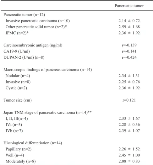
Table 2 shows the relationship between the Vs of the re-
sected pancreatic tumors and the tumor-related factors. Tu- mors included solid pancreatic carcinomas, other pancreatic solid tumors, and IPMN in 14 patients. The Vs of the tumor was not significantly associated with tumor markers, macro- scopic findings, tumor size, tumor stage, or histological dif- ferentiation of the resected pancreas.
Table 2. Relationship between ARFI-Vs and resected tumor-related parameters.
Pancreatic tumor Pancreatic tumor (n=12)
Invasive pancreatic carcinoma (n=10) Other pancreatic solid tumor (n=2)#
IPMC (n=2)*
Carcinoembryonic antigen (ng/ml) CA19-9 (U/ml)
DUPAN-2 (U/ml) (n=8)
Macroscopic findings of pancreas carcinoma (n=14) Nodular (n=4)
Invasive (n=8) Cystic (n=2)
Tumor size (cm)
Japan TNM stage of pancreatic carcinoma (n=14)**
I, II, III(n=4) IVa (n=3) IVb (n=7)
Histological differentiation (n=14) Papillary (n=2)
Well (n=4) Moderately (n=8)
2.14±0.72 2.59±1.68 2.36±1.92
r=-0.139 r=-0.141 r=-0.424
2.34±1.31 2.25±0.76 2.36±1.92
r=0.121
2.33±1.67 2.28±0.36 2.39±1.07
2.26±1.52 2.45±1.00 2.08±0.83 IPMN; intraductal papillary mucin-producing neoplasm, DUPAN-2; pancreatic cancer associated antigen
#; pancreatic neuroendocrine tumor in one and pancreatic metastasis
*; Intraductal papillary mucin-producing carcinoma with mucin production and solid component
**; According to the Classification of Pancreatic carcinoma (17)
Discussion
Recent ultrasonic technology has allowed for precise
measurement of organ stiffness (8-12). By applying this mo- dality, the diagnostic accuracy of
fibrosis can be examinedwithout invasive biopsy. In recent years, elastography has been widely used for diagnosing stiffness or tumors in solid organs (18-20), and ultrasonic elastography to diagnose pan- creas stiffness or pancreatic tumor has been increasingly ap- plied (13-15, 21-23). Göya et al. reported the usefulness of ARFI for diagnosis of acute pancreatitis (24).
Three modalities of ultrasonic elastography have been
primarily reported, and each mechanism of measuring stiff- ness is different, with specific advantages (9-11, 19). A se- lection of ARFI imaging in the pancreas is clinically useful (13-15). Determination of the measuring point was impor- tant in this procedure, and we examined the Vs of ARFI in the target ROI in the pancreatic head or body regions in this study. ARFI imaging may allow for examining deep tissues.
In the present series, the Vs of the pancreas was similar to that in previous reports (13). Determination could not be
fixed in a specific part of the pancreas because of physicalconditions, such as the thickness of the pancreas or distur- bances by intestinal air. This is a limitation to determine con- trol or reference data of ARFI-Vs in the pancreas. By com- paring the Vs between the pancreas head and body, there was no significant difference; however, the gap of the mean value was 0.17m/sec. based on our results. A standard method for measuring the pancreas itself is necessary. Even with this limitation, diagnostic sensitivity of acute or chronic pancrea- titis was high in comparison with conventional US or other imaging modalities (14, 15) We examined several factors, including patient demographics, status of background pan- creas, preoperative laboratory data, pancreatic function, sur- gical records, and outcomes after pancreatectomy in the present study. However, there were no significant correla- tions between the Vs in the pancreas and these factors. Al- though we hypothesized that the Vs would be correlated with the hardness or stiffness of the pancreas by palpation or measurement using a durometer, the Vs was not correlated at all, in contrast to previous reports (14, 15). Harada et al. or Lee et al. reported that ARFI was useful to estimate patho- logical fibrosis and related pancreatic fistula (23, 25). How- ever, on the other hand, postoperative mortality was not cor- related with the Vs in the present study and our aim to predict morbidity would be difficult at this stage. As the Vs was cor- related with histological
fibrosis in liver by our previousstudy (26), it is necessary to examine in a larger number of patients in the next step to clarify correlation with between
the Vs and pancreatic fibrosis.
An intra-pancreatic solid mass lesion hypothetically in-
creases stiffness or decreases elasticity. Previous studies on elastography clarified the usefulness of this method for diag- nosing pancreatic tumor regions (21, 26-28). Park et al. re- cently reported the diagnostic usefulness to define malignant solid pancreatic lesion using ARFI as well (29). The Vs of intrahepatic solid malignancies would increase in compari- son with non-cancerous pancreas, and the present result showed a significant increase of Vs in the tumors. By com- paring pancreatic carcinoma, neuroendocrine tumor, metas- tasis, and IPMN, the Vs was not significantly different.
D'Onofrio et al. also reported the Vs as measured by ARFI was increased in highly mucinous cystic tumors of the pan- creas and, however, reliable value of the VS might not be always obtained. (30-32). In the present study, although the high Vs was measured in both two cases of IPMN because of solid component with mucin production, which was more increased in comparison with the non-tumorous lesion. In general, however, it is still difficult to accurately measure the reliable Vs in various cystic tumor of the pancreas as above previous reports (30-32). The Vs of the all tumor lesion was not correlated with tumor-related factors. We also applied ARFI-Vs to diagnose the existence of pancreatic solid tu- mors; however, this was not useful to determine malignant behaviors of the pancreatic carcinomas.
Endoscopic ultrasonography-elastography has recently
been applied for diagnosing pancreatitis or pancreas tumors (19, 27, 33). As this procedure may be more sensitive for determining pancreatic elasticity or tumor locations, we plan to apply this latest technique in future studies. Furthermore, Yin et al. reported the usefulness of magnetic resonance im- aging elastography (34). In the future, it will be important to compare other modalities to measure organ elasticity.
In conclusion, we demonstrated the usefulness of a newly
developed elastography, ARFI image ultrasonography, in
patients with peri-pancreatic diseases. Measuring the Vs of
intra-pancreatic malignancies may allow to define malignant
tumor lesion in the pancreas. We found that measuring the
Vs in non-tumorous pancreas does not reflect the pancreatic
function or status, and that the Vs of the pancreas tumor can-
not be used for the differential diagnosis of tumors or malig-
nant behaviors of tumors. This modality is only useful to
diagnose pancreatic tumors.
References
1. Petzel MQ, Parker NH, Valentine AD, et al. Fear of cancer recurrence after curative pancreatectomy: a cross-sectional study in survivors of pancreatic and periampullary tumors. Ann Surg Oncol. 19:4078-4084, 2012.
2. Shaib Y, Davila J, Naumann C, et al. The impact of curative intent surgery on the survival of pancreatic cancer patients: a U.S. Popula- tion-based study. Am J Gastroenterol. 102:1377-1382, 2007.
3. Vollmer CM Jr, Sanchez N, Gondek S, et al; Pancreatic Surgery Mor- tality Study Group. A root-cause analysis of mortality following major pancreatectomy. J Gastrointest Surg. 16:89-102, 2012
4. Callery MP, Pratt WB, Kent TS, et al. A prospectively validated clini- cal risk score accurately predicts pancreatic fistula after pancreatodu- odenectomy. J Am Coll Surg. 216:1-14, 2013.
5. Gaujoux S, Cortes A, Couvelard A, et al. Fatty pancreas and increased body mass index are risk factors of pancreatic fistula after pancreati- coduodenectomy. Surgery. 148:15-23, 2010.
6. Belyaev O, Munding J, Herzog T, et al. Histomorphological features of the pancreatic remnant as independent risk factors for postoperative pancreatic fistula: a matched-pairs analysis. Pancreatology. 11:516- 524, 2011.
7. Tajima Y, Kuroki T, Tsutsumi R, et al. Risk factors for pancreatic anastomotic leakage: the significance of preoperative dynamic mag- netic resonance imaging of the pancreas as a predictor of leakage. J Am Coll Surg. 202:723-731, 2006.
8. Shrikhande SV, Barreto SG, Goel M, et al. Multimodality imaging of pancreatic ductal adenocarcinoma: a review of the literature. HPB (Oxford). 14:658-668, 2012.
9. Nguyen-Khac E, Capron D. Noninvasive diagnosis of liver fibrosis by ultrasonic transient elastography (Fibroscan). Eur J Gastroenterol He- patol. 18:1321-1325, 2006.
10. Han KH, Yoon KT. New diagnostic method for liver fibrosis and cir- rhosis. Intervirology. 51:11-16, 2008.
11. Baranova A, Lal P, Birerdinc A, et al. Non-invasive markers for he- patic fibrosis. BMC Gastroenterol.11:91, 2011.
12. Yashima Y, Sasahira N, Isayama H, et al. Acoustic radiation force impulse elastography for noninvasive assessment of chronic pancrea- titis. J Gastroenterol. 47:427-432, 2012.
13. Gallotti A, D'Onofrio M, Pozzi Mucelli R. Acoustic Radiation Force Impulse (ARFI) technique in ultrasound with Virtual Touch tissue quantification of the upper abdomen. Radiol Med. 115:889-897, 2010.
14. Mateen MA, Muheet KA, Mohan RJ, et al. Evaluation of ultrasound based acoustic radiation force impulse (ARFI) and eSie touch sono- elastography for diagnosis of inflammatory pancreatic diseases. JOP.
13:36-44, 2012.
15. Yashima Y, Sasahira N, Isayama H, et al. Acoustic radiation force impulse elastography for noninvasive assessment of chronic pancrea- titis. J Gastroenterol. 47:427-432, 2012.
16. Osaki A, Kubota T, Suda T, et al. Shear wave velocity is a useful mark- er for managing nonalcoholic steatohepatitis. World J Gastroenterol.
16:2918-2925, 2010.
17. The Japanese edition of the General Rules for the Study of Pancreatic Cancer by the Japan Pancreas Society (JPS). The third English edi- tion, Tokyo: Kanehara:2011
18. Kinney TP, Freeman ML. Recent advances and novel methods in pan- creatic imaging. Minerva Gastroenterol Dietol. 54:85-95, 2008.
19. Garra BS. Imaging and estimation of tissue elasticity by ultrasound.
Ultrasound Q. 23:255-268, 2007.
20. Arda K, Ciledag N, Aktas E, et al. Quantitative assessment of normal soft-tissue elasticity using shear-wave ultrasound elastography. Am J Roentgenol. 197:532-536, 2011.
21. Uchida H, Hirooka Y, Itoh A, et al. Feasibility of tissue elastography using transcutaneous ultrasonography for the diagnosis of pancreatic diseases. Pancreas. 38:17-22, 2009.
22. Pei Q, Zou X, Zhang X, et al. Diagnostic value of EUS elastography in differentiation of benign and malignant solid pancreatic masses: a meta-analysis. Pancreatology. 12:402-408, 2012.
23. Harada N, Ishizawa T, Inoue Y, Aoki T, Sakamoto Y, Hasegawa K, Sugawara Y, Tanaka M, Fukayama M, Kokudo Net al. Acoustic radia- tion force impulse imaging of the pancreas for estimation of patho- logic fibrosis and risk of postoperative pancreatic fistula. J Am Coll Surg. 219:887-894, 2014
24. Göya C, Hamidi C, Hattapoğlu S, et al Use of acoustic radiation force impulse elastography to diagnose acute pancreatitis at hospital admis- sion: comparison with sonography and computed tomography. J Ul- trasound Med. 33:1453-1460, 2014
25. Lee TK, Kang CM, Park MS, et al. Prediction of postoperative pancre- atic fistulas after pancreatectomy: assessment with acoustic radiation force impulse elastography. J Ultrasound Med. 33:781-786, 2014.
26. Săftoiu A, Vilmann P, Gorunescu F, et al. Neural network analysis of dynamic sequences of EUS elastography used for the differential di- agnosis of chronic pancreatitis and pancreatic cancer. Gastrointest Endosc. 68:1086-1094, 2008.
27. Hirche TO, Ignee A, Barreiros AP, et al. Indications and limitations of endoscopic ultrasound elastography for evaluation of focal pancreatic lesions. Endoscopy. 40:910-917, 2008.
28. Schrader H, Wiese M, Ellrichmann M, et al. Diagnostic value of quan- titative EUS elastography for malignant pancreatic tumors: relation- ship with pancreatic fibrosis. Ultraschall Med. 33:E196-201, 2012.
29. Park MK, Jo J, Kwon H, et al. Usefulness of acoustic radiation force impulse elastography in the differential diagnosis of benign and ma- lignant solid pancreatic lesions. Ultrasonography. 33:26-33, 2014.
30. D'Onofrio M, Gallotti A, Falconi M, et al. Acoustic radiation force impulse ultrasound imaging of pancreatic cystic lesions: preliminary results. Pancreas. 39:939-940, 2010.
31. D'Onofrio M, Gallotti A, Mucelli RP. Pancreatic mucinous cystade- noma at ultrasound acoustic radiation force impulse (ARFI) imaging.
Pancreas. 39:684-685, 2010.
32. D'Onofrio M, Crosara S, Canestrini S, et al. Virtual analysis of pan- creatic cystic lesion fluid content by ultrasound acoustic radiation force impulse quantification. Ultrasound Med. 32:647-651, 2013.
33. Săftoiu A, Vilmann P, Gorunescu F, et al; European EUS Elastogra- phy Multicentric Study Group. Efficacy of an artificial neural network- based approach to endoscopic ultrasound elastography in diagnosis of focal pancreatic masses. Clin Gastroenterol Hepatol. 10:84-90, 2012.
34. Yin M, Chen J, Glaser KJ, et al. Abdominal magnetic resonance elas- tography. Top Magn Reson Imaging. 20:79-87, 2009.