構造活性相関研究
著者 高橋 典子
雑誌名 星薬科大学紀要
号 54
ページ 105‑114
発行年 2012
URL http://id.nii.ac.jp/1240/00000148/
1. はじめに
ビタミンA (レチノール) は体内で種々の重要な役割 を果たしているが、 十分な量を合成することができない ため、 -カロチンとして食餌から摂取しなくてはならな い。 このような微量栄養素であるビタミンAの欠乏は 夜盲症を引き起こすことから、 ビタミンAは視覚に深 く関与している。 この作用以外にも、 ビタミンAは皮 膚粘膜形成、 成長促進、 細胞分化誘導、 免疫調節、 抗癌 などの作用も発揮する。
癌 (悪性腫瘍) はわが国の死因の第一位を占め、 現在、
副作用の少ない抗癌剤の開発が社会から強く要請されて いる。 ビタミンA自身の抗癌作用は非常に弱いものの、
ビタミンAの代謝物であるレチノイン酸 (RA) は急性 前骨髄性白血病患者 (APL) の治療薬として使用され ている。 RAは分化終末細胞 (成熟血球) まで分化する ことのないまま増殖状態にある未分化な白血病細胞を、
成熟した顆粒球様細胞に分化誘導する1)。 このように RAは癌細胞を正常細胞に誘導・変換させる能力をもつ。
RAによる白血病治療は分化誘導療法と呼ばれ、 現在 RAは臨床で用いられており、 約95 %のAPL患者を完 全寛解させている2)。 分化誘導療法は化学療法と異なり 癌細胞にのみ作用し、 正常細胞に大きな影響を与えない ことから、 副作用が少なく治癒率が非常に高いという利 点がある。 しかしながら、 再発した患者にRAを投与し ても効かなくなり、 見かけ上RA耐性となる。 この問題 を克服するため、 RAの投与濃度を低くし他の薬剤を併 用して、 RA単独で用いる場合と同等の効果を得ること を目指した薬剤併用療法が考えられている。
癌が転移しなければ癌で命を落とすことはほとんど無 くなる。 それほど癌転移を阻止することには、 大きな意 味がある。 癌転移を規定する要因に、 細胞の解離、 浸潤、
接着、 増殖などがある。 特に癌巣で発生する活性酸素種 (ROS) が、 癌細胞の運動、 転移を促進する重要な鍵物 質として認知されている3-5)。 ROSのうちスーパーオキ シド (O2-) が癌転移に促進的に作用することが知られ ている。 一方、 DNA傷害を引き起こす発癌には、 ROS のヒドロキシラジカル (・OH) が深く関わる。 ヒドロ キシラジカルは、 強い酸化力をもち、 酵素蛋白質や細胞 骨格蛋白質、 脂質、 糖質、 核酸 (DNA、 RNA) と反応
する6, 7)。 このように、 ROSが発癌、 癌転移に促進的に
働くことから、 これを消去する抗酸化物質は抗癌剤、 癌 予防剤となりうるのである。
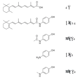
Fenretinide (N-(4-hydroxyphenyl)retinamide, 4- HPR, Fig. 1) は RAの末端のカルボキシル基にヒド ロキシルフェニルアミノ基が結合した合成レチノイドで ある。 4-HPRはRAの誘導体として合成されたが、 そ
総 説
抗酸化作用をもつ抗癌剤の創製:Fenretinideの構造活性相関研究
高 橋 典 子
星薬科大学 医薬品化学研究所 病態機能制御学研究室
Development of anticancer agents exhibiting antioxidative activities : Structure-activity relationship studies on Fenretinide
Noriko TAKAHASHI
Laboratory of Physiological Chemistry, Institute of Medicinal Chemistry, Hoshi University, Shinagawa, Tokyo 142-8501, Japan
RA
4-HPR
p-AAP
p-MAP 4-AP
Fig. 1. Chemical structures of RA, 4-HPR, and aminophenols (p-MAP, 4-AP, and p-AAP).
の抗癌作用は強く、 有効な癌化学予防剤、 癌細胞増殖抑 制剤として知られている8-11)。 4-HPRはRAと異なり核 内RAレセプターと結合しないことから、 その作用機構 はよく分かっていない。 ひとつの作用機構として、 4- HPRが細胞内で活性酸素を発生させることによって、
アポトーシス (programmed cell death) を誘導する ことが提唱されている12-17)。 現在、 4-HPRの臨床試験が、
膀胱癌、 肺癌、 前立腺癌、 リンパ腫、 神経芽腫に対して 行われている11, 13, 18-25)。 4-HPRは一般的に忍容性におい ては良好であったが、 低用量で血漿レチノールレベルの 枯渇による可逆性暗順応障害、 乾燥肌、 発疹、 高用量で 可逆性肝機能障害、 高グリセリド血症、 小児特異偽脳腫 瘍、 嘔吐、 軽度の血小板減少症が観察されている。 この ように、 4-HPRは有効な治療薬ではあるものの副作用 を現す。
合成レチノイドFenretinide、 4-HPRの抗癌剤とし ての作用と問題点を踏まえて、 副作用のない新規の抗癌 剤と抗酸化剤の創製について述べる。 また、 抗癌作用 (細胞増殖抑制作用) と抗酸化作用 (ROSの消去) との 関連性を考察する。
2. Fenretinideとその誘導体の癌細胞に対する作用
(1) FenretinideとRAの細胞増殖、 分化、 生存率に 与える影響26)
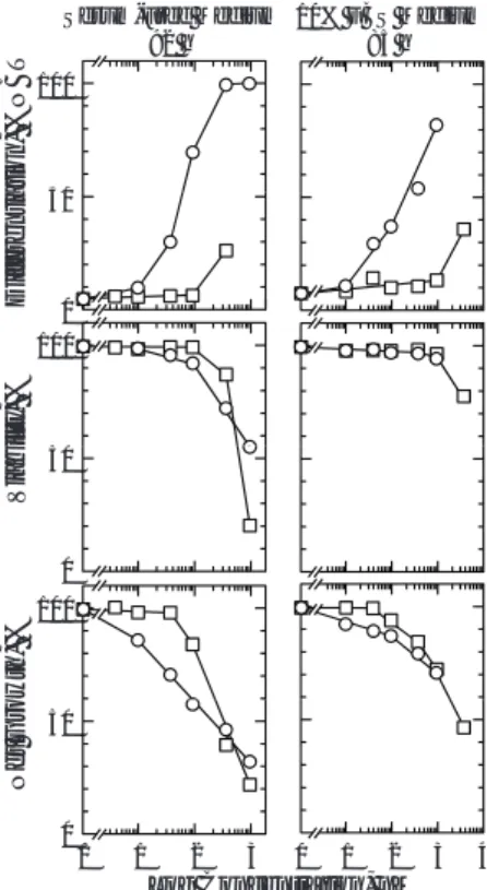
4-HPRの前骨髄性白血病細胞株HL60における分化 誘導作用は、 RAと比較して非常に弱かった (Fig. 2)。
また、 RAによる分化誘導作用は培養液中の血清の有無 に関わらず、 細胞増殖抑制作用と負の相関を示した。 高 濃度で化合物処理を行なった場合、 無血清培地でのみ細 胞の生存率の低下が見られた。 4-HPRによる細胞増殖 と生存率の低下は、 RAに比べて細胞の分化と連動 (相 関) しなかった。 血清含有培地を使用した時、 4 M 4- HPRは約40 %の細胞の分化を誘導したが、 その際の 細胞生存率は約80 %、 細胞増殖抑制率は約50 %であっ た。 4-HPRは細胞増殖抑制作用を示すが、 RAと比べ 非常に弱い細胞分化誘導作用しか持たなかった。
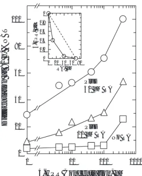
(2) FenretinideとRAの併用による細胞分化26) 4-HPRは単独では非常に弱い分化誘導能しか持たな いことから、 次に4-HPRのRAによるHL60細胞の分 化誘導作用に与える影響について調べた。 単独で分化を ほとんど誘導しない濃度のRA (10 nM or 40 nM) と 4-HPR (4~400 nM) を併用することで、 4-HPRはRA による細胞分化を増大させた (Fig. 3)。 これらのデー タをアイソボログラムで解析したところ、 Combination Indexは約0.6であった。 これらのことから、 4-HPR はRA によるHL60細胞分化誘導作用を相乗的に増強 させることが判った。 即ち、 4-HPRは細胞の増殖を抑
制させアポトーシスを誘導するのに対し、 RAは細胞の 分化を誘導することから異なる作用点で両化合物が作用 し合うことによって、 より効果的に抗癌作用を発揮した。
以上の結果から、 単独で細胞分化誘導能が弱い薬剤でも RAと併用することによりRAの作用を増強することが できれば、 臨床的に使用できる可能性を示唆した。
(3) Fenretinideの構造活性相関27)
4-HPRが暗順応障害、 乾燥肌などの副作用を示すこ とから、 4-HPRの活性を保持し、 且つ、 副作用を持た ない化合物の創製を試みた。 4-HPRの副作用は内在性 のレチノイド作用を阻害したことに起因していることか
Serum-Free Medium 92 h
10% FBS Medium 85 h 100
ation, %NBT 50
100
lity, %
50
Differentia 0
100
owth, %
50
0
Viabi
50
0
Net Gro
0 1 2 3 0 1 2 3 4
Log Concentration, nM
Fig. 2. Comparison of the effects of RA and 4-HPR on differentiation, viability, and growth of HL60 cells. Cells (2×10⁵/ml) were grown with various concen- trations of RA (○) or 4-HPR (□) in serum-free medium (left panels) or in medium containing 10 % FBS (right panels). Measurements were made at 92 h for cells growing in serum-free medium and at 85 h for cells growing in medium containing serum. Each point is the mean of at least 4 measurements. The SE of each data point was 8 % of the mean. Control cultures had a cell density of 1.1×10⁶/ml in serum-free medium and 1.02×10⁶/ml in serum-containing medium. The values for % net growth were calculated with the following for- mula : [(cell concentration of experimental culture) - (initial cell concentration) / (cell concentration of control culture) - (initial cell concentration)]×100.
ら、 副作用の原因となる4-HPRの構造はレチノイド構 造 (サイクロヘキセン環) であると考えられた。 しかし ながら、 この構造を欠失した化合物が4-HPRと同等の 抗癌活性を示すとは限らない。 そこで先ず、 4-HPRの 抗癌作用を現す構造を見出すため、 4-HPRの部分構造 を持つ4種の化合物: RA、 アミノフェノール (4-AP)、
アセトアミノフェノール (p-AAP, アセトアミノフェン)、
メチルアミノフェノール (p-MAP) について (Fig. 1)、
細胞増殖抑制・アポトーシス誘導作用を調べた。
Fig. 4に示すように、 HL60細胞に対する細胞増殖抑 制作用を調べたところ、 1 M p-MAPは約33 %を抑え たのに対し、 同じ濃度のRA、 4-HPRは順に約29 %、
約28 %を抑制した。 化合物10 Mの濃度では、 p-MAP がRAと3種のアミノフェノールのうち最も強く、 4-
HPRと同等の細胞増殖抑制作用を示した。 細胞増殖は、
p-MAPで約99.7 %、 4-HPRで約99.6 %、 RAで約39
%、 4-APで約21.8 %、 p-AAPで約4 %の割合で阻害さ れた。 さらに、 4-HPRは薬剤耐性癌細胞の増殖を抑制 す る が 、 p-MAP も4-HPRと 同 様 にRA 耐 性HL60 (HL60R) 細胞、 RA耐性乳癌 (MCF-7/AdrR) 細胞に 対し増殖抑制活性を示した27)。 また、 p-MAPは4-HPR よりも若干弱いものの、 4種の化合物のうち最も効果的 に、 乳癌 (MCF-7) 細胞、 前立腺癌 (DU-145) 細胞、
肝癌 (HepG2) 細胞の増殖を抑制した27)。 これらの結 果から、 p-MAPが4種の化合物の中で最も強力な抗細 胞増殖化合物であることが明らかとなった。 またメチル 基が鍵構造であると考えられた。
次に、 これら化合物がHL60細胞のアポトーシスを 誘導させるかを、 DNAフラグメンテーション-アガロー ス解析により調べた。 コントロールと1 M p-MAPで 処理した細胞から抽出したDNAはフラグメンテーショ ン し て い な か っ た が 、 10 Mの 濃 度 のp-MAPと4- HPRではフラグメントラダーが認められた (Fig. 5)。
100
B T
1 0.8 R, µM0.6
80
60
Plus 40 nM RA
tion at 92 h, %N B
0.4 0.2 0
0 20 40 60 80 RA, nM
4-HPR
40
20
No RA Plus
10 nM RA 40 nM RA
Dif ferentia t
4-HPR Concentration, nM
0 10 100 1000
0
● ●
●
●
Fig. 3. HL60 cell differentiation in serum-free me- dium induced by increasing concentrations of 4- HPR in the presence of fixed concentrations of RA. Cells (2×10⁵/ml) were grown for 92 h in serum-free medium with the indicated concentrations of RA and 4- HPR. Each point is the mean of at least 4 measure- ments. The SE of each data point was 8 % of the mean. The isoboles (inset) are for combinations of 4- HPR with RA that are isoeffective (ED50) for differentia- tion of HL60 cells. When there is additivity the experi- mental values for a combination fall on the dashed line connecting the values for each agent alone. The ED50
value for 4-HPR alone was estimated by extrapolation.
This value could not be determined experimentally be- cause of low viability (see Results). The viabilities were
> 85 % for each of the other isoeffective conditions shown in the isobologram.
HL60
120
94 h
h, % of Control
60 80 100
94 h
Net Growth
0 20 40
0
Fig. 4. Growth of HL60 cells in the presence of aminophenols (p-MAP, 4-AP, p-AAP), RA, and 4- HPR. Cells (2×10⁵/ml) were grown without or with aminophenols (p-MAP, 4 AP, and p-AAP), RA, or 4-HPR at 1 M or 10 M concentration in medium containing 10 % FBS for 94 hours. The SD of each data point was 8 % of the mean. Experiments were repeated at least four times. RA was used as an internal standard for growth inhibition. Values for percent net growth were calculated with the following formula :[(cell concentra- tion of experimental culture) - (initial cell concentration) / (cell concentration of control culture) - (initial cell con centration)]×100.
以上の結果から、 p-MAPはアポトーシスを誘導し、 細 胞増殖を抑制することが明らかとなった。
(4) Fenretinideからp-Alkylaminophenol28)
以上の研究からp-MAPが4-HPRと同程度の抗癌活 性を示し、 作用発現にp-MAPのメチル基が重要である ことが明らかになった。 そこで、 メチル鎖を伸長させ、
より脂溶性を高くした化合物p-アルキルアミノフェノー ル[p-ブチルアミノフェノール (p-BAP)、 p-ヘキシル アミノフェノール (p-HAP)、 p-オクチルアミノフェノー ル (p-OAP)、 p-メトキシベンジルアミノフェノール (p-MBAP)](Fig. 6) を創製し、 癌細胞に対する増殖 抑制・アポトーシス誘導作用を検討した。
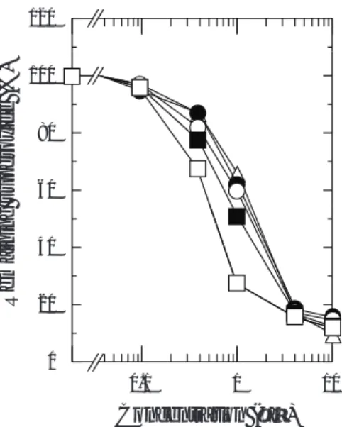
その結果、 4-HPRはHL60細胞とRA耐性である HL60R細胞の増殖を濃度依存的に抑制した (Fig. 7A)。
HL60細胞において、 p-OAPは0.1〜1 Mといった低 濃度で4-HPRよりも強い細胞増殖阻害作用を示したが、
HL60R細胞では逆転した。 化合物4 Mの濃度での HL60細胞とHL60R細胞のnet増殖%を、 Fig. 7Bに 表した。 HL60細胞では、 p-OAP (約60 %抑制) は4- HPR (約62 %抑制) と同等の活性を示した。 これに対 し、 HL60R細胞において、 p-OAP (約70 %抑制) の 作用は4-HPR(約90 %抑制) より弱かった。 アミノフェ Fig. 5. Agarose gel analysis of DNA fragmentation of HL60 cells grown in the presence of p-MAP and 4- HPR. Cells (2×10⁵/ml) were grown in the presence of DMSO (Control), 1 M p-MAP, 10 M p-MAP, and 10 M 4-HPR in medium containing 10 %. FBS for 24 h.
DNA was extracted from cells and DNA fragmentation was measured by agarose gel electrophoresis. These ex- periments were repeated at least three times.
4-HRP
p-MAP p-BAP p-HAP p-OAP p-MBAP pMBAP
HL60
80 100 120
of control
HL60R
80 100 120
of control
(A)
0 20 40 60
0.1 1 10
0
NET Growth, %
Concentration (µM)
0 20 40 60
0.1 1 10
0
NET Growth, % o
Concentration (µM)
(B)
40 60 80 100 120
Growth, % of control
40 60 80 100 120
Growth, % of control
0 20 40
NET G
0 20 40
NET G
Fig. 6. Chemical structures of p-alkylaminophenols (p-MAP, p-BAP, p-HAP, p-OAP, and p-MBAP) and 4-HPR.
Fig. 7. Growth of HL60 and HL60R cells in the pres- ence of p-alkylaminophenols and 4-HPR. (A) Cells (2 for HL60 or 1 for HL60R×105 cells/ml) were grown without (□) or with various concentration of 4-HPR (◇) and p-alkylaminophenols (p-MAP (○), p-BAP (●), p-HAP (△), p-OAP (■), and p-MBAP (▲)) in RPMI me- dium containing 10 % FBS. Growth was measured at 113 h for HL60 or 67 h for HL60R. (B) Net growth % of control was shown for each compounds at 4 M con- centration. Each point is the mean of at least four measurements. The SD of each point was 8 % of the mean.
ノール基に繋がるアルキル鎖の伸長に伴い、 抗増殖活性 は増加した (Fig. 7B)。 また、 p-OAPはアルキル鎖の 代わりにメトキシベンジル基を持つ化合物p-MBAPよ りも強力であった。 さらに、 p-アルキルアミノフェノー ルの中でp-OAPは4-HPRよりも強力にRA耐性乳癌 (MCF-7/AdrR) 細胞に対し増殖抑制活性を示した28)。 ま た、 p-OAPは4-HPRと比べ、 肝癌 (HepG2) 細胞に 対して同等、 或いは、 乳癌 (MCF-7) 細胞に対して若 干弱い作用を現すものの、 アルキル鎖長に依存して細胞 増殖を抑制した。 前立腺癌 (DU-145) 細胞の増殖阻害 においては、 p-BAP、 p-HAP、 p-OAPは4-HPRより も強力であった28)。 これらの結果は、 p-OAPが5種の p-アルキルアミノフェノールの中で最も有効であり、 ま たアミノフェノール基に結合するオクチル基の脂溶性が 抗癌作用に重要であることが示された。
次に、 これら化合物がHL60細胞のアポトーシスを 誘導させるかを、 DNAフラグメンテーション-アガロー ス解析により調べた。 コントロール細胞から抽出した DNAにはフラグメンテーションが見られなかったが、
10 Mの濃度のp-アルキルアミノフェノールではフラ グメントラダーが認められた (Fig. 8)。 ラダー強度は アルキル鎖長に依存して増加した。 以上の結果から、 p- アルキルアミノフェノールはアポトーシスを誘導し、 細 胞増殖を抑制することが明らかとなった。
3. Fenretinideとその誘導体の抗酸化作用
(1) DPPHラジカル消去作用27-29)
ビタミンEの1分子は2分子のDPPHラジカルを消
去するのに対して、 システインの1分子は1分子の DPPHラジカルを消去することは、 以前の研究から明 らかにされている。 これらの結果を基にして、 各化合物 のDPPHラジカル消去能を測定したところ、 RAは DPPHラジカル消去能を持たなかった27, 29)。 これに対し、
1分子のp-AAPは0.5分子の、 1分子の4-HPRは1分 子の、 1分子の4-AP、 p-MAP、 p-BAP、 p-HAP、 p- OAPは2分子のDPPHラジカルを消去した (Fig. 9)。
また、 p-MBAPの1分子は4分子のDPPHラジカルを 除去した (Fig. 9B)。
以上のことから、 DPPHラジカル消去能はRAには 無いが、 調べた全てのアミノフェノールには有り、 p- AAP以外のアミノフェノールはビタミンEと同等ある いはそれ以上の効力を有することが明らかとなった。 特 にp-MBAPは最も効率よくDPPHラジカルを消去し、
ビタミンE よりも2倍強力であった。
(2) 脂質の過酸化抑制作用27-29)
脂質の過酸化によってマロンジアルデヒド (MDA) が生成される。 ラットの肝臓ミクロソームを用いて、 ビ タミンE、 RA、 4-HPR、 p-AAP、 4-AP、 p-MAPによ る脂質過酸化の阻害実験を、 残存するMDA量を測定す Fig. 8. DNA fragmentation of HL60 cells grown in
the presence of p-alkylaminophenols. HL60 cells (4
×105 cells/ml) were grown in the presence of p-MAP, p-BAP, p-HAP, p-OAP, and p-MBAP at the concentra- tion of 10 M or DMSO (Control) in medium containing 10 % FBS for 48 h. DNA was extracted from the cells and DNA fragmentation was measured by agarose gel electrophoresis.
(A) (B)
100
ty, % of Vitamin E ty, % of Vitamin E
150 200 250
50
Antioxidant Activi Antioxidant Activit
50 100
0 0
Fig. 9. Effects of aminophenols and 4-HPR on DPPH radicals. Ethanol (2 ml) and a 500 M solution of DPPH radicals in ethanol (1 ml) were added to 0.1 M acetic acid buffer, pH 5.5 (2 ml). Each compound was added, and mixtures were incubated at room tempera- ture for 30 min. Absorbance was measured spectrophotometrically at 517 nm. Antioxidant activity in the presence of compounds[(A) p-MAP, 4-AP, p- AAP, 4-HPR, vitamine E, or cysteine; (B) vitamine E, p-MAP, p-BAP, p-HAP, p-OAP, or p-MBAP]at a con- centration of 20 M, expressed as % of vitamin E calcu- lated according to the following formula: (control absorbance - absorbance in the presence of compound) / (control absorbance - absorbance in the presence of vita- min E)×100. The means ± SD of at least four meas- urements are shown.
ることにより検討した。 その結果、 MDA生成はRA、 4- HPR、 4-AP、 p-MAPの処理によって濃度依存的に抑制 されたが、 ビタミンEと p-AAPではほとんど影響しな かった (Fig. 10)。 IC50値はRAでは約2〜3 M、 p- MAP と4-HPRでは約4.5 M、 4-APでは約20 Mで あった。 これに対し、 p-AAPは0〜100 Mの範囲で不 活性であった。 これらの結果は、 アミノフェノールに結 合している残基の性質が脂質の過酸化阻害に大きく影響 することを示唆した。
p-MAPのメチル鎖を伸長した化合物p-BAP、 p-HAP、
p-OAPについて、 脂質の過酸化抑制作用を検討したと ころ、 p-OAP、 p-HAP、 p-BAP、 p-MAPの順にMDA 生成を強く抑制した (Fig. 11)。 IC50値はp-OAP (C8) では約0.0014 M、 p-HAP (C6) では約0.033 M、 p- BAP (C4) では約0.3 M、 p-MAP (C1) では約4.6 M、 p-MBAPでは約0.4 Mであった。 p-OAP (C8) による抗酸化活性は、 p-MAP (C1) の約330倍、 RA の約229倍、 4-HPRの約357倍程度強かった。 以上の 結果から、 アミノフェノールに結合しているアルキル鎖 の伸長に依存して、 脂質の過酸化抑制作用を増強し、
p-OAPは最も強い抗酸化力を有することが明らかとなっ た。
(3) スーパーオキシド消去作用30)
スーパーオキシド消去活性は、 残存したスーパーオキ シドにより黄色のニトロテトラゾリウムから生成した青 色のホルマザンの吸収を測ることによって行なった。
RAと4-HPRは10 Mの濃度ではスーパーオキシドを 消去しなかった30)。 これに対して、 p-アルキルアミノフェ ノールは濃度依存的にスーパーオキシドを消滅させた (Fig. 12)。 驚くべきことに、 これらのp-アルキルアミ ノフェノールの内、 最もアルキル鎖の短いp-MAPが最 も強力なスーパーオキシド消去剤であった。 化合物1 Mの濃度では、 p-MAPは約72 %、 p-BAPは約49 %、
p-HAPは約40 %、 p-OAPは約38 %、 p-MBAPは約34
%のスーパーオキシドを減少させた。 IC50値はp-MAP (C1) では約0.6 M、 p-BAP (C4) では約1 M、 p- HAP (C6)、 p-OAP (C8)、 及びp-MBAPでは約1.5 M であった。 これらの結果は、 p-アルキルアミノフェノー 80
100
40 60
A (nmol/mg)
60 80
MDA (%)
20
MDA
20 40
M
Concentration (µM)
0
1 10 100
0
Fig. 10. Inhibition of ascorbate-dependent lipid peroxidation by aminophenols (p-MAP, 4-AP, p- AAP), 4-HPR, vitamin E, and RA. Microsomes (0.5 mg protein/ml) without (□) or with various concentra- tions of p-MAP (■), 4-AP (●), p-AAP (◆), 4-HPR (△), vitamin E (○), or RA (◇) in 100 mM Tris-HCl (pH 7.5) containing 15 M FeCl3, and 4 mM ADP, were preincubated at 37 ℃ for 1 min. To reaction mixtures was added 1 mM ascorbic acid and incubation was con- tinued at 37 ℃ for 20 min. TBA reagent was added, then the mixtures were heated in a boiling waterbath for 15 min, and centrifuged. Supernatant absorbance was measured at 535 nm ( = 156,000 cm-1M-1). The means ± SD of at least four measurements are shown.
Experiments were repeated at least four times.
100
100
DA (%)
A(nmol/mg)
80
60
50 M
MDA
40
20
0
0 0.01 0.1 1 10
Concentration (µM)
0
Fig. 11. Marked reduction of lipid peroxidation by p-MAP, p-BAP, p-HAP, p-OAP, and p-MBAP.
Microsomes (0.5 mg protein/ml) and various concentra- tions (0.01 ~ 10 M) of p-MAP (○), p-BAP (●), p-HAP (□), p-OAP (■), and p-MBAP (◇) in 100 mM Tris-HCl (pH 7.5) containing 15 M FeCl3, and 4 mM ADP, were preincubated at 37 ℃ for 1 min. To reaction mixtures was added 1 mM ascorbic acid and incubation was con- tinued at 37 ℃ for 20 min. TBA reagent was added, then the mixtures were heated in a boiling waterbath for 15 min, and centrifuged. Supernatant absorbance was measured at 535 nm ( = 156,000 cm-1M-1). The means ±SD of at least four measurements are shown.
Experiments were repeated at least three times.
ルはスーパーオキシド消去作用を示し、 アルキル鎖の伸 長はスーパーオキシドを捉える能力を減弱させることを 明らかにした。
4. おわりに
合成レチノイド4-HPRの抗癌剤としての作用と問題 点を踏まえ、 4-HPRの構造活性相関研究から新規抗癌 剤を創製し、 抗癌作用と抗酸化作用について検討を行っ た。 その結果、 抗癌作用 (細胞増殖抑制・アポトーシス 誘導作用) はp-MAP構造が最も強く27)、 メチル鎖をオ クチル鎖へと伸長すると作用が増強することが明らかに なった30)。 この抗癌作用は抗酸化作用のうち脂質の過酸 化抑制作用と正の相関[p-OAP>p-HAP>p-BAP>p- MAP]を示した28)。 また、 抗癌作用は弱いが、 4-HPR に比べて強いDPPHラジカル消去能をもつ化合物[2 分子消去:p-MAP、 p-BAP、 p-HAP、 p-OAP、 4分子 消去:p-MBAP (最強)]28)、 及び、 スーパーオキシド 消 去 能 を も つ 化 合 物 [p-MAP>p-BAP>p-HAP>p- OAP>p-MBAP]を見出すことができた30)。
4-HPRの患者への投与は血中レチノール濃度を減少 させることによる暗順応障害、 乾燥肌、 発疹といった副 作用を引き起こした。 本研究により得られたp-アルキ ルアミノフェノールは、 内在性のレチノールに影響を与 えないことから31)、 4-HPRのもつ副作用は解消される
と考えられる。 また、 p-OAPは癌細胞 (HL60細胞、
MCF-7/AdrR 細 胞 、 或 い はDU-145細 胞 ) に 対 し て 4-HPRよりも強力な細胞増殖抑制作用を示すことから、
4-HPRの副作用を克服した抗癌剤となりうる可能性が 示唆された。
癌転移は癌患者にとって脅威であり、 癌転移を阻止す ることは命題である。 癌細胞の運動、 転移を促進するオ キシラジカル、 特にスーパーオキシド (O2-) の消去酵 素であるスーパーオキシドジスムターゼ (SOD) が癌 転移において注目されている3-5)。 本研究において、 RA と4-HPRは全くO2- 消去作用を示さないが、 p-MAP を初めとする全てのp-アルキルアミノフェノールはO2- を消去することが明らかとなった30)。 これらの化合物の 転移抑制に対する有効性を、 in vitro、 in vivoで検討 する予定である。 一方、 強い酸化力をもつヒドロキシラ ジカル (・OH) はDNA傷害を起こすことから、 発癌に 深く関わるとされている6, 7)。 RAと4-HPRは弱い・OH 消去作用しか現さないのに対し、 p-OAP、 p-HAP、 p-BAPは・OHを低濃度で効率よく消去することが可能
である27-29)。 これらの新規化合物による発癌抑制という
観点からのin vivo研究が期待される。
生体の酸化ストレスは癌に留まらず、 アルツハイマー
型認知症32-34)、 パーキンソン病34, 35)、 関節リュウマチ36-37)、
糖尿病合併症38-40)、 神経変性4, 34, 41) などの広い病気の進 行に寄与していると考えられている。 また、 心血管疾患 については特に酸化ストレスが関連していることがよく 知られている42)。 これらの疾患に対する治療薬、 予防薬
としてp-アルキルアミノフェノールの適用についても
検討することは意義深い。 また、 抗酸化剤は医薬品に留 まらず、 老化予防剤、 食品添加剤 (抗変色、 抗酸敗、 抗 品質劣化)、 化粧品、 栄養補助食品など利用されている。
p-アルキルアミノフェノールの有益な機能を発揮する新 規素材としての有効利用についても検討し、 ライフサイ エンスの発展と人々の健康と安全に寄与していきたい。
抗癌作用と抗酸化作用は必ずしもパラレルではなかっ たが、 抗癌剤開発の過程で、 強力な抗酸化剤も見出すこ とができた。 これらの活性をうまく組み合わせることに より、 効果的に癌細胞の増殖と転移を阻止することので きる処方見出すことも興味深い。 今後もp-アルキルア ミノフェノールの知見を基にして、 より有効な新規抗癌 剤の創製を目指していく。
120
60 80 100
superoxide (%)
0 20 40
Remaining
0
0.1 1 10
Concentration (µM)
Fig. 12. Superoxide scavenging activity by various concentrations of p-alkylaminophenols. Reaction mixture containing various concentrations of p- alkylaminophenols (p-MAP (□), p-BAP (■), p-HAP (○), p-OAP (●), and p-MBAP (△)) were mixed, and then xanthin oxidase was added. After the incubation at 25
℃ for 20 minutes, CuCl2was added to the reaction mix- ture. The subsequent extent of NBT reduction was de- termined at 560 nm by spectrophotometer. The SD of each data point was 8 % of the mean.
参 考 文 献
1) Breitman, T. R., Selonick, S. E. and Collins, S. J. Induction of differentiation of the human promyelocytic leukemia cell line (HL-60) by retinoic acid. Proc. Natl. Acad. Sci. U.S.A. 77, 2936-2940 (1980).
2) Huang, M. E., Ye, Y. C., Chen, S. R., Chai, J. R., Lu, J. X., Zhoa, L., Gu, L. J. and Wang, Z. Y. Use of all-trans retinoic acid in the treatment of acute promyelocytic leukemia. Blood 72, 567-572 (1988).
3) Yoshizaki, N., Mogi, Y., Muramatsu, H., Koike, K., Kogawa, K. and Niitsu, Y. Suppressive effect of recombinant human Cu, Zn-superoxide dismutase on lung metastasis of murine tumor cells. Int. J. Cancer 57, 287-292 (1994).
4) Hempel, N., Carrico, P. M. and Melendez, J. A. Manganese superoxide dismutase (Sod2)and redox-control of signal- ing events that drive metastasis. Anticancer Agents Med. Chem. 11, 191-201 (2011).
5) Ramya, D., Siddikuzzaman, M. A. and Berlin Grace, V. M. Chemoprotective effect of all-trans retinoic acid (ATRA) on oxidative stress and lung metastasis induced by benzo(a)pyrene. Immunopharmacol. Immunotoxicol. 34, 317-325 (2012).
6) Dizdaroglu, M. and Jaruga, P. Mechanisms of free radical-induced damage to DNA. Free Radic. Res. 46, 382-419 (2012).
7) Dizdaroglu, M. Chemical determination of free radical-induced damage to DNA. Free Radic. Biol. Med. 10, 225-242 (1991).
8) Moon, R. C., Metha, R. G. and Rao, K. V. N. Retinoids and cancer in experimental animals. (ed. Goodman, D. S.) (Raven Press, Ltd., New York, 1994).
9) Abou-Issa, H., Webb, T. E., Minton, J. P. and Moeschberger, M. Chemotherapeutic evaluation of glucarate and N-(4- hydroxyphenyl)retinamide alone and in combination in the rat mammary tumor model. J. Natl. Cancer. Inst. 81, 1820-1823 (1989).
10) Meyskens, F., Jr., Alberts, D. S. and Salmon, S. E. Effect of 13-cis-retinoic acid and 4-hydroxyphenyl-all-trans- retinamide on human tumor colony formation in soft agar. Int. J. Cancer 32, 295-299 (1983).
11) Pienta, K. J., Nguyen, N. M. and Lehr, J. E. Treatment of prostate cancer in the rat with the synthetic retinoid fenretinide. Cancer Res. 53, 224-226 (1993).
12) Oridate, N., Suzuki, S., Higuchi, M., Mitchell, M. F., Hong, W. K. and Lotan, R. Involvement of reactive oxygen spe- cies in N-(4-hydroxyphenyl)retinamide-induced apoptosis in cervical carcinoma cells. J. Natl. Cancer Inst. 89, 1191- 1198 (1997).
13) Reynolds, C. P. Differentiating agents in pediatric malignancies: retinoids in neuroblastoma. Curr. Oncol. Rep. 2, 511-518 (2000).
14) Delia, D., Aiello, A., Lombardi, L., Pelicci, P. G., Grignani, F., Grignani, F., Formelli, F., Menard, S., Costa, A. and Veronesi, U., et al. N-(4-hydroxyphenyl)retinamide induces apoptosis of malignant hemopoietic cell lines including those unresponsive to retinoic acid. Cancer Res. 53, 6036-6041 (1993).
15) Delia, D., Aiello, A., Formelli, F., Fontanella, E., Costa, A., Miyashita, T., Reed, J. C. and Pierotti, M. A. Regulation of apoptosis induced by the retinoid N-(4-hydroxyphenyl) retinamide and effect of deregulated bcl-2. Blood 85, 359- 367 (1995).
16) Lovat, P. E., Ranalli, M., Bernassola, F., Tilby, M., Malcolm, A. J., Pearson, A. D., Piacentini, M., Melino, G. and Redfern, C. P. Synergistic induction of apoptosis of neuroblastoma by fenretinide or CD437 in combination with chemotherapeutic drugs. Int. J. Cancer 88, 977-985 (2000).
17) Lovat, P. E., Ranalli, M., Bernassola, F., Tilby, M., Malcolm, A. J., Pearson, A. D., Piacentini, M., Melino, G. and Redfern, C. P. Distinct properties of fenretinide and CD437 lead to synergistic responses with chemotherapeutic rea- gents. Med. Pediatr. Oncol. 35, 663-668 (2000).
18) Decensi, A., Fontana, V., Fioretto, M., Rondanina, G., Torrisi, R., Orengo, M. A. and Costa, A. Long-term effects of fenretinide on retinal function. Eur. J. Cancer 33, 80-84 (1997).
19) Rotmensz, N., De Palo, G., Formelli, F., Costa, A., Marubini, E., Campa, T., Crippa, A., Danesini, G. M., Delle Grottaglie, M. and Di Mauro, M. G., et al. Long-term tolerability of fenretinide (4-HPR) in breast cancer patients.
Eur. J. Cancer 27, 1127-1131 (1991).
20) Veronesi, U., De Palo, G., Costa, A., Formelli, F. and Decensi, A. Chemoprevention of breast cancer with fenretinide.
IARC Sci. Publ. 136, 87-94 (1996).
21) Decensi, A., Bruno, S., Costantini, M., Torrisi, R., Curotto, A., Gatteschi, B., Nicolo, G., Polizzi, A., Perloff, M. and Malone, W. F., et al. Phase IIa study of fenretinide in superficial bladder cancer, using DNA flow cytometry as an intermediate end point. J. Natl. Cancer Inst. 86, 138-140 (1994).
22) Sabichi, A. L., Lerner, S. P., Atkinson, E. N., Grossman, H. B., Caraway, N. P., Dinney, C. P., Penson, D. F., Matin, S., Kamat, A., Pisters, L. L., Lin, D. W., Katz, R. L., Brenner, D. E., Hemstreet, G. P., 3rd, Wargo, M., Bleyer, A., Sanders, W. H., Clifford, J. L., Parnes, H. L. and Lippman, S. M. Phase III prevention trial of fenretinide in patients with resected non-muscle-invasive bladder cancer. Clin. Cancer Res. 14, 224-229 (2008).
23) Schneider, B. J., Worden, F. P., Gadgeel, S. M., Parchment, R. E., Hodges, C. M., Zwiebel, J., Dunn, R. L., Wozniak, A. J., Kraut, M. J. and Kalemkerian, G. P. Phase II trial of fenretinide (NSC 374551) in patients with recurrent small cell lung cancer. Invest. New Drugs 27, 571-578 (2009).
24) Moore, M. M., Stockler, M., Lim, R., Mok, T. S., Millward, M. and Boyer, M. J. A phase II study of fenretinide in patients with hormone refractory prostate cancer: a trial of the Cancer Therapeutics Research Group. Cancer Chemother. Pharmacol. 66, 845-850 (2010).
25) Villablanca, J. G., London, W. B., Naranjo, A., McGrady, P., Ames, M. M., Reid, J. M., McGovern, R. M., Buhrow, S. A., Jackson, H., Stranzinger, E., Kitchen, B. J., Sondel, P. M., Parisi, M. T., Shulkin, B., Yanik, G. A., Cohn, S.
L. and Reynolds, C. P. Phase II study of oral capsular 4-hydroxyphenylretinamide (4-HPR/fenretinide) in pediatric patients with refractory or recurrent neuroblastoma: a report from the Children’s Oncology Group. Clin. Cancer Res.
17, 6858-6866 (2011).
26) Takahashi, N., Sausville, E. A. and Breitman, T. R. N-(4-hydroxyphenyl)retinamide (Fenretinide) in combination with retinoic acid enhances differentiation and retinoylation of proteins. Clin. Cancer Res. 1, 637-642 (1995).
27) Takahashi, N., Ohba, T., Togashi, S. and Fukui, T. Biological Activities of p-Methylaminophenol, Essential Structure of N-(4-Hydroxyphenyl)retinamide, Fenretinide. J. Biochem. 132, 767-774 (2002).
28) Takahashi, N., Tamagawa, K., Kubo, Y., Fukui, T., Wakabayashi, H. and Honda, T. Enhancement of antioxidant ac- tivity of p-alkylaminophenols by alkyl chain elongation. Bioorg. Med. Chem. 11, 3255-3260 (2003).
29) Takahashi, N. Antioxidant properties of N-(4-hydroxyphenyl)retinamide (fenretinide). Biol. Pharm. Bull. 23, 222-225 (2000).
30) Takahashi, N., Honda, T. and Ohba, T. Anticancer and superoxide scavenging activities of p-alkylaminophenols hav- ing various length alkyl chains. Bioorg. Med. Chem. 14, 409-417 (2006).
31) Takahashi, N., Watanabe, Y., Maitani, Y., Yamauchi, T., Higashiyama, K. and Ohba, T. p-Dodecylaminophenol de- rived from the synthetic retinoid, fenretinide: antitumor efficacy in vitro and in vivo against human prostate cancer and mechanism of action. Int. J. Cancer 122, 689-698 (2008).
32) Christen, Y. Oxidative stress and Alzheimer disease. Am. J. Clin. Nutr. 71, 621S-629S (2000).
33) Nunomura, A., Castellani, R. J., Zhu, X., Moreira, P. I., Perry, G. and Smith, M. A. Involvement of oxidative stress in Alzheimer disease. J. Neuropathol. Exp. Neurol. 65, 631-641 (2006).
34) Melo, A., Monteiro, L., Lima, R. M., Oliveira, D. M., Cerqueira, M. D. and El-Bacha, R. S. Oxidative stress in neurodegenerative diseases: mechanisms and therapeutic perspectives. Oxid. Med. Cell Longev. 2011, Article ID 467180 (2011).
35) Wood-Kaczmar, A., Gandhi, S. and Wood, N. W. Understanding the molecular causes of Parkinson’s disease. Trends Mol. Med. 12, 521-528 (2006).
36) Hitchon, C. A. and El-Gabalawy, H. S. Oxidation in rheumatoid arthritis. Arthritis. Res. Ther. 6, 265-278 (2004).
37) Phillips, D. C., Dias, H. K., Kitas, G. D. and Griffiths, H. R. Aberrant reactive oxygen and nitrogen species genera- tion in rheumatoid arthritis (RA): causes and consequences for immune function, cell survival, and therapeutic inter- vention. Antioxid. Redox. Signal 12, 743-785 (2010).
38) Davi, G., Falco, A. and Patrono, C. Lipid peroxidation in diabetes mellitus. Antioxid. Redox. Signal 7, 256-268 (2005).
39) Giugliano, D., Ceriello, A. and Paolisso, G. Oxidative stress and diabetic vascular complications. Diabetes Care 19, 257-267 (1996).
40) Sedeek, M., Montezano, A. C., Hebert, R. L., Gray, S. P., Di Marco, E., Jha, J. C., Cooper, M. E., Jandeleit-Dahm, K., Schiffrin, E. L., Wilkinson-Berka, J. L. and Touyz, R. M. Oxidative stress, nox isoforms and complications of dia- betes-potential targets for novel therapies. J. Cardiovasc. Transl. Res. 5, 509-518 (2012).
41) Cookson, M. R. and Shaw, P. J. Oxidative stress and motor neurone disease. Brain Pathol. 9, 165-186 (1999).
42) Van Gaal, L. F., Mertens, I. L. and De Block, C. E. Mechanisms linking obesity with cardiovascular disease. Nature 444, 875-880 (2006).
Development of anticancer agents exhibiting antioxidative activities : Structure-activity relationship studies on Fenretinide
Noriko TAKAHASHI
Laboratory of Physiological Chemistry, Institute of Medicinal Chemistry, Hoshi University, Shinagawa, Tokyo 142-8501, Japan
Fenretinide, N-(4-hydroxyphenyl)retinamide (4-HPR) is an aminophenol-containing synthetic retinoid derivative of all-trans-retinoic acid (RA) that is a chemopreventive and antiproliferative agent against various cancers. 4-HPR is a po- tent inducer of apoptosis in several cancer cells. Clinical studies with 4-HPR have shown side effects consisting of night blindness and ocular toxicity. In order to maintain potent anticancer activity without such side effects, we investigated the anticancer actions of 4-HPR and designed new compounds based on its structure-activity relationships. 4-HPR alone was a poor inducer of differentiation of HL60 human leukemia cells as compared to RA. 4-HPR was an effective anti- oxidant that scavenged , -diphenyl- -picrylhydrazyl radicals and inhibited lipid peroxidation. Combinations of 4-HPR and RA synergistically induced differentiation of HL60 cells. To elucidate those structural components of 4-HPR, which contribute to these effects, we examined the biological activities of RA, 4-aminophenol, p-methylaminophenol (p-MAP) and p-acetaminophen. Both anticancer and antioxidative activities shown by 4-HPR were due to the p-MAP moiety.
Elongation of alkyl chains in the p-alkylaminophenols increased anticancer and lipid peroxidation inhibitory activities.
Anticancer efficacy of p-alkylaminophenols correlated with the inhibitory activity of lipid peroxidation, but not with the superoxide scavenging activity. p-Alkylaminophenol showed no effect on blood retinol concentrations, in contrast to re- ductions observed following 4-HPR administration. New p-alkylaminophenols, particularly p-octhylaminophenol, appear to represent the most effective anticancer agents and antioxidants without side effects. These may have clinical utility for the treatment of diseases, such as cancers, diabetes, cardiovascular disease and neurodegenerative diseases including Alzheimer and Perkinson’s disease.