Review J Tokyo Wom Med Univ 89(Extra1): E4-E65, 2019 Jul
Mysteries in Lymph, Lymphatics, and Lymphoid Tissues
Taichi Ezaki1
and Masahiko Kotani2
1Department of Anatomy and Developmental Biology, School of Medicine, Tokyo Women s Medical University, Tokyo, Japan 2
Emeritas Professor, Kumamoto University, Kumamoto, Japan (Accepted March 8, 2019)
Lymphology has made remarkable progress in last two decades. Various techniques to identify lymphatic vessels in the body have been developed and brought about great changes in this fields including both basic and clinical research. Although multiple achievements have been made by our predecessors over time, however, many fun-damental questions in this field of lymphology still remain unresolved. We recently described these issues in our review articles, titled Seven Mysteries of the Lymphatics and Some Remaining Mysteries in Lymphology: Re-lationships between Lymph and Fat . In this review, we described several particularly important topics and in-teresting points being considered from various perspectives by emphasizing the unresolved mysteries.
Key Words: lymph, lymphatic vessel, lymphoid tissue, lymphatic system, fat
Foreword
Lymphology research has greatly progressed in re-cent years, with the discovery of specific lymphatic endothelial markers and the various factors and genes involved in their proliferation and reproduc-tion, as well as various advancements in diagnostic and clinical technologies. However, while research-ers tend to be drawn to discoveries using these new technologies and techniques, would this not lead to overlooking and neglecting older, better-known data and biological phenomena? Although multiple achievements have been made by our predecessors over time, many fundamental questions in this field of lymphology still remain unresolved.
We recently described these issues in our review articles, titled Seven Mysteries of the Lymphatics1 and Some Remaining Mysteries in Lymphology : Relationships between Lymph and Fat .2
While
an-swers to some outstanding questions have been partially answered, new information may be ob-tained by retracing the flow of our research.
In Parts 1-4 of this paper, we will describe sev-eral particularly important topics and interesting points being considered from various perspectives. This discussion will present these topics using more familiar phenomenology, as well as by using older data, while maintaining a short distance from the latest findings at the molecular and genetic levels. We invite younger researchers and clinicians to join us in understanding the lymphatic system.
Part 1: What is the Lymphatic System?
The lymphatic (vessel) system comprises of the fol-lowing elements:
i) The entire vascular system lined with charac-teristic walls through which the lymph flows
Corresponding Author: Taichi Ezaki, Department of Anatomy and Developmental Biology, School of Medicine, Tokyo Women s Medical University, 8-1 Kawada-cho, Shinjuku-ku, Tokyo 162-8666, Japan. [email protected]
doi: 10.24488/jtwmu.89.Extra1_E4
Copyright Ⓒ 2019 Society of Tokyo Women s Medical University. This is an open access article distributed under the terms of Creative Commons Attribution License (CC BY), which permits unrestricted use, distribution, and reproduction in any medium, provided the original source is properly credited.
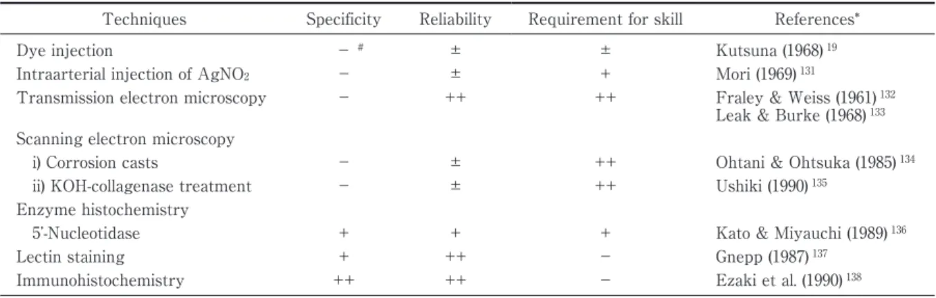
Table 1 Techniques mainly used for the observation of lymphatic vessels in the conventional histological methods.
Techniques Specificity Reliability Requirement for skill References*
Dye injection − # ± ± Kutsuna (1968) 19
Intraarterial injection of AgNO2 − ± + Mori (1969) 131
Transmission electron microscopy − ++ ++ Fraley & Weiss (1961) 132 Leak & Burke (1968) 133 Scanning electron microscopy
i) Corrosion casts
ii) KOH-collagenase treatment
− − ± ± ++ ++
Ohtani & Ohtsuka (1985) 134 Ushiki (1990) 135
Enzyme histochemistry
5 -Nucleotidase + + + Kato & Miyauchi (1989) 136
Lectin staining + ++ − Gnepp (1987) 137
Immunohistochemistry ++ ++ − Ezaki et al. (1990) 138 * Listed only representative references.
# − : negative, ± : weakly positive, +: positive, ++: strongly positive.
( lymph trunks, collecting lymphatics, lymphatic capillaries, lymph sinuses, and lymph sacs)
ii) Supplementary organs for this system include a) lymphoid tissues (lymph nodes, lymph nodules, and lymph invasions) and b) motile organs (propul-sor: lymph heart)
iii) The contents of the system (lymph, chyle) Depending on the species and its level of phyloge-netic differentiation, some of the components listed above may not be present.
1.Structure and functions of the lymphatics
1)Lymphatic capillaries
Lymphatic capillaries are the origins of lymph vessels and begin in blind ends that absorb and drain surrounding interstitial fluid. They are func-tionally the most important part of the lymphatic system and can absorb almost anything: molecules too large to be taken up by the blood vessels, fats (particularly long-chain fatty acids with 12 or more carbon chain length), cells and foreign bodies. De-spite their name, lymphatic capillaries have diame-ters ranging from few to several times more than those of normal blood capillaries (15-75 and 5-10μm, respectively). However, the thin walls of the lym-phatic capillaries and the transparency of the lymph itself make lymphatic capillaries difficult to distinguish from blood capillaries and tissue spaces by direct observation or under a normal micro-scope. This is one of the reasons for the delayed re-search on lymphatic system as compared to vascu-lar system. Moreover, it becomes exceedingly
diffi-cult for an inexperienced individual to identify lymph vessels (Table 1). In recent years, however, specific markers and genes expressed in the lym-phatic endothelium3
as well as specific lymphatic growth factors have been discovered, and then lym-phatic research has undergone great progress, nearly reaching up to the level of vascular re-search.4
Endothelial cells of the lymphatic capillaries are flattened except for their nuclei, which protrude into the lumen with slightly higher abundance of cytoplasm and organelles. Unlike blood capillaries, lymphatic capillaries show extremely poor develop-ment of basedevelop-ment membrane, and lack both a con-tinuous basement membrane and adventitial cells. This makes them suitable for the absorption of in-terstitial fluid, particularly material particulates. When not performing their absorptive function, neighboring endothelial cells shorten the narrow gaps between themselves and overlap with each other. These cells are not bound by tight junctions. Once absorption begins, the spaces between these cells widen to varying degrees. Material particu-lates such as carbon particles can be taken up, and during periods of intense absorption, these gaps can widen enough to accept cells such as erythrocytes. During intense absorption,5
lymphatic endothelial cells change their shape: stretching, shrinking, flat-tening, thickening, and sometimes even bending. The intense absorption is either caused spontane-ously or by such active deformation of the endothe-lial cells. But here is the mystery. What has brought
about these morphological and functional changes between the absorbing and non-absorbing states ? Inside these endothelial cells, superfine cytoplasmic filaments form bundles of various sizes, most of which run parallel to the cells major axis.6
Identify-ing the contractile ability of these filaments may ex-plain this activity. Baluk et al.,7
identified button-like structures unique to the beginnings of lymph ves-sels at the boundaries of lymphatic capillary endo-thelial cells. According to their study, interstitial fluid can be freely absorbed in the space inside these structures. Additionally, lymphatic endothe-lial cells contain numerous pinocytic vesicles, and interstitial fluid is absorbed via encapsulation into these vesicles. Fat droplets containing chylomi-crons (0.1-0.3μm) are absorbed only by lymph ves-sels located at the center of intestinal villi.8
Again, the process occurs via the separation of endothelial cells and uptake of pinocytotic vesicles.9
Uptake by pinocytotic vesicles is a basic form of phagocytosis that has existed since the era of unicellular life. Fur-ther, bundles of cellular fibers emerge from the ba-sal sides of lymph capillary endothelial cells, which serve to connect the cells to the surrounding con-nective tissue and are known as anchoring fila-ments. These filaments are thought to help prevent the lymphatic lumen from collapsing when tissue pressure increases. Nerve endings contact endothe-lial cells of the lymph vessels central to the villi, and some studies10
show that by stimulating smooth muscle aligned with the major axis of the central lymph vessel, neuropeptides from these nerve end-ings regulate absorption and transport of the lym-phatics.
2)Collecting lymphatics
Like blood vessels, collecting lymphatics are thor-oughly covered with a complete basement mem-brane and contain valves in their lumen and smooth muscles in their walls. Special characteristics of these muscle layers include the lack of a clear boundary between the tunica intima and tunica me-dia (muscular layer) because of their frequent con-nection with the sub-endothelium, an abundance of connective tissue between the fibers, diagonal and spiral arrangements, and a mixture of left- and
right-wrapping elements. Nevertheless, in lymph vessels of the lower limbs, these fibers are clearly separated into three layers: the inner longitudinal layer, middle circular layer, and outer longitudinal layer. Lymph vessels of the upper limbs have only two layers and lack the outer longitudinal layer. Muscle fiber development of lymph vessels of the head and the neck are quite poor, and many either only have weak diagonal longitudinal fibers or com-pletely lack muscle fibers altogether. Lymph ves-sels from internal organs lack muscle fibers when they run through organs, but away from organs, nearly diagonal longitudinal fibers and occasionally nearly diagonal circular fibers are detected. Most lymph vessels are hence not just simply cylindrical. Instead, areas between valves have better-developed musculature and thicker walls, while ar-eas near the valves are thin-walled, and show either very poor musculature or lack fibers entirely. Thus, the portion of a vessel between two valves has been traditionally regarded as a single unit, called the lymphangion.11
The spontaneous contractions of lymph vessels12-14
occur unit by unit in these lym-phangia to carry lymph towards the trunk of the body. Increased lymph volume and intralymphatic pressure are thought to cause spontaneous contrac-tion. Additionally, the smooth musculature runs longer than one lymphangion node.15
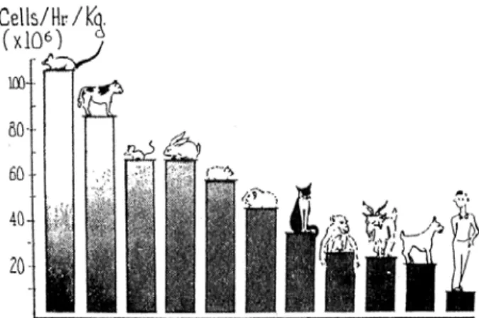
Lymph vessels of the mesentery are often used to observe sponta-neous lymphatic contraction. In cows, sheep, guinea pigs, rats, and mice, contractions occur at a high fre-quency, while in cats, dogs, and rabbits, they occur at a much lesser frequency. All animals with low contractile frequency exhibit poor development of lymphatic vessel muscle fibers. Spontaneous lym-phatic contraction has also been studied in humans; the vessels in the lower limbs contract approxi-mately 4-5 times per minute, while those of the tho-racic duct contract 1-4 times per minute. When col-lecting lymph from the thoracic duct through can-nulation, if either respiration or peristalsis stops, lymph flow will also stop. At such times, a light per-cussion of the abdomen can help resume lymph flow. Additionally, when cannulating lymph ducts in the limbs, it is nearly impossible to collect lymph if
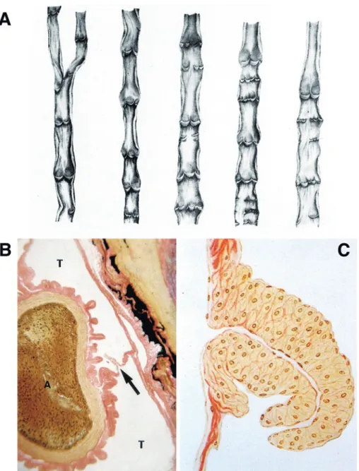
Figure 1 Lymphatic valves in various species.
A: Various types of valves in human lymphatic vessels of the lower extremities. Most of human lymphatic valves are semilunar, each consisting of 2 valves. (from Ogo, 1933) 129 B: The thoracic duct (T) enveloping the aorta (A) of a snake. A valve equipped with smooth muscle fibers is indicated by an arrow. Van Gieson staining. (from Kotani, 1959) 17 C: A craterform valve with smooth muscles in the septum between subcutaneous lymph sacs of toad. Van Gieson staining. (from Kihara and Nose, 1931) 130
the patient is under anesthesia, but a light massage of the legs or fingers can resume lymph flow. These observations indicate that lymph circulation is pre-dominantly passive, and massage and muscular contractions in the surrounding area are important for lymph flow.
In the collecting lymphatics, multiple valves are present and occur much more frequently compared to valves in the veins. These valves are particularly
numerous in the subcutaneous vessels of the limbs and trunk, and in some places are present every 1-2 mm apart. These valves are normally bicuspid semilunar valves (Figure 1A). In the shallow envi-ronment of the lymph sacs, the valves can be crescent-shaped. Unicuspid, tricuspid and tetracus-pid valves are the ones occasionally observed. These valves are composed of folds of lining mem-branes; lymphatic endothelium covers both sides of
a thin flap of connective tissue membrane. Fibers radiate outward from valve attachment points and interlace at the center of the valve cusps. The valves of lymph vessels are well-developed, ensur-ing that lymph flows uni-directionally without any backflow. As a result, the functional range of lym-phatic visualization at the extremities following in-jection of dye or contrast agent is, by definition, lim-ited. However, if lymph vessels or nodes become ob-structed, lymph congestion occurs, causing the lym-phatics to swell. This swelling eventually prevents lymphatic valves from closing completely. If the swelling spreads to the peripheral circulation, lymph backflow can occur. Retrograde metastasis is unlikely, but it could occur if lymph vessels or nodes become blocked by cancerous tissues be-cause of incomplete valve closure.
Some aspects of the muscular layer of lymph ves-sels and valves are a mystery. First, looking at the muscular layer, only birds and mammals have smooth musculature in the walls of their lymphat-ics, while reptiles, amphibians, and fish do not. Al-though the lymphatics of snakes, turtles, frogs, and toads are very well-developed, and contain large lymph sacs and sinuses, the walls of these struc-tures contain no musculature. Next, looking at the valves, the valves of birds and mammals are semilu-nar, while those in the barrier walls of large lymph sacs and wide lymph sinuses of reptiles and anuran amphibians are funnel-shaped. Further, the semilu-nar valves of birds and mammals are not muscled, but in snakes and toads, their lymphatic funnel valves are the only part of the system in which smooth muscle is found (Figure 1B & C). But why is that? Are these differences due to the sudden nar-rowing of lymph ducts in birds? Or are they due to differences in the strength of lymph flow?
The skin of anuran amphibians like frogs and toads can be grasped and pulled far away from their bodies16
because the space underneath their skin functions as a large lymph sac. The area of this sac corresponds to the subcutaneous tissues in a mammal. Below this and between their organs, there is a large lymph sinus, through which arteries and veins run. Reptiles like snakes and turtles also
have large lymph sinuses through which arteries and veins run (Figure 1B). In fact, the Japanese rat snake, Elaphe climacophora, not only has this type of vasculature, but some organs, such as the thyroid, thymus and liver, are also present within the lymph cavities.17
These animals which cannot produce their own heat are referred to as cold-blooded; not because their blood is cold, but because their heat is derived from external sources. They must use heat from the sun to regulate their body temperature. The lymph sinuses and sacs, filled with lymph, which is a poor conductor of heat, are thought to be necessary for these organisms to maintain their body heat. Lymph is also thought to be useful for water storage. In hot regions of Australia, there is a type of frog that stores a large amount of water in the lymph sac beneath its skin, burrows into the soil, and estivates. This water-holding frog is said to utilize this behavior to avoid heat.18
In birds, how-ever, which can produce their own heat, the lym-phatics are very thin, but their heat production is still not sufficient to ensure survival. Thus, their bodies are covered in feathers, and some or all of the blood vessels in many species are surrounded by lymphatics. The lymphatics of mammals are quite thin.
In contrast, caudate amphibians, which belong to the same class as those discussed above, have a completely different configuration. Although they spend almost all their time in the water, caudates such as newts and salamanders have a very well-developed but nevertheless thin, vessel-like lym-phatics. Even among frogs, completely aquatic or-ganisms such as tadpoles have thin vessel-like lym-phatics. As they begin to emerge onto land, the lymphatics fuse together to form one large lymph sac. Ostariophysi such as carp and crucian carp, also have well-developed vascular lymphatics. In general, the configuration of an organism s lymphat-ics changes based on whether it is homeothermic or poikilothermic, and whether it lives in water or on land. Here, too, is another mystery. There is little variation in the blood circulatory systems of these animals compared to that of their lymphatics.
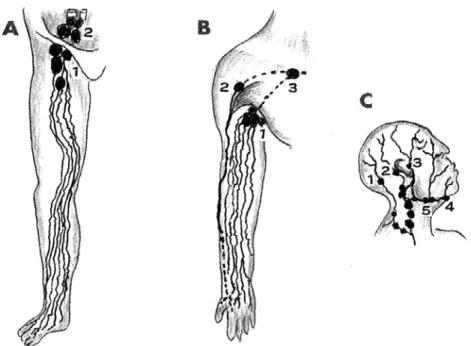
Figure 2 Illustrations of main lymphatic flows in the extremities, head and neck. A: Lower extremity; superficial inguinal lymph nodes (A1), inferior iliac lymph nodes (A2). B: Upper extremity; central axillary lymph nodes (B1), interdeltoidpectoral lymph nodes (B2), subclavian lymph nodes (B3).
C: Head and neck; occipital lymph nodes (C1), postauricular lymph nodes (C2), superficial auricular lymph nodes (C3), submental lymph nodes (C4), submandibular lymph nodes (C5). (from Kotani, 2012) 1
3 ) Lymph trunks : lymph trunk and lymphatic duct
Longitudinal smooth muscle fibers often form in the tunica intima of the thoracic duct, the largest lymph trunk inside the body ( on the left side ) .16 These fibers become more developed further down the length of the duct. The tunica media is made up of inner longitudinal, middle circular, and outer lon-gitudinal layer, all of which have a near-diagonal ori-entation. The development of the near-diagonal in-ner longitudinal layer is most advanced. A unique characteristic of this layer is the abundance of con-nective tissues between muscle fibers. Elastic fibers run subintimally and outside of the muscular layer to form a fiber network. The right lymphatic duct and lumbar trunks have walls thinner than the
tho-racic duct but have much more muscle fibers, and three clear layers of musculature can also be seen. The bronchomediastinal and the intestinal lymph trunks lack the outer longitudinal musculature. The jugular and the subclavian lymph trunks have even thinner walls, with only diagonal longitudinal mus-culature. Multiple valves are also present in the tho-racic duct.
2.Route and size of the lymphatics
Since ancient times, the distribution of lymphat-ics throughout the body has been traced by inject-ing ink, dye or other pigmented liquids into the con-nective tissue of a specific region.19* Footnote
Injecting dye between the toes causes up to 10 or so superfi-cial lymphatics to become visible on the dorsum of the foot, enabling clear visualization of these struc-*Footnote: Visualization of lymphatics upon dye injection is possible in humans, animals, and cadavers. It may be even
eas-ier to inject these dyes several hours after death because, by that time, blood in the vasculature will have clotted and the dye will be injected selectively into the lymphatics. There are no red blood cells or platelets in the lymph, making it virtu-ally incapable of clotting. Another method involves direct injection of dye into the lymph nodes to visualize the lymphatics and nodes downstream of the injection site, closer to the trunk of the body. This method is still widely used today as a clini-cal diagnosis technique, as lymphangiography.
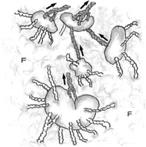
Figure 3 An illustration of human superficial inguinal lymph nodes.
Both afferent and efferent lymphatic system in lymph nodes are present. In general, several afferent lymphatic vessels enter a lymph node; however, efferent lymphatic vessels (gray) from the node, even if two or three efferent vessels initially leave the node, soon converge and leave as only one with unidirectional lymph flow (arrows). Thus, although there may be several afferent vessels, there is eventually never more than one efferent lymphatic vessel from the node. Note all lymph nodes are located in the adi-pose tissue (F). (with modification from Kotani, 2012) 1
tures as they ascend the lower leg ( Figure 2 A ) . They pass the knee, rapidly ascend the thigh, and flow into the superficial inguinal nodes.19
Interest-ingly, the superficial lymphatics do not follow the same course as the blood vessels in the area (the two or three superficial lymphatics that originate in the heel enter the superficial nodes of the popliteal fossa). Lymphatics that exit the superficial inguinal nodes become deep lymphatics, which then pass through the lower iliac lymph nodes along the ex-ternal iliac artery, through the upper iliac lymph nodes along the common iliac artery, and through the paraaortic lymph nodes along the abdominal aorta. They form the left and right lumbar lymph trunks and ultimately empty into the cisterna chyli. From here, the thoracic duct begins. The intestinal lymph trunk, from the intestinal lymphatics, also flows into the cisterna chyli. When dye is injected between the fingers, up to 10 or so lymphatics ap-pear on the dorsal and palmar surfaces of the hand
and ascend the forearm (Figure 2B). They pass the elbow, rapidly ascend the upper arm, and after passing either through the deltopectoral nodes or central axillary node, they flow into the subclavian lymph node. Here too, they do not follow the course of the blood vessels ( the two or three superficial lymphatics that originate on the ulnar side of the palm enter the cubital lymph nodes). After exiting the subclavian nodes, the lymphatics become the subclavian trunk. On the right, they converge with the cervical lymph trunk and then empty into the right angulus venosus, while on the left, they con-verge with the cervical trunk and thoracic duct and empty into the left angulus venosus. The approxi-mately 10 superficial lymphatics that become vis-ible after injecting the scalp or center of the face with dye trace the following paths. From the head, the lymphatics pass through the occipital nodes, posterior auricular nodes and superficial parotid nodes; from the center of the face, the lymphatics pass through the superficial parotid nodes, submen-tal nodes, and submandibular nodes (Figure 2 C ) . Both sets of lymphatics then flow into the superior and inferior deep cervical lymph nodes, which run along the common carotid artery and internal jugu-lar vein. The efferent lymph vessels of the inferior deep cervical nodes form the cervical lymph trunk. On the right, these vessels then converge with the right subclavian lymph trunk, and empty into the right angulus venosus (venous angle), while on the left, they converge with the left subclavian trunk and the thoracic duct, and empty into the left angu-lus venosus.
Here is another mystery. There is little to no change in the size of the lymphatics at their origins in the periphery compared to those in the trunk of the body.20
Even when superficial lymphatics be-come deep lymphatics as well as when deep lym-phatics become lymph trunks or ducts, there is al-most no change in their size. The diameter of even the largest lymph trunks of the body, such as the thoracic duct, does not exceed a few millimeters. This consistency is not found in the blood vascula-ture. Like a large tree, the thin branches of the veins converge and gradually fuse into one large,
thick vein. In contrast, the arteries become thinner and narrower with each division. According to Kutsuna,19
the lymphatics not only function to ab-sorb and drain lymph from the body, but also leak lymph partway through their route to outside the vessels. The leaked lymph spreads into the sur-rounding tissue via the paralymphovascular fluid pathway.20
The arrangement of the afferent and efferent lymphatics of a lymph node also raises some ques-tions (Figure 3). Normally, several afferent lymph vessels enter a lymph node ; however, if two or three efferent vessels initially leave the node, these lymphatics soon converge, leaving only one efferent lymphatic from the node. Even when several nearby lymph nodes form a group, the same phe-nomenon occurs. Thus, although there may be sev-eral afferent vessels, there is eventually never more than one efferent lymphatic vessel from the node. Additionally, there is essentially no difference in the size of afferent and efferent vessels. It may be possi-ble to explain this mystery to a certain extent. By temporarily slowing and congesting the flow of lymph from afferent lymphatics into the lymph si-nus of the node, antigens, and cells carried by the lymph are better able to diffuse into the paren-chyma of the node, allowing lymph nodes to func-tion as a lymph flow checkpoint and contribute to biological defense mechanisms.
A further mystery lies in the relationship be-tween lymph vessels, nodes, and adipose tissue ( Figure 3 ) . A special characteristic of superficial lymphatics is that they do not follow blood vessels but follow individual courses. The superficial lym-phatics from the dorsa of the foot and hand and those of the abdominal wall travel, without excep-tion, through subcutaneous adipose tissues above the superficial fascia towards their associated lymph nodes. Nearly all associated lymph nodes are embedded in adipose tissue, and lymph vessels and nodes have a very close relationship with adipose tissue (as explained in Part4). These factors help preserve the functions of lymph vessels and nodes through the flexibility and low heat conductivity of adipose tissue, which not only protect these
struc-tures from external pressures but also maintain their thermostability.
3 . Lymphatic distribution and the flow of body fluids
1)Basic configuration of lymphatic distribution Lymphatics are not distributed throughout the entire body. Epithelial tissues neither have blood nor lymph vessels. However, the cells of these tis-sues carry out critical functions and must divide rapidly, and thus cannot afford to be starved of oxy-gen and nutrients. Because of this, blood capillaries are distributed immediately beneath the basement membranes of the epithelia. These capillaries form a dense network in the papilla of the outer surface of the dermis in the skin and outer surface of the lamina propria of the mucosa. The lymph capillary network is situated slightly apart from this blood vascular network and is distributed on a deeper layer. This allows it to perform its original function of absorbing fluids that permeate outside the blood vessels. This is why the lymphatics were tradition-ally known as absorbing vessels. Lymphatics are distributed in the interlobular connective tissue and capsules of parenchymal organs such as the liver, pancreas, and salivary glands, but not within the lobules themselves. The lobules of the lungs also lack lymphatic capillaries. Here is another mystery. Why do lymphatics not enter the lobules of these parenchymal organs? Blood vessels are abundantly distributed throughout this area. Do lymphatics fol-low a principle that they must be distributed away from the epithelia and the blood capillaries ? Per-haps this arrangement allows for maximal use of limited intra-organ space for bile, pancreatic juice, and saliva secretory ducts or gas-exchanging alve-oli.
The exceptions among parenchymal organs are the gonads (ovaries and testes) and thyroid, where lymphatics are present. However, even in these or-gans the same principle is observed in which blood capillaries are distributed close to the glandular epi-thelium, whereas lymphatic capillaries are distrib-uted away from both. In the ovaries, blood capillar-ies are densely distributed in the theca interna, the membrane covering an ovarian follicle, while
lym-phatic capillaries are distributed in the theca ex-terna, the outer layer. In the corpus luteum, larger lymph vessels penetrate alongside blood vessels into the center of the structure, but unlike the blood capillaries, they are not finely distributed all the way up to the luteal cells themselves. In the testes, blood capillaries envelop each seminiferous tubule, while lymphatic capillaries are distributed away from these tubules in the spaces where connective tissue is relatively abundant. Additionally, in the thyroid, blood capillaries form dense networks that envelop follicular cells, while lymphatic capillaries are distributed away from these cells in the spaces between where connective tissue is abundant. As a result, testosterone secreted by the testes and, thy-roxine (T4) and triiodothyronine (T3) secreted by the thyroid are released not only into blood vessels but also into the lymphatics.21,22
However, a compari-son of blood flow volume to lymph flow volume re-veals that the blood vasculature is the primary route through which these hormones are released into the body. However, an unexpected result was obtained in a study of thyroid lymph collection from dogs. In thyroid follicular cells, thyroglobulin ( 650 kDa) is the primary component of the intracellular follicular colloid. Normally, thyroglobulin does not exit the follicle. If this does occur, thyroglobulin is recognized as an antigen, despite being an endoge-nous protein, and causes autoimmune disease (Hashimoto s disease). However, when cannulating the draining lymphatics of a dog s thyroid, light per-cutaneous massage of the thyroid easily causes thy-roglobulin to appear in the cannulated lymph.23
Ad-ministering thyroid stimulating hormone (TSH) fur-ther increases its levels. When a high-molecular weight substance such as thyroglobulin leaves a fol-licular cell, only lymphatics can absorb it. It is now known that thyroglobulin can easily be released through a lymphatic route into the general circula-tion. This finding may provide new perspectives on the cause of Hashimoto s disease.
So, what about tissues other than epithelial tis-sue? If we look at lymphatic distribution in the mus-cular tissue, blood capillaries tightly encircle each muscle fiber and lymphatic capillaries are absent.
Lymphatic capillaries are distributed in the connec-tive tissue sheaths that bundle together many mus-cle fibers (perimysium) and in the connective tissue membrane that surrounds the entire muscle (epimysium, also known as fascia). In adipose tissue, blood capillaries tightly encircle each adipocyte, but lymphatic capillaries are absent. Lymphatic capil-laries only begin to appear in the thick connective tissue sheaths that separate groups of adipocytes into many-celled lobular structures. In terms of the lymphatic distribution in bone tissue, blood capillar-ies are rather abundant in Haversian canals, while lymphatic capillaries are absent. Marrow also con-tains an abundance of blood capillaries but lacks lymphatic capillaries. Lymphatics are instead dis-tributed in the periosteum, the connective tissue membrane that covers bones. This may be because in metabolically active tissues such as muscle, adi-pose, and bone tissues, in addition to an abundance of blood capillaries, the reticular fiber network that facilitates the circulation of interstitial fluid is thickly distributed. Thus, even without abundant lymphatic distribution, these tissues receive ade-quate nutrients and the interstitial fluid can ensure proper circulation. Unlike the examples described above, the blood vessels and lymphatics do not penetrate cartilage tissue. Thus, in cartilage tissue, both blood and lymph vessels are distributed only in the perichondrium.
2)Arteries and the heart
There are surprising differences in the distribu-tion of lymphatics in the arteries and heart. Large arteries branch off occasionally to form smaller ar-teries to supply themselves, whereas large veins re-ceive their nutritional supply from neighboring ar-teries. Vessels that supply other vessels in this way are collectively known as the vasa vasorum . The vasa vasorum splits into many small branches in the tunica externa of arteries, some of which move towards the tunica media and form a capillary net-work between the smooth muscle fibers. The capil-lary network becomes sparser along the tunica me-dia and ends before reaching the tunica intima. The tunica intima does not contain blood vessels and consequently the tunica intima and part of the
tu-nica media close to the intima can only be supplied by fluids leaked from the arterial endothelium and capillary network of the tunica media. The elastic lamina, located at the border of the tunica intima and tunica media, is fenestrated and therefore aids the permeation of interstitial fluid. Lymphatics are distributed in the deep parts of the tunica externa of large arteries, but do not reach the tunica media or tunica intima. Thus, there is no way to eliminate waste products from the tunica intima and intimal region of the tunica media. Particularly when hy-pertension damages arterial endothelial cells and when this is accompanied by hyperlipidemia, lipids seep into the tunica intima. Cholesterol deposits form, but they cannot be removed due to the lack of lymphatics in these areas. If lymphatics specialized for the removal of high-molecular weight lipids were distributed in the inner layers of the arteries, arteriosclerosis might never occur or become a critical condition.
However, here is another mystery. Unlike in the arteries, lymphatics are well-developed in the heart.24-26
Lymphatic capillaries are distributed im-mediately beneath the endocardium and at the bor-der with the myocardium. The draining lymphatics that run off the lymphatic network of the endocar-dium travel through the thick bundles of connec-tive tissue that pass through the myocardium and connect to the lymphatic network of the epicar-dium. There are no lymphatics inside bundles of cardiac muscle fiber, but lymphatic capillaries do exist inside the connective tissue between bundles and drain to the lymphatic network of the epicar-dium. In the epicardium, two networks of lymphat-ics are present: deep and superficial. The deep lym-phatic network of the epicardium receives draining lymphatics from the myocardium and then ascends towards the coronary sulcus.19
The lymph vessels accompanying the right coronary artery ascend the front face of the ascending aorta, front face of the pulmonary artery, or between the two, and, in hu-mans, empty into the aortic arch lymph nodes at the upper anterior aortic arch. The lymph vessels accompanying the left coronary artery ascend along the back of the pulmonary trunk following its
long axis and empty into upper right tracheobron-chial lymph nodes. Lymphatic capillaries also form a network in the superficial layer of the epicardium. Experimentally injecting dye into the pericardial space of rabbits and other organisms enables visu-alization of this network.27,28
Lymphatics are also ob-served in the connective tissue between special myocardial fibers of the impulse conducting system such as the sinus node and atrioventricular node. Both the arteries and the heart were originally one tubular structure. However, the heart twisted and turned upon itself, developed contractility, and was eventually enveloped by a serosal cavity. Then, why are the lymphatics of the heart so well-developed? Even within the same pericardial space, the heart-supplying coronary arteries function as any other artery: their tunica intima do not possess blood or lymphatic capillaries. Thus, this high level of lymphatic development in the heart is very mys-terious indeed!
3)Extravascular fluid pathways
Upon injection of dye into subcutaneous tissue or serosal cavities such as the peritoneum or the pleura, before the dye enters the lumens of the vari-ous lymphatics, it has been noted that the dyed fluid flows more easily in some areas compared to others. Kihara20
found that these areas have special gaps in the connective tissue that are abundant in reticular fibers, which not only allow bodily fluids to flow continuously, but also contain pathways for cells and other material particulates to pass through. He named these pathways as
extravascu-lar fluid pathways ( or, the extravascuextravascu-lar
fluid-path-system).
There are three pathways as follows. (1) the pre-lymphovascular fluid pathway : the inlet pathway that brings fluid from serosal cavities to lymphatic capillaries; (2) the paralymphovascular fluid path-way: the drainage pathway that accommodates ex-tralymphatic leakage; and (3) the paravenular fluid pathway : the absorbing pathway situated around the venules. Normally, these fluid pathways receive little attention, but in various disease states closely related to the vascular system, such as pleural, peri-toneal, or other localized edemas, inflammatory
re-sponses, and even tumor metastasis, these struc-tures may play very critical roles.
4)Lymphatics and serosal cavities (prelymphatic spaces)
Dye injected into the peritoneal cavity is quickly absorbed by lymphatic capillaries of the diaphragm tendon.5,16,29
As the mesothelial cells of the perito-neum open underneath, the shape of the lymphatic endothelium changes drastically along the fiber bundles of the peritoneum; forming large canal-like passageways between the two structures. These passageways are characteristic of the prelympho-vascular fluid pathway. They form a connective tis-sue absorption route from the serosal cavities (pleura and peritoneum) to the lymphatic capillar-ies, and are known as the macula cribriformis.30
Ab-sorption is carried out not only by the formation of passageways through the separative transforma-tion of lymphatic endothelia, but also through up-take by pinocytotic vesicles composed of lymphatic endothelial cells. Carbon dye (Pelican inkⓇ
: Günther Wagner, Hanover) injected into the pleural cavity is quickly absorbed by the lymphatic capillaries of the costal pleura. Canal-like passageways are formed between peritoneal mesothelial cells and lymphatic endothelial cells. Carbon dye injected into the peri-cardial cavity is quickly absorbed by the lymphatic capillaries of the visceral pericardium. It is well-known that in the central nervous system,16,31
dye in-jected into the cerebrospinal meninges follows the olfactory nerve and leaves the cranial cavity into the nasal mucosa, where it is absorbed by lym-phatic capillaries. Later, one of Kihara s students16 demonstrated that carbon dye is absorbed by lym-phatic capillaries distributed at all sites at which cranial and spinal nerves exit the cranial cavity or intervertebral foramina. In the peripheral nerves, the carbon dye appears in the lymphatics of the epineurium. The subepineural space continues into the subdural space and the intraneural space con-tinues into the subarachnoid space. Thus, carbon dye travels through the intraneural space to the subepineural space and finally appears in the lym-phatics of the epineurium. Here is another mystery. Serosal cavities are sealed spaces covered in their
own mesothelia. Similarly, lymphatics are sealed spaces lined with endothelia. Nevertheless, dye in-jected into serosal cavities does not appear from the entire serosa, instead it quickly appears in the lym-phatic capillaries from specific locations. Thus, all serosal cavities, including the peritoneal cavity, pleural cavity, pericardial cavity, and meningeal cavity could be referred to as prelymphatic spaces or lymphatic pre-cavities.
In frogs and toads, countless small, crater-like holes are present in the barrier wall between the pleuroperitoneal cavity and the structure corre-sponding to the mammalian thoracic duct, the sub-vertebral sinus, and dye injected into the pleurop-eritoneal cavity passes through these holes before emptying into the lymphatic sinus.16
Experimental phylogenetic verification of the presence of this or similar structures in fish, caudate amphibians, rep-tiles, and birds is needed. Phylogenetic research into lymphatic absorption from the pericardial and meningeal spaces should also be conducted.
When carbon dye is injected into the abdominal cavity of a rabbit, the lymphatics of the diaphragm are clearly rendered in black. However, many black spots also appear on the omentum, which are known as milky spots32
and are congregations of a large number of macrophages that phagocytosed the carbon. Endothelial cells of the lymphatic capil-laries that begin here join the peritoneal mesothe-lium and appear to participate in the absorption of dye at the milky spots. Further, these lymphatics pass through the omentum and empty into the nodes along the greater curvature of the stomach. Omental milky spots have been known for a long time as the site of peritoneal macrophage produc-tion and macrophages that escape into the perito-neal cavity take up the dye and enter the lymphat-ics of the diaphragm. However, some macrophages may return to the omental milky spots. Many other questions regarding the relationship between sero-sal cavities and the lymphatics remain unanswered. Even when focusing on the peritoneal cavity a large number of issues remain a mystery, including re-tention of peritoneal fluid due to peritonitis or cir-rhosis, transperitoneal dialysis, and impeded
perito-Figure 4 The thoracic ducts in various species.
A: Carp; the aorta (A1), posterior cardinal vein (A2), propulsor (A3).
B: Duck; the aorta (B1), cervico-pectoral lymph nodes (B2), lumbar lymph nodes (B3). C: Human; the aorta (C1), cisterna chyli (C2). *: The anglus venosus and thoracic duct-associated Virchow s lymph nodes.
(with modification from Kotani, 2012) 1
neal lymphatic absorption due to radiation expo-sure.
Part 2: Development, Differentiation, Regeneration and Pathologies
of the Lymphatic System 1.Lymphatic system development
1)Phylogenetics of the lymphatics
Next, we will describe differentiation of the lym-phatic system from a phylogenetic perspective.16,33 First, the lamprey, a cyclostome, has a cavity on the ventral side of the aorta and posterior cardinal vein known as the subspinal sinus, which opens into the posterior cardinal vein through a structure with nu-merous small holes, each with a funnel-shaped valve. Based on its location, this sinus could be ex-pected to correspond to the mammalian thoracic duct, but the intestine of the lamprey lacks the cen-tral lacteal present in mammals. Instead, chyle is absorbed by the veins of the intestinal mucosa, and as these veins empty into the subspinal sinus, it would be more apt to refer to this structure as a hemolymph vessel20
or hemocoel. In cartilaginous fishes like sharks, a pair of lymphatic trunks
corre-sponding to the thoracic duct extend down the length of the aorta, with one on either side. At the rostral end of the shark, each trunk meets a cervi-cal trunk and empties into either the left or right angulus venosus, while at the caudal end, they empty into the posterior cardinal vein. These struc-tures are not independent lacteals and are compara-ble to hemolymph vessels. The lymphatic system is completely independent of the blood vascular sys-tem only in carp and crucian carp ( osteichthyes ) . These animals have a pair of thoracic ducts
(Fig-ure 4A) that ascends along both sides of the aorta.
Each trunk converges with the lateral lymphatic trunks (Truncus lymphaticus lateralis) that travel along the center of the lateral side of the body wall and opens into the left and right angulus venosus.33 This pair of trunks becomes a single vessel after passing through the anus of the fish, and follows the dorsal side of the aorta, descending as far as the root of the caudal fin, where it converges with the lateral lymphatic trunks (which descend along the body wall) and empties into the posterior cardinal vein. Caudate amphibians like newts and salaman-ders also have a pair of thoracic trunks; they ascend
both sides of the aorta and empty into the left and right angulus venosus; past the anus, they become a single vessel that empties into the posterior cardi-nal vein. Beneath the vertebrae of anuran amphibi-ans like frogs and toads, the thoracic duct forms a large lymph sinus ( subvertebral sinus, Sinus sub-vertebralis). However, this is a fused version of the pair of thoracic ducts that travels along the aorta of these animals during their tadpole stage. The tho-racic ducts of birds like ducks, geese, and chickens begin at the base of the celiac artery and appear to surround the aorta, which then follows up the body (Figure 4B). Near the third or fourth thoracic verte-brae, the duct splits, with each half emptying into either the left or right angulus venosus.33
In humans, the intestinal lymphatic trunk, and left and right lumbar lymphatic trunks converge, typically in front of the second lumbar vertebra, and on the right dorsal side of the celiac artery, they form the cisterna chyli, which can vary in size. The thoracic duct begins at the cisterna chyli, fol-lows the right dorsal surface of the celiac artery up the body, passes through the aortic hiatus of the diaphragm, enters the thoracic cavity, and contin-ues ascending the body between the aorta and azy-gos vein. However, around the fifth thoracic verte-bra, this duct gradually begins to bend to the left, passes behind and emerges on the left side of the esophagus at the third thoracic vertebra, continues to ascend, bends forward in an arch at the seventh cervical vertebra, receives the left cervical lym-phatic trunk and left subclavian lymlym-phatic trunk between the left common carotid artery and left subclavian artery, and then empties into the left an-gulus venosus.
Here is another mystery. Phylogenetically, all ani-mals that lack completely independent lymphatic and vascular systems, such as cartilaginous fishes, and all animals with independent systems, such as osteichthyes, amphibians, reptiles and birds, have a pair of thoracic ducts with one on either side of the aorta, which together ascend rostrally and empty into the left and right angulus venosus. This princi-ple, however, breaks down in humans and many other mammals. In effect, the venous opening of the
right thoracic duct and most of the left thoracic duct disappear (Figure 4C), while most of the right thoracic duct and venous opening of the left duct remain, as does one of the anastomotic branches that once connected the left and right ducts, linking these structures. The right thoracic duct uses the remaining venous opening of the left thoracic duct to empty into the left angulus venosus. Why does the thoracic duct follow this route in mammals ? This raises the question of whether taking this route provides some sort of benefit. The degenera-tion or disappearance of the right venous opening of the right thoracic duct may already be occurring in certain birds.33
Further, in the very early stages of the embryo, humans have both left and right tho-racic ducts, each of which empties into the left or right angulus venosus.33,34
In the venous system, a similar pattern is observed. The venous system first develops as a pair of vessels that is bilaterally symmetrical; however, midway through the proc-ess, regression of a part of the system occurs asym-metrically on one side, and thus the final system takes a bilaterally asymmetric course (the inferior vena cava and its branches, such as the azygos vein, hemiazygos and accessory hemiazygos vein). Per-haps a very similar or even related process can be observed in the lymphatic system.
2)Venous openings and lymph heart
Osteichthyes or bony fishes such as carps and crucian carps have structures at the left and right venous openings of their thoracic ducts called pro-pulsors.33
A similar propulsor is also on the ventral surface of their final vertebra, near the root of the caudal fin, where the movement of this fin sends lymph into the posterior cardinal vein. The propul-sor contains valves. In zebrafish, each body seg-ment contains the intersection of intersegseg-mental lymphatics and intersegmental veins.35,36
In amphibi-ans and reptiles, lymphatic venous openings have lymph hearts.16,33
Caudate amphibians such as newts and salamanders have one pair of lymph hearts per body segment, totaling to approximately 10 pairs of lymph hearts throughout their entire body. In anu-ran amphibians such as frogs and toads, the num-ber of lymph hearts is reduced to merely one
ante-Table 2 Molecular markers of lymphatic and blood vessel endothelial cells.
Marker Species* Location Molecular size
(kDa) Function Representative references# Lymphatic vessel
LYVE-1 H, M, R cell surface 60 hyaluronan receptor Banerji et al. (1999) 139 Prevo et al. (2001) 140 Podoplanin
D2-40
H, M, R cell surface 43 40
adhesion? Breiteneder-Geleff et al. (1999) 141 Kahn et al. (2002) 142
Schacht et al. (2005) 143 VEGFR-3 (Flt4) H, M, R cell surface 170 VEGF-C receptor Kaipainen et al. (1995) 40 Prox-1 H, M, R nuclear 85-95 transcription factor Wigle and Oliver (1999) 144 Foxc-2 H, M, R nuclear 54-63 transcription factor Petrova et al. (2004) 54 D6 H, M, R cell surface 43 chemokine receptor Nibbs et al. (2001) 145 CD73 H, M, R cell surface 69 adhesion, 5 -Nucleotidase Thompson et al. (1989) 146 B27 R cytoplasm ? ? Ezaki et al. (1990) 138 LA102 M cell surface 25-27 mouse-Thy1 ? Ezaki et al. (2006) 110 Blood vessel
CD31 H, M, R cell surface 110-120, 130-140 PECAM-1 Albelda et al. (1991) 147 DeLisser et al. (1993) 148 CD34 H, M, R cell surface 90, 105-120 GlyCAM-2, mucosialin Baumhueter et al. (1994) 149
Pusztaszeri et al. (2006) 150 CD105 H, M, R cell surface 90, 180 TGF-β receptor, endoglin Gougos and Letarte (1990) 151
Duff et al. (2003) 152 MECA-32 M cell surface 50-55, 100-120 diaphragm protein
(mouse PV-1)
Hallmann et al. (1995) 153 Stan (2005) 154
PAL-E H cell surface 60-120 diaphragm protein (human PV-1)
Schlingemann et al. (1985) 155 OX-43 R cell surface 90 ? Robinson et al. (1986) 156 LA5 M cell surface 12-13 ? Ezaki et al. (2006) 110 * H, human; M, mouse; R, rat.
# Only original or representative papers are listed.
rior and one posterior pair. In reptiles such as snakes and lizards, the number is reduced yet again: they have one posterior pair of lymph hearts. Anterior lymph hearts have disappeared in snakes, instead these animals have one large lymphatic lu-men located above the heart, which uses the beat-ing motion of the heart to propel lymph into the veins.16,17
The walls of lymph hearts are composed of striated muscles and are capable of spontaneous contraction ( Supplemental Figure : Attached Im-ages. https://doi.org/10.24488/jtwmu.89.Extra1_E 4). The rice grain-sized lymph heart at the root of a lizard s hind leg contracts approximately 40 times per minute. The posterior lymph heart of a frog contracts approximately 60-80 times per minute in summer; this rate is reduced to 20-30 times per min-ute in winter.33
The inlets and outlets of amphibian and reptile lymph hearts are equipped with smooth muscled valves. Birds temporarily have one pair of posterior lymph hearts, which disappears within one month of hatching.37
To sum up the characteristics of lymphatic ve-nous openings across multiple species, fewer and fewer lymphatic venous openings are observed as we move from fish towards mammals. Ultimately, in humans, there is only one venous opening of the thoracic duct into the left angulus venosus. Why is that? Does this show that lymphatic systems might have gradually differentiated themselves away from vascular systems during evolution?
2.Morphogenesis (ontogeny) of the lymphatics
The lymphatic system is first visible in the hu-man fetus two months after conception.19
Lym-phatic ontogeny has been widely debated for many years. There are two competing theories: the cen-trifugal theory 38
which states that the lymphatics elongate towards the periphery through budding of the venous endothelium, and the centripetal the-ory39
which states that cells in the mesenchymal in-terstitium around veins flatten to form small lu-mens which then connect towards the center of the body, first forming primitive lymphatic sacs and
eventually lymphatic vessels. Linking of the lym-phatics and veins occurs secondarily. There is also a compromise theory , in which the thoracic duct is thought to arise from fetal veins and the rest from the interstitium, or that the cervical lymph sac is venous in origin, while the rest arises from the in-terstitium.
The primary reason that debates surrounding lymphangiogenesis have continued for more than 100 years is that for much of that time, there was no method for rigorously identifying tissues as part of either the lymphatic system or blood vascular sys-tem. Blood vascular biology has progressed greatly since the discovery of specific markers of vascular endothelia. In contrast, it has been more than 20 years since markers specific to lymphatic endothe-lia were discovered, but lymphatic research has still been significantly delayed. Despite VEGFR3 (FLT4) being present specifically in lymphatic endothelia was discovered in 1995,40
other molecular markers are only recently being reported (Table 2). These specific lymphatic markers enable identification of lymphatic vessels, revealing the relatedness of the lymphatics and the blood vessels. In recent years, there have been an increasing number of molecular biology findings41
that provide further support to the centrifugal theory. However, very recently, live imaging analysis of zebrafish revealed evidences42,43 supporting the centripetal theory and provided in-sight into the origin of lymphatic endothelial cells and that the mechanisms controlling their differen-tiation differ in the head and trunk of the organism. However, these theories require further investiga-tion.
With regards to the ontogeny of the lymphatic system, if we consider the development of the lym-phatics in a human embryo from the perspective of the centrifugal theory, we can broadly categorize this process into two subprocesses: lymphvasculo-genesis; the creation of lymph vessels from the bud-ding of veins, and lymphangiogenesis ; the elonga-tion of new lymph vessels from existing vessels. Buttler et al.44
reported venous budding lymphvas-culogenesis and simultaneous peripheral lymphatic formation which recruits macrophage-like cells
de-scended from mesenchymal tissues in the embry-onic stage. Further, Kato and his colleagues45,46
used enzyme histochemistry to detect a process in which primitive lymphatic sac-like structures ( lymphatic islands) often appear in the interstitium of various organs and grow small protuberances which then link and fuse together, gradually forming a valvular lymphatic network. Either way, during formation of new lymphatic vessels in adults, lymphangiogenesis is thought to be the primary process involved. Evi-dence of a formative process similar to vasculogene-sis observed in the vasculature, such as the pres-ence of lymphatic endothelial progenitor cells in pe-ripheral blood, is not well understood at the present time.
3.Lymphatic regeneration
Lymph vessels regenerate while lymph nodes do not, but the reason for this is unclear. Lymphatics are structures suited for the absorption of high-molecular weight substances and cells. Conse-quently, when foreign bodies, including bacteria and antigens invade the body s tissues, they are taken up by the lymphatics. Lymph nodes provide a biological defense mechanism against these invad-ers. Primitive development of lymph nodes followed that of the lymphatics, occurring only after the lym-phatics achieved a certain level of complexity, and only then in specific locations. In the age of primi-tive lymph nodes, two patterns of development oc-curred: nodes of vascular origin developed from the mesenchyme of lymph vessel walls and nodes of sac origin developed from the mesenchyme of lym-phatic sacs, derived from immature lymlym-phatic plex-uses. The deep cervical nodes, aortic nodes. and iliac nodes, among others are the latter sort of sac-derived nodes, whereas the rest of the nodes, such as superficial cervical nodes, submandibular nodes, axillary nodes, popliteal nodes, and inguinal nodes are of the former sort, i.e. vascular-derived nodes19 . In humans, primitive lymph nodes begin to appear in the fetus from two (axillary, inguinal nodes, etc.) to three months (mesenteric lymph nodes, etc.). At the latest, they begin to appear by the end of the third month (popliteal nodes, etc.) and the beginning of the fourth month.47
at which lymph nodes appear and the number in each location is virtually predetermined. Each lymph node or lymph node group is an organ that has been predetermined both in number and loca-tion for biological defense over the long course of mammalian evolution, in a similar way as the spleen. Like the spleen, lymph nodes do not regen-erate when removed, and this is due to the same mechanism.
However, the lymphatics can regenerate. If an entire lymph node or a group of nodes are removed, new lymphatics eventually begin to grow and join the severed ends of the existing afferent and effer-ent lymphatics. It is thought that increased intra-lymphatic pressure caused by ligation-induced lymph congestion triggers lymphatic regeneration. It is also possible that the tissue gaps at the excised portions link and form a coupled passageway. This line of thinking is quite similar to that of the cen-tripetal theory16,33
of lymphatic ontogeny. Experi-mentally ligating the lymphatics causes acute edema in areas drained by these vessels, affecting tissue and organ function. If the cervical lymph trunk which drains from the lymph vessels of the thymus, is ligated in guinea pigs, the cell division of cortical thymocytes is reduced by 50% within one day and medullary thymocyte division is reduced by 50% within five days.48
Following lymphatic re-generation, the cell division recovers after approxi-mately 10 days. Ligating the draining lymphatics of the testes in rabbits causes edema in the interstitial tissue of the testes, severely affecting sperm pro-duction in the convoluted seminiferous tubules.22
Se-cretion of androgens is also completely halted. The lymphatics regenerate within 12-15 days after liga-tion, and organ function returns to normal after 30-60 days. However, a point that requires clarification here, is that out of the 10 cases studied, lymphatic regeneration failed completely in 2-3 cases. In these rabbits, sperm production did not recover even af-ter 90 days, resulting in widening of the basement membrane of the convoluted seminiferous tubules, and residual edema and fibrosis in the interstitium. Next, when the thoracic ducts of these rabbits were ligated, their extremities became extremely swollen
and their lymph was completely congested. After 10-14 days, a very thin lymph vessel had formed ex-tending from the extremities towards the animals core. The regeneration of this single thin lymph vessel prolonged the life span of these rabbits. Clini-cally, when performing surgery on esophageal can-cer, etc., if the surgeon accidently injures the tho-racic duct or performs some other action capable of causing chylothorax, it is essential that the thoracic duct be ligated immediately. However, it must be reiterated, that in this experiment, 1 to 2 of the 10 rabbits where the thoracic ducts were ligated failed to show any regeneration. This is also a mystery. Why did a small number of rabbits fail to show lym-phatic regeneration in both experiments, involving ligation of the draining lymphatics of the testes and the thoracic duct ? This does not appear to have been caused by errors in experimental technique.
Even if the surgical intervention or external in-jury result in severance of lymphatics, the resulting condition is never fatal, and because the lymphatics are transparent, the severance is often not even no-ticed. The exudate is absorbed by the surrounding lymphatics to some degree, albeit at low levels, sub-sequently causing it to accumulate in the area sur-rounding the injury, giving the injured site the fluid-filled appearance. This encourages fiber prolif-eration and granulation tissue formation. Eventu-ally, when new lymphatics are regenerated at the site of the injury, the local edema abates and fibro-sis ceases. When pleurisy causes adhesion of the visceral and the parietal pleura, lymphatics are newly generated at the site of adhesion. Although the lymphatics on the lungs surface flow towards the hilum, those generated at the site of adhesion carry lymph down the chest wall and towards the diaphragm.49
As a result, the intercostal and paraaortic lymph nodes of the chest cavity can be-come stained with black by the dust they absorb from the lungs. Whether lymphatics can link graft and host tissue together is a very important ques-tion related to the graft rejecques-tion response. In both autografts and homografts, the graft and the host are linked by lymphatics 10-14 days post-operation. Antigen information for the graft tissue is carried
Figure 5 Illustrations of typical patients of elephantiasis. A: A woman suffering from elephantiasis in her right leg. B: A typical foot skin lesion caused due to chronic elephantiasis. (from Esmarch and Kulenkampff, 1885) 51
even more quickly than that by host lymphatics to associated lymph nodes. If a rejection response oc-curs, 3-4 days before the grafted tissue succumbs to necrosis, the lymphatics between the graft and the host are destroyed and the two are no longer linked.
If lymph flow is obstructed, lymphatic neogenesis not only enables resumption of flow but also pro-mote the formation of alternate collateral routes. The diseased state most characteristic of this proc-ess is chyluria.50
Infection by the parasite Wuchereria bancrofti can occlude the thoracic duct. In an at-tempt to return the chyle-containing thoracic duct lymph to general circulation, many lymphatic ves-sels extending from the origin of the duct begin to grow retroperitoneally towards the abdominal wall. The urinary tracts, including the kidneys, are along this route, and occasionally certain lymph vessels will terminate in one of these tracts. The mecha-nism for how this occurs is unknown, but the exis-tence of this process demonstrates the amazing ten-dency for formation of lymphatic drainage routes.
4 . Lymphatic pathologies and their manage-ment
When comparing the pathologies of the lymphat-ics to those of the cardiovasculatures, there are gen-erally no severe conditions that are directly life-threatening. For this reason, it is inevitable that clinical awareness of the lymphatics is compara-tively lacking. However, certain severe problems
can be surprisingly frequent if the diseased states progress unchecked.
1)Edema
Lymphedema ( swelling ) is a disease caused by dysfunction due to insufficient formation of lym-phatics or valve failure/blockage in which the tis-sue fluid congests or accumulates in the intersti-tium of the periphery. Acute and chronic types of lymphedema exist.
Acute lymphedema occurs when the axillary or the pelvic nodes are completely excised from a pa-tient such as when a radical surgical operation is performed for cancer. However, this eventually re-solves through lymphatic regeneration or formation of collateral pathways. Why removal of the axillary nodes causes edema of the upper limbs, but re-moval of the pelvic nodes causes less edema of the lower limbs, was unclear for many years. One rea-son suggested is that the pelvic nodes are located in the loose connective tissue of the retroperitoneum, where lymphatic regeneration and formation of col-lateral pathways are quite easily accomplished. If the surgical areas are expanded in the pelvis by such as total hysterectomy, however, serious edema of the lower limb may occur. Anatomically, the pelvic nodes are located along large arteries and veins, therefore the surgery rarely results in vascular invasion. It is presumed that lateral chest and upper limb edema caused by axillary node exci-sion often involves venous edema. Further
anatomic examination reveals that the superficial lymphatics originating from the cephalic vein as-cend the upper arm, enter the deltopectoral groove, and empty into the subclavian lymph nodes via the deltopectoral lymph nodes (Figure 2B). This route has no direct connection with the lymphatics of the breast, and therefore it is speculated that if these lymphatics are not damaged during excision of the axillary nodes, it may be possible to minimize upper limb edema. Even if the subclavian nodes are re-moved, postoperative edema is unlikely to develop if a collateral lymph route is newly generated.
Chronic lymphedema can either be primary or secondary. The causes of secondary edema are well-understood and include infections that cause inflammation of the lymphatics, particularly tricho-phyton (tinea) or filaria infections that cause lymph vessel or node obstruction. Although this condition is now uncommon, filariasis is an endemic disease of the tropics, and is known as elephantiasis in its final stages51
( Figure 5 ) . Problems arise with primary chronic lymphedema of unknown etiology. Up to 10% of these cases are congenital. Most of the re-maining 90% of cases begin in puberty and are known as lymphedema praecox.12,13
Seventy to ninety percent of these cases occur in women. Nor-mally, one lower limb is affected, but in some cases only one forearm or one upper limb may be af-fected. This disease does not involve obstruction of the lymphatics but rather cause convolution or twisting of the lymphatics, and swelling or narrow-ing of drainnarrow-ing lymphatics.12,13
Here is a mystery. Autoimmune diseases52
such as systemic lupus erythematosus ( SLE ) , Hashimoto s disease, Sjögren s syndrome and myasthenia gravis are more common in women and often begin in pu-berty. The patient profiles of these diseases are quite similar (explained further in Part 4) and many patients experience changes in their symptoms during their menstrual cycles. However, this does not imply that primary lymphedema is an autoim-mune disease. Although sex hormones appear to be involved in the pathology of this condition, the symptoms are often far too localized to suggest hor-monal involvement. Recently, VEGFR3 and
FOXC253,54
were reported to be the causative genes of certain congenital lymphedemas such as Milroy s disease and lymphedema-distichiasis ( LD ) syn-drome.
2)Malignant tumors and lymphogenous metas-tasis
Clinically, it has often been said that carcinomas are lymphogenous and sarcomas are hematoge-nous. The mystery here is, why do the metastatic routes for carcinomas and sarcomas differ? Carcino-mas are malignant tumors of epithelial origin, whereas sarcomas are non-epithelial malignant tu-mors. Various mechanisms involving these patholo-gies are poorly understood, including the mecha-nism where tumor cells break off the main site and permeate the surrounding tissues and also the mechanism involving formation of tumor blood ves-sels and lymphatics. Here, we will focus on re-examining and comparing the differences in the dis-tribution of blood and lymph vessels in normal epi-thelial and non-epiepi-thelial tissues.
If the lymphatics are distributed in the same way as blood vessels, tumor cells that invade surround-ing tissues should enter the lymphatics. This is be-cause structurally, lymphatic capillaries lack adven-titia, almost completely lack a basement membrane, and the endothelial cell gaps open very easily. In epithelial tissue, the connective tissue is present be-tween the lymphatics and the blood capillary net-work, and outside of this the lymphatic capillaries form a dense network, regardless of whether the tissue in question is stratified squamous epithelium, monolayer columnar epithelium or glandular tissue. Furthermore, the lymphatic capillaries extend sev-eral times wider than blood capillaries. In contrast, in non-epithelial muscle tissue, lymphatics are only distributed in the connective tissue sheaths that bundle together many muscle fibers or the pe-rimysium that envelops the entire muscle. In adi-pose tissue, lymphatic distribution begins only in the thick connective tissues that separate many adi-pocytes into lobule-like structures. Thus, in epithe-lial tissues, lymphatic capillaries are located far closer to epithelial cells and are far more densely distributed than in non-epithelial tissues. Logically,
organs prone to lymphogenous metastasis such as the thyroid and the ovaries have lymphatics distrib-uted abundantly throughout their parenchyma, as previously explained.
In contrast, when comparing the distribution of blood capillaries in epithelial and non-epithelial tis-sues, epithelial tissues do not contain blood capillar-ies, while in non-epithelial tissues each muscle fiber and each adipocyte is enveloped in an extremely rich supply of blood capillaries. Even in bone tissue where the lymphatics are confined exclusively to the periosteum, blood capillaries are relatively abundantly distributed. While osteosarcomas, which are derived from osteoblasts, have a long path to travel before reaching the lymphatic-containing periosteum, they have an abundance of blood capillaries distributed very close nearby. As compared to older men and women, osteosarcomas occur preferentially in younger individuals, who have blood capillaries more abundantly distributed. Nevertheless, if osteosarcomas develop for long enough, they reach the periosteum and then un-dergo lymphogenous metastasis. Regardless of the tissue in which the tumor occurs, to affect hemato-genous metastasis, the blood vessels must be de-stroyed. This, however, is not true for lymphoge-nous metastasis.
Whether the metastasis is lymphogenous or he-matogenous, each tumor cell type has a different preference for the organ into which they prefer to metastasize, and metastasis therefore occurs in a variety of different organs. Surprisingly, the spleen is almost never the site for metastasized cancer. The spleen functions primarily as a filtration device for the blood. Tumor cells that enter the spleen via the splenic artery may not be permitted to attach themselves and are either passed out of the spleen via the splenic vein or killed by macrophages in the splenic tissue.
In tumor lymph node metastasis, the general con-sensus until recently was that proliferating tumor cells coincidentally enter a nearby, existing lymph vessel and eventually arrive at a lymph node. Addi-tionally, tumor tissues were thought to be very rich in blood vessels but lacked lymphatics55
. However,
there have been reports of cases where tumors spread lymphogenously by expressing and secret-ing VEGF-C56
and VEGF-D57
, thereby inducing lym-phangiogenesis. Further, the correlation between VEGF-C expression and lymphogenous metastasis has been demonstrated in stomach, colon, lung, prostate, thyroid and melanocyte cancers. The lym-phatics generated in and around the tumor have been called tumor lymphatics . Trials with anti-lymphangiogenetic therapies, including administer-ing VEGFR3 antibodies have begun to combat this phenomenon.58
3)Obesity
As we explained previously, fat and lymph ves-sels are inextricably related. However, many details of this relationship remain unknown.2
Many stud-ies59
have reported about the relationship between the blood vessels and the adipocytes, including those detailing the existence of adipokines. Harvey et al.60
was the first to specifically report that obe-sity can be caused by lymphatic dysfunction. In summary, in Prox 1+ / −
mice, lymph leakage from lymph vessels was found to promote adipocyte en-largement, thus causing obesity. Additionally, Nurmi et al.61
showed that even with consumption of a high-fat diet in VEGF-C knockdown mice, the lacteals of the small intestine retract and smooth muscle function in the villi decreases, causing fat absorption to decrease and preventing obesity from developing.
In contrast, if lymphedema becomes chronic, the fat can be irreversibly and locally stored alongside tissue fluids, leading to adipocyte increase and pro-gressive lipedema . If allowed to progress for a long period of time, this can cause fibrosis, tissue hardening, and in its final stages, elephantiasis
(Fig-ure 5). Consequently, controlling fat metabolism will
not only prevent obesity but also preserve sound lymphatic function. Lymphatic exercise62
has been proposed as a method for preventing and improv-ing edema as well as preventimprov-ing lifestyle illnesses such as obesity.
While obesity itself is not a life-threatening condi-tion, as a lifestyle disease it is a serious condition that progresses steadily, escaping early diagnosis.