ORIGINAL INVESTIGATION
Prognostic value of non-alcoholic fatty
liver disease for predicting cardiovascular
events in patients with diabetes mellitus
with suspected coronary artery disease:
a prospective cohort study
Keishi Ichikawa
1, Toru Miyoshi
1*, Kazuhiro Osawa
1,2, Takashi Miki
1, Hironobu Toda
1, Kentaro Ejiri
1,
Masatoki Yoshida
1, Yusuke Nanba
1, Masashi Yoshida
1, Kazufumi Nakamura
1, Hiroshi Morita
3and Hiroshi Ito
1Abstract
Background: Risk stratification of cardiovascular events in patients with type 2 diabetes mellitus (T2DM) has not been established. Coronary artery calcium score (CACS) and non-alcoholic fatty liver disease (NAFLD) are indepen-dently associated with cardiovascular events in T2DM patients. This study examined the incremental prognostic value of NAFLD assessed by non-enhanced computed tomography (CT) in addition to CACS and Framingham risk score (FRS) for cardiovascular events in T2DM patients.
Methods: This prospective pilot study included 529 T2DM outpatients with no history of cardiovascular disease who underwent CACS measurement because of suspected coronary artery disease. NAFLD was defined on CT images as a liver:spleen attenuation ratio < 1.0. Cardiovascular events were defined as cardiovascular death, nonfatal myocardial infarction, late coronary revascularization, nonfatal stroke, or hospitalization for heart failure.
Results: Among 529 patients (61% men, mean age 65 years), NAFLD was identified in 143 (27%). Forty-four cardio-vascular events were documented during a median follow-up of 4.4 years. In multivariate Cox regression analysis, NAFLD, CACS, and FRS were associated with cardiovascular events (hazard ratios and 95% confidence intervals 5.43, 2.82–10.44, p < 0.001; 1.56, 1.32–1.86, p < 0.001; 1.23, 1.08–1.39, p = 0.001, respectively). The global χ2 score for pre-dicting cardiovascular events increased significantly from 27.0 to 49.7 by adding NAFLD to CACS and FRS (p < 0.001). The addition of NAFLD to a model including CACS and FRS significantly increased the C-statistic from 0.71 to 0.80 (p = 0.005). The net reclassification achieved by adding CACS and FRS was 0.551 (p < 0.001).
Conclusions: NAFLD assessed by CT, in addition to CACS and FRS, could be useful for identifying T2DM patients at higher risk of cardiovascular events.
Keywords: Cardiovascular disease, Computed tomography, Coronary artery calcium, Non-alcoholic fatty liver disease, Risk stratification
© The Author(s) 2021. This article is licensed under a Creative Commons Attribution 4.0 International License, which permits use, sharing, adaptation, distribution and reproduction in any medium or format, as long as you give appropriate credit to the original author(s) and the source, provide a link to the Creative Commons licence, and indicate if changes were made. The images or other third party material in this article are included in the article’s Creative Commons licence, unless indicated otherwise in a credit line to the material. If material is not included in the article’s Creative Commons licence and your intended use is not permitted by statutory regulation or exceeds the permitted use, you will need to obtain permission directly from the copyright holder. To view a copy of this licence, visit http://creat iveco mmons .org/licen ses/by/4.0/. The Creative Commons Public Domain Dedication waiver (http://creat iveco mmons .org/publi cdoma in/ zero/1.0/) applies to the data made available in this article, unless otherwise stated in a credit line to the data.
Background
The prevalence of type 2 diabetes mellitus (T2DM) has been increasing rapidly worldwide [1]. T2DM is associated with a two- to four-fold increased risk of
Open Access
*Correspondence: [email protected]
1 Department of Cardiovascular Medicine, Dentistry and Pharmaceutical
Sciences, Okayama University Graduate School of Medicine, 2-5-1 Shikata-cho, Kita-ku, Okayama 700-8558, Japan
cardiovascular events compared with non-T2DM sub-jects [2, 3]. The prevention of cardiovascular events in T2DM patients has thus become a major concern. Although several clinical risk scores for predicting car-diovascular events have been proposed, there is currently no widely used risk stratification for T2DM patients. Pre-vious studies showed that coronary artery calcium score (CACS) determined by coronary computed tomography (CT) provided additional information on cardiovascular events in T2DM patients beyond that provided by the commonly used Framingham risk score (FRS) [4, 5]. The latest American Heart Association and American College of Cardiology (AHA/ACC) guidelines for the primary prevention of atherosclerotic cardiovascular disease allowed the use of CACS in intermediate-risk patients if the risk level was uncertain [6]. Thus, CACS could be a useful factor for determining cardiovascular risk in patients with T2DM.
Growing evidence suggests that non-alcoholic fatty liver disease (NAFLD) is associated with cardiovascu-lar events independently of established cardiovascucardiovascu-lar risk factors [7–9]. NAFLD is a frequent comorbidity of metabolic syndrome and T2DM [10]. Previous stud-ies showed that metabolic syndrome and T2DM might be predictors of vascular damage [11, 12], and complex and bidirectional associations have been demonstrated between NAFLD and metabolic syndrome and T2DM [13]. We recently reported on the prognostic value of NAFLD assessed by non-enhanced CT in patients with suspected coronary artery disease who underwent cor-onary CT angiography [14], highlighting the benefits
of concomitant assessment of liver fat content during the acquisition of coronary CT angiography to detect patients at higher risk of cardiovascular events.
We therefore hypothesized that the assessment of NAFLD using non-enhanced CT, in addition to CACS and FRS, might improve risk stratification for cardiovas-cular events in T2DM patients. We tested this hypoth-esis in a cohort of patients with suspected coronary artery disease who underwent CACS measurement, with the aim of evaluating the additional prognostic value of NAFLD compared with CACS and FRS in T2DM patients.
Methods
Study population and data collection
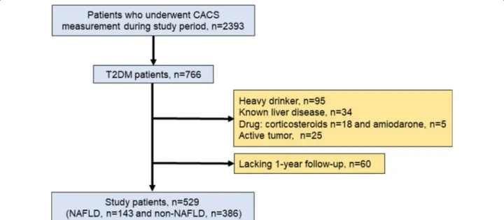
Patient enrollment in this study is shown in Fig. 1. This prospective study enrolled 529 Japanese outpatients with T2DM from August 2011 to December 2016. The patients had no history of cardiovascular disease but had been referred to our hospital with suspected coronary artery disease. CACS was measured in 172 asymptomatic patients for risk stratification by evaluating coronary atherosclerosis, and in the remaining 357 patients with atypical symptoms [n = 44] or no symptoms [n = 313] because of abnormalities in rest or stress electrocardio-grams. No patients had any history of cardiovascular dis-eases, including coronary artery disease, heart failure, or cerebrovascular disease. Patients were excluded if they consumed > 20 g of alcohol per day, had known liver dis-ease, were using oral corticosteroids or amiodarone, or had a coexisting active tumor. All the patients underwent
blood tests, measurement of CACS, and abdominal CT on the same day.
Assessment of coronary calcification
CT imaging was performed using a Somatom Defini-tion Flash scanner (Siemens Medical SoluDefini-tions, Erlan-gen, Germany). CACS was measured using the following parameters: 120 kV, 150 mA, and 3-mm thickness. CACS was calculated using an automated computerized sys-tem (Virtual Place, Raijin; AZE Inc., Tokyo, Japan) and the Agatston method, which involved multiplying the area of each calcified plaque by a density factor deter-mined by the peak pixel intensity within the plaque. The plaque-specific scores for all the slices were added together. The density factor was 1, 2, 3, or 4 for plaques with peak intensities of 130–199, 200–299, 300–399, and ≥ 400 Hounsfield units (HU), respectively [15]. In addi-tion, patients were divided into three groups according to CACS: CACS 0, CACS (1–99), and CACS (≥ 100). Assessment of visceral adipose tissue and NAFLD
Abdominal non-contrast CT scans were carried out alongside cardiac CT, at a level including the liver, spleen, and umbilicus. The visceral adipose tissue area was assessed using a semi-automatic segmentation technique at the umbilical level [16]. Hepatic and splenic Hounsfield attenuations were measured using the maximum circular regions of interest in the liver and spleen (at least 1 cm2) [17]. Regions of interests in the liver were located at two segments (right anterior and right posterior) by avoiding the inclusion of large vessels or biliary structures [17]. The liver:spleen attenuation ratio was calculated using the mean HU measurement of the two right liver lobe regions of interest. In this study, we defined hepatic stea-tosis as a liver:spleen attenuation ratio < 1.0 [18]. NAFLD was finally diagnosed after other causes of hepatic steato-sis were ruled out.
Assessment of other risk factors
Hypertension was defined as a seated blood pres-sure > 140/90 mmHg or current treatment with antihy-pertensive medication. Dyslipidemia was defined as one or more of the following: serum triglycerides ≥ 150 mg/ dL, high-density lipoprotein cholesterol < 40 mg/dL, low-density lipoprotein cholesterol ≥ 140 mg/dL, or current treatment with a lipid-lowering drug. Smoking status was defined as currently smoking or not smoking. Obe-sity was defined as a body mass index ≥ 25 kg/m2. FRS was calculated according to the algorithm presented by Wilson et al. to estimate the 10-year risk of a coronary heart disease event [19]. In addition, patients were classi-fied into three groups according to the European Society
of Cardiology recommendations as very-high, high, or moderate risk [20].
Outcomes and follow‑up
The patients were followed up prospectively from the date of CT. Clinical follow-up information was obtained by review of medical records or telephone interviews by attending physicians. The study endpoint was cardiovas-cular events defined as cardiovascardiovas-cular death, nonfatal myocardial infarction, late coronary revascularization, nonfatal stroke, or hospitalization due to heart failure. The diagnosis of myocardial infarction was based on the criteria of typical acute chest pain and persistent ST-segment elevation or positive cardiac enzymes. Late coronary revascularization was defined as percutaneous coronary intervention or coronary artery bypass grafting as indicated by the treating physician due to stable angina with a newly positive functional test. Patients with sched-uled revascularization within 90 days of the CACS meas-urement were not counted as events and were censored at the time of first revascularization. Hospitalization for heart failure was defined as any unplanned stay overnight or longer in a hospital environment, for which heart failure was the principal reason for admission. Nonfatal stroke was defined as sudden-onset non-convulsive and focal neurological deficit persisting for > 24 h.
Statistical analysis
Continuous variables were expressed as mean ± standard deviation or median with interquartile range. Dichoto-mous variables were expressed as number and percent-age. Differences in continuous variables between two groups were analyzed by Student’s t-test or Mann–Whit-ney U-test, as appropriate. Categorical data were com-pared by χ2 tests. In subsequent analysis, triglyceride data were log-transformed because they did not show a nor-mal distribution. Similarly, the distribution of Agatston score data was also highly skewed, and CACS was there-fore log-transformed after adding 1 to all calcium scores to manage values of 0 (log[CACS + 1]). Kaplan–Meier curves were used to estimate cumulative rates of car-diovascular events. Differences between time-to-event curves were compared by log-rank tests. Annual event rates were calculated by dividing the 4-year Kaplan– Meier event rates by 4 and comparing them. The effects of variables on cardiovascular events were analyzed by Cox proportional hazard analysis, and the results were reported as hazard ratios (HR) with 95% confidence intervals (CI). The incremental value of NAFLD was assessed by global χ2 tests and receiver operating charac-teristic (ROC) curve analysis. C-statistics were calculated from the ROC curves and compared using the Delong test. The category-free net reclassification improvement
was also calculated. All reported p values were two-sided and p < 0.05 was considered statistically significant. Sta-tistical analyses were performed using SPSS staSta-tistical software (version 24; IBM Corp., Armonk, NY, USA) and the R statistical package (version 3.5.2; R Foundation for Statistical Computing, Vienna, Austria).
Results
Patient characteristics
The mean age of the study population was 65 years, 324 (61%) patients were men, and the median CACS was 63. Overall, 143 (27%) patients had CT evidence of NAFLD. The baseline characteristics of patients with and without
NAFLD are shown in Table 1. Patients with NAFLD were younger (p < 0.001) and had a higher body mass index (p < 0.001) and visceral adipose tissue area (p < 0.001), and higher prevalences of dyslipidemia (p = 0.021) and obe-sity (p < 0.001) compared with patients without NAFLD. The proportion of very-high-risk patients was greater in patients with NAFLD than in patients without NAFLD (p = 0.003). Oral anti-hyperglycemic drugs were more frequent among patients with NAFLD. Patients with NAFLD also had higher levels of liver enzymes (p < 0.001) and triglycerides (p < 0.001), and lower levels of high-den-sity lipoprotein cholesterol (p < 0.001). There was a sig-nificant difference in CACS between the two groups with
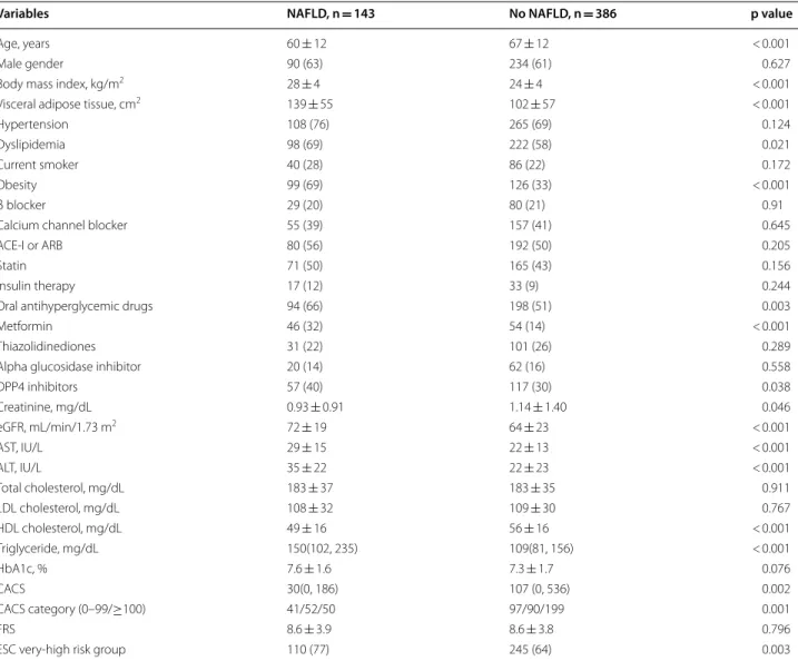
Table 1 Patient characteristics according to presence or absence of NAFLD
Data presented as mean ± standard deviation, number (%), or median [25th–75th percentile]
NAFLD non-alcoholic fatty liver disease, ACE-I angiotensin-converting enzyme inhibitors, ARB angiotensin receptor blockers, eGFR estimated glomerular filtration rate, AST aspartate aminotransferase, ALT alanine aminotransferase ratio, LDL low-density lipoprotein, HDL-C high-density lipoprotein, HbA1c glycated hemoglobin A1c, CACS coronary artery calcium score, FRS Framingham risk score, ESC European Society of Cardiology, DPP4 dipeptidyl peptidase-4
Variables NAFLD, n = 143 No NAFLD, n = 386 p value
Age, years 60 ± 12 67 ± 12 < 0.001
Male gender 90 (63) 234 (61) 0.627
Body mass index, kg/m2 28 ± 4 24 ± 4 < 0.001
Visceral adipose tissue, cm2 139 ± 55 102 ± 57 < 0.001
Hypertension 108 (76) 265 (69) 0.124
Dyslipidemia 98 (69) 222 (58) 0.021
Current smoker 40 (28) 86 (22) 0.172
Obesity 99 (69) 126 (33) < 0.001
β blocker 29 (20) 80 (21) 0.91
Calcium channel blocker 55 (39) 157 (41) 0.645
ACE-I or ARB 80 (56) 192 (50) 0.205
Statin 71 (50) 165 (43) 0.156
Insulin therapy 17 (12) 33 (9) 0.244
Oral antihyperglycemic drugs 94 (66) 198 (51) 0.003
Metformin 46 (32) 54 (14) < 0.001
Thiazolidinediones 31 (22) 101 (26) 0.289
Alpha glucosidase inhibitor 20 (14) 62 (16) 0.558
DPP4 inhibitors 57 (40) 117 (30) 0.038 Creatinine, mg/dL 0.93 ± 0.91 1.14 ± 1.40 0.046 eGFR, mL/min/1.73 m2 72 ± 19 64 ± 23 < 0.001 AST, IU/L 29 ± 15 22 ± 13 < 0.001 ALT, IU/L 35 ± 22 22 ± 23 < 0.001 Total cholesterol, mg/dL 183 ± 37 183 ± 35 0.911 LDL cholesterol, mg/dL 108 ± 32 109 ± 30 0.767 HDL cholesterol, mg/dL 49 ± 16 56 ± 16 < 0.001 Triglyceride, mg/dL 150(102, 235) 109(81, 156) < 0.001 HbA1c, % 7.6 ± 1.6 7.3 ± 1.7 0.076 CACS 30(0, 186) 107 (0, 536) 0.002 CACS category (0–99/≥100) 41/52/50 97/90/199 0.001 FRS 8.6 ± 3.9 8.6 ± 3.8 0.796
higher CACS among non-NAFLD patients (p = 0.002). The mean dose-length product for abdominal CT was 251 mGy cm, and the effective dose for each imaging modality was 3.77 mSv, using a conversion coefficient of 0.015.
Outcome data
Forty-six patients (11 NAFLD, 35 non-NAFLD) with scheduled revascularization within 90 days of the index CT were censored at the time of revascularization. Forty-four cardiovascular events were documented during
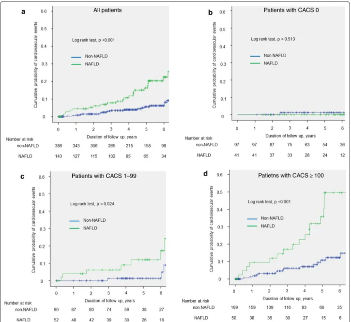
a median follow-up of 4.4 years (23 events in NAFLD patients: 1 cardiovascular death, 5 stroke, 5 myocar-dial infarction, 10 late revascularization, 2 heart failure; 21 events in non-NAFLD patients: 3 stroke, 7 myocar-dial infarction, 8 late revascularization, 3 heart failure). Kaplan–Meier curves showed the cumulative event-free survivals for cardiovascular events in patients stratified by CACS (0, 1–100 or ≥ 100), with or without NAFLD. The annual cardiovascular event rate in patients with NAFLD was significantly higher than that in patients without NAFLD (2.95% vs. 0.98%; p < 0.001) (Fig. 2a). In
Fig. 2 Kaplan–Meier curves showing cumulative incidence of cardiovascular events. Kaplan–Meier curves of cardiovascular events presented according to a presence or absence of NAFLD, and b–d presence or absence of NAFLD according to CACS categories. a All patietnts. b Patients wigh CACS 0. c Patients with CACS 1–99. d Patients with CACS ≥ 100. CACS coronary artery calcium score, NAFLD non-alcoholic fatty liver disease
addition, patients were divided into three groups accord-ing CACS and compared in relation to the presence or absence of NAFLD (Fig. 2b–d). Patients with NAFLD and CACS ≥ 100 had a significantly higher incidence of cardiovascular events. The annual rates of cardiovas-cular events in patients with CACS 0 with and without NAFLD were very low and similar in both groups (0.00% vs. 0.30%; p = 0.513) (Fig. 2b). The annual rates of cardio-vascular events in patients with CACS 1–99 and ≥ 100 were significantly higher in NAFLD compared with non-NAFLD patients (2.25% vs. 0.33%; p = 0.024, Fig. 2c; and 6.40% vs. 1.78%; p < 0.001, Fig. 2d, respectively). As shown in Table 2, univariate Cox regression analysis identified presence of NAFLD, CACS, and FRS as factors associated with cardiovascular events. Furthermore, mul-tivariate Cox regression analysis identified the presence of NAFLD, CACS, and FRS as associated with cardio-vascular events (HR, 95% CI 5.43, 2.82–10.44, p < 0.001;
1.56, 1.32–1.86, p < 0.001; 1.23, 1.08–1.39, p = 0.001, respectively).
Comparison of predictive performances for cardiovascular events
We investigated the incremental value of adding NAFLD compared with CACS and FRS alone for predicting car-diovascular events. The global χ2 score and ROC curve analysis were calculated to assess the incremental predic-tive value of NAFLD. Adding NAFLD to log (CACS + 1) and FRS significantly increased the global χ2 score from 27.0 to 49.7 (p < 0.001) (Fig. 3a). The results of ROC analy-sis comparing the area under the curve for each group are shown in Fig. 3b. Adding NAFLD significantly increased the C-statistic of Model 1 (FRS + log [CACS + 1]) from 0.71 to 0.80 (p = 0.005). The net reclassification index achieved by adding log (CACS + 1) and FRS was 0.551 Table 2 Univariate and multivariate predictors of cardiovascular events
ACE-I angiotensin-converting enzyme inhibitors, ARB angiotensin receptor blockers, NAFLD non-alcoholic fatty liver disease, LDL low-density lipoprotein, HDL-C high-density lipoprotein, HbA1c glycated hemoglobin A1c, CACS coronary artery calcium score, FRS Framingham risk score, ESC European Society of Cardiology, DPP4 dipeptidyl peptidase-4
Variables Univariate Multivariate
Hazard ratio (95% CI) p value Hazard ratio (95% CI) p value
Age 1.04 (1.01–1.07) 0.010 1.00 (0.96–1.04) 0.938 Male, gender 1.64 (0.87–3.10) 0.127 2.50 (1.01–6.18) 0.047 Hypertension 1.28 (0.63–2.60) 0.496 0.77 (0.38–1.57) 0.467 Dyslipidemia 1.28 (0.69–2.36) 0.439 1.14 (0.59–2.22) 0.695 Current smoker 1.21 (0.61–2.39) 0.592 0.75 (0.35–1.62) 0.467 Obesity 1.12 (0.62–2.03) 0.704 0.96 (0.49–1.88) 0.912 β blocker 0.56 (0.22–1.43) 0.229
Calcium channel blocker 1.46 (0.82–2.67) 0.189
ACE-I or ARB 1.20 (0.66–2.17) 0.554
Statin 1.05 (0.58–1.90) 0.872
Insulin therapy 0.87 (0.44–1.73) 0.690
Oral antihyperglycemic drugs 0.81 (0.45–1.47) 0.488
Metformin 0.36 (0.13–1.00) 0.050
Thiazolidinediones 1.95 (0.90–4.19) 0.089
Alpha glucosidase inhibitor 0.50 (0.18–1.40) 0.189
DPP4 inhibitors 1.33 (0.72–2.45) 0.368
Visceral adipose tissue
(> 100 cm2) 1.91 (1.01–3.60) 0.046 NAFLD 2.91 (1.61–5.25) < 0.001 5.43 (2.82–10.44) < 0.001 Total cholesterol 1.00 (0.99–1.01) 0.934 LDL cholesterol 1.00 (0.99–1.01) 0.944 HDL cholesterol 0.99 (0.96–1.01) 0.151 Log (triglyceride) 1.48 (0.84–2.60) 0.178 HbA1c 0.93 (0.77–1.12) 0.424 Log (CACS + 1) 1.48 (1.27–1.72) < 0.001 1.56 (1.32–1.86) < 0.001 FRS 1.14 (1.05–1.24) 0.002 1.23 (1.08–1.39) 0.001
(p < 0.001). The addition of NAFLD to CACS and FRS thus improved the predictability of cardiovascular events. Discussion
This study demonstrated that the presence of NAFLD in non-enhanced CT images, in addition to CACS and FRS, improved the risk classification of cardiovascular events in T2DM patients. However, this study was conducted in a cohort of T2DM patients with suspected coronary artery disease, and further studies are needed to deter-mine if the results apply to all T2DM patients.
Several lines of evidence have shown that NAFLD is associated with an increased risk of cardiovascular events in T2DM patients. Lee et al. showed that NAFLD was independently associated with progression of carotid intima-media thickness, as a well-established surrogate marker of subclinical atherosclerosis, in T2DM patients [21]. In an observational study of 2103 T2DM patients, NAFLD was associated with an increased incidence of cardiovascular events after adjustment for multiple risk factors (HR 1.96, 95% CI 1.4–2.7) [22]. However, the mechanisms by which NAFLD increases the risk of car-diovascular events are complex and not fully understood. A previous study showed that the histological severity of NAFLD was associated with increased arterial stiffness
and endothelial dysfunction [23]. In addition, inflamma-tory cytokines increased in line with the severity of liver disease in NAFLD patients [24]. The presence of systemic inflammation promoted by cytokines secreted from the liver leads to endothelial dysfunction, altering vascu-lar tone and enhancing vascuvascu-lar plaque formation. This mechanism was supported by a clinical study that found a significant association between the severity of NAFLD and both surrogate markers of atherosclerosis and an increased risk of cardiovascular mortality in NAFLD patients [25–28].
CACS is a well-established surrogate marker of sub-clinical coronary artery atherosclerotic plaque burden, which can predict risk beyond the established cardio-vascular risk score. Budoff et al. reported that CACS was independently and strongly associated with cardiovas-cular events, and CACS > 100 signified at least a 7.5% 10-year risk of cardiovascular events regardless of age, sex, or ethnicity among 6814 subjects from the general population [29]. CACS is also used to predict cardio-vascular risk in T2DM patients, with elevated CACS in T2DM compared with non-T2DM subjects [30]. The Diabetes Heart Study comprising 1123 T2DM patients demonstrated that CACS predicted cardiovascular events more accurately than FRS [4]. In addition, the
Fig. 3 Incremental prognostic value of NAFLD in addition to CACS and FRS. a Global χ2 test. b Receiver operating characteristic curve
2019 AHA/ACC guidelines for the primary prevention of atherosclerotic cardiovascular disease included the measurement of CACS for patients in intermediate-risk groups [6]. These data support the possible use of CACS as a means of assessing risk for cardiovascular outcomes in T2DM patients.
Hepatic steatosis has been reportedly associated with the presence of coronary artery calcium in some stud-ies [31–33]. In addition, Sung et al. reported that hepatic steatosis was independently associated with coronary artery calcium progression [34]. However, the associa-tion between NAFLD and CACS has been inconsistent across studies, especially in T2DM patients. In a study of 213 participants with T2DM, NAFLD was not asso-ciated with CACS in patients with glycated hemoglobin A1c (HbA1c) < 7%, but was significantly associated with CACS in patients with HbA1c ≥ 7% [35]. In contrast, McKimme et al. reported no significant association between hepatic steatosis and CACS in T2DM patients [36]. Kim et al. reported an association between NAFLD and the prevalence of CACS, but this association was attenuated and was no longer significant after adjust-ing for insulin resistance [37]. The current study also found no association between NAFLD and higher CACS. Given that our results indicated that NAFLD and CACS were independent factors, the combination of NAFLD and CACS might improve the identification of T2DM patients at higher risk of cardiovascular events.
Ultrasonography is commonly used to assess liver fat infiltration in clinical practice; however, non-enhanced CT can also be useful for diagnosing liver fat. Previous studies showed that a liver:spleen ratio < 1.0 could be used effectively to diagnose the presence of liver fat with high reproducibility [18, 38, 39]. However, the preva-lence of NAFLD in T2DM in this study was lower than that reported in other studies [40], in which NAFLD was mostly diagnosed by ultrasonography and magnetic res-onance imaging. On the other hand however, our study applied CT, which was reported to have a lower sensitiv-ity for diagnosing hepatic steatosis compared with ultra-sonography and magnetic resonance imaging, especially in cases with mild steatosis (< 30% steatosis) [41]. The present study may thus have included patients with mod-erate to severe hepatic steatosis.
NAFLD is closely associated with obesity [22], and the prevalence of NAFLD has been reported to increase in parallel with increasing severity of obesity [42]. Recently, severe obesity among children and adolescents has recently become a significant public health concern [43]. Furthermore, pediatric fatty liver disease clustered with cardiometabolic risk factors, associated with an increase in subsequent adult cardiovascular mortality among ado-lescents with severe obesity [44]. A healthier diet and
physical activity should thus be promoted among adoles-cents with obesity to mitigate the cardiometabolic risk.
This study had several limitations that need to be addressed. First, the number of patients and cardiovas-cular events were relatively small. In addition, our study population only consisted of Japanese patients with sus-pected coronary artery disease, and the results therefore cannot be applied directly to the T2DM population or to other ethnic groups. Second, the histological severity of liver damage was not confirmed in this study. However, CT is a useful tool for diagnosing NAFLD, without the complications associated with invasive methods. The association between CACS and histologic findings of NAFLD, such as ballooning grade, need to be evaluated in future studies. Third, we have no data about the dura-tion of T2DM, which has been reported to increase the risk of cardiovascular events [45]. Fourth, longitudinal information on changes in medications, risk factor con-trol, and changes in body mass index and lifestyle during the follow-up period was not available. Finally, FRS was originally developed in Western societies and is therefore not accurate in Asians. The Suita score has been pro-posed and validated as an alternative score for predict-ing coronary heart disease in Japanese populations [46]. The application of the Suita score instead of FRS did not affect the findings of this study (data not shown).
Conclusions
This study demonstrated the potential incremental prog-nostic value of NAFLD assessed by non-enhanced CT, in addition to CACS, for risk stratification of cardiovas-cular events in T2DM patients with suspected coronary artery disease. Further studies are needed to validate the applicability of NAFLD and CACS examination by non-enhanced CT to all T2DM patients.
Abbreviations
CACS: Coronary artery calcium score; CT: Computed tomography; FRS: Framingham risk score; HbA1c: Glycated hemoglobin A1c; HU: Hounsfield unit; NAFLD: Non-alcoholic fatty liver disease; ROC: Receiver operating charac-teristic; T2DM: Type 2 diabetes mellitus.
Acknowledgements
We thank Edanz Group (https ://en-autho r-servi ces.edanz group .com/ac) for editing a draft of this manuscript.
Authors’ contributions
KI and TM designed the study, analyzed the data, and wrote the manuscript. KO, TM, HT, KE, MY, YN, MY and KN collected the data. MH and HI assisted in data interpretation and manuscript revision. All authors read and approved the final manuscript.
Funding
No funding to declare.
Availability of data and materials
The datasets used and/or analyzed during the current study are available from the corresponding author on reasonable request.
Ethics approval and consent to participate
The study protocol was approved by the Institutional Review Board of Okayama University Hospital and conducted in accordance with the principles contained within the Declaration of Helsinki. All patients enrolled in the study provided written informed consent.
Consent for publication
Not applicable.
Competing interests
We declare that we have no conflict of interest.
Author details
1 Department of Cardiovascular Medicine, Dentistry and Pharmaceutical
Sci-ences, Okayama University Graduate School of Medicine, 2-5-1 Shikata-cho, Kita-ku, Okayama 700-8558, Japan. 2 Department of Cardiovascular Medicine,
Japanese Red Cross Okayama Hospital, Okayama, Japan. 3 Department of
Car-diovascular Therapeutics, Dentistry, and Pharmaceutical Sciences, Okayama University Graduate School of Medicine, Okayama, Japan.
Received: 11 September 2020 Accepted: 9 December 2020
References
1. Zimmet P, Alberti KG, Shaw J. Global and societal implications of the diabetes epidemic. Nature. 2001;414(6865):782–7.
2. Gu K, Cowie CC, Harris MI. Mortality in adults with and without diabetes in a national cohort of the U.S. population, 1971–1993. Diabetes Care. 1998;21(7):1138–45.
3. Emerging Risk Factors C, Sarwar N, Gao P, Seshasai SR, Gobin R, Kaptoge S, Di Angelantonio E, Ingelsson E, Lawlor DA, Selvin E, et al. Diabetes mellitus, fasting blood glucose concentration, and risk of vascular disease: a collaborative meta-analysis of 102 prospective studies. Lancet. 2010;375(9733):2215–22.
4. Agarwal S, Cox AJ, Herrington DM, Jorgensen NW, Xu J, Freedman BI, Carr JJ, Bowden DW. Coronary calcium score predicts cardiovascular mortality in diabetes: diabetes heart study. Diabetes Care. 2013;36(4):972–7. 5. Mielke CH, Shields JP, Broemeling LD. Coronary artery calcium, coronary
artery disease, and diabetes. Diabetes Res Clin Pract. 2001;53(1):55–61. 6. Arnett DK, Blumenthal RS, Albert MA, Buroker AB, Goldberger ZD,
Hahn EJ, Himmelfarb CD, Khera A, Lloyd-Jones D, McEvoy JW, et al. 2019 ACC/AHA guideline on the primary prevention of cardiovascular disease: a report of the American College of Cardiology/American Heart Association Task Force on clinical practice guidelines. Circulation. 2019;140(11):e596-646.
7. Targher G, Day CP, Bonora E. Risk of cardiovascular disease in patients with nonalcoholic fatty liver disease. N Engl J Med. 2010;363(14):1341–50. 8. Hamaguchi M, Kojima T, Takeda N, Nagata C, Takeda J, Sarui H, Kawahito
Y, Yoshida N, Suetsugu A, Kato T, et al. Nonalcoholic fatty liver disease is a novel predictor of cardiovascular disease. World J Gastroenterol. 2007;13(10):1579–84.
9. Stepanova M, Younossi ZM. Independent association between nonalco-holic fatty liver disease and cardiovascular disease in the US population. Clin Gastroenterol Hepatol. 2012;10(6):646–50.
10. Non-alcoholic Fatty Liver Disease, Study G, Lonardo A, Bellentani S, Argo CK, Ballestri S, Byrne CD, Caldwell SH, Cortez-Pinto H, Grieco A, Machado MV, et al. Epidemiological modifiers of non-alcoholic fatty liver disease: focus on high-risk groups. Dig Liver Dis. 2015;47(12):997–1006. 11. Tuttolomondo A, Di Raimondo D, Di Sciacca R, Pecoraro R, Arnao V, Butta
C, Licata G, Pinto A. Arterial stiffness and ischemic stroke in subjects with and without metabolic syndrome. Atherosclerosis. 2012;225(1):216–9. 12. Tuttolomondo A, Maida C, Pinto A. Diabetic foot syndrome: immune-inflammatory features as possible cardiovascular markers in diabetes. World J Orthop. 2015;6(1):62–76.
13. Lonardo A, Ballestri S, Guaraldi G, Nascimbeni F, Romagnoli D, Zona S, Targher G. Fatty liver is associated with an increased risk of diabetes and cardiovascular disease-evidence from three different disease models: NAFLD, HCV and HIV. World J Gastroenterol. 2016;22(44):9674–93.
14. Ichikawa K, Miyoshi T, Osawa K, Miki T, Nakamura K, Ito H. Prognostic Value of coronary computed tomographic angiography in patients with nonalcoholic fatty liver disease. JACC Cardiovasc Imaging. 2020;13(7):1628–30.
15. Agatston AS, Janowitz WR, Hildner FJ, Zusmer NR, Viamonte M Jr, Detrano R. Quantification of coronary artery calcium using ultrafast computed tomography. J Am Coll Cardiol. 1990;15(4):827–32.
16. Yoshizumi T, Nakamura T, Yamane M, Islam AH, Menju M, Yamasaki K, Arai T, Kotani K, Funahashi T, Yamashita S, et al. Abdominal fat: standardized technique for measurement at CT. Radiology. 1999;211(1):283–6. 17. Ma X, Holalkere NS, Kambadakone RA, Mino-Kenudson M, Hahn PF,
Sahani DV. Imaging-based quantification of hepatic fat: methods and clinical applications. Radiographics. 2009;29(5):1253–77.
18. Zeb I, Li D, Nasir K, Katz R, Larijani VN, Budoff MJ. Computed tomography scans in the evaluation of fatty liver disease in a population based study: the multi-ethnic study of atherosclerosis. Acad Radiol. 2012;19(7):811–8. 19. Wilson PW, D’Agostino RB, Levy D, Belanger AM, Silbershatz H, Kannel WB.
Prediction of coronary heart disease using risk factor categories. Circula-tion. 1998;97(18):1837–47.
20. Cosentino F, Grant PJ, Aboyans V, Bailey CJ, Ceriello A, Delgado V, Federici M, Filippatos G, Grobbee DE, Hansen TB, et al. 2019 ESC guidelines on diabetes, pre-diabetes, and cardiovascular diseases developed in collabo-ration with the EASD. Eur Heart J. 2020;41(2):255–323.
21. Lee HH, Cho Y, Choi YJ, Huh BW, Lee BW, Kang ES, Park SW, Cha BS, Lee EJ, Lee YH, et al. Non-alcoholic steatohepatitis and progression of carotid atherosclerosis in patients with type 2 diabetes: a Korean cohort study. Cardiovasc Diabetol. 2020;19(1):81.
22. Targher G, Bertolini L, Rodella S, Tessari R, Zenari L, Lippi G, Arcaro G. Non-alcoholic fatty liver disease is independently associated with an increased incidence of cardiovascular events in type 2 diabetic patients. Diabetes Care. 2007;30(8):2119–21.
23. Tuttolomondo A, Petta S, Casuccio A, Maida C, Corte VD, Daidone M, Di Raimondo D, Pecoraro R, Fonte R, Cirrincione A, et al. Reactive hyperemia index (RHI) and cognitive performance indexes are associated with his-tologic markers of liver disease in subjects with non-alcoholic fatty liver disease (NAFLD): a case control study. Cardiovasc Diabetol. 2018;17(1):28. 24. du Plessis J, Korf H, van Pelt J, Windmolders P, Vander Elst I, Verrijken A,
Hubens G, Van Gaal L, Cassiman D, Nevens F, et al. Pro-inflammatory cytokines but not endotoxin-related parameters associate with disease severity in patients with NAFLD. PLoS ONE. 2016;11(12):e0166048. 25. Mangla N, Ajmera VH, Caussy C, Sirlin C, Brouha S, Bajwa-Dulai S,
Madamba E, Bettencourt R, Richards L, Loomba R. Liver stiffness severity is associated with increased cardiovascular risk in patients with type 2 diabetes. Clin Gastroenterol Hepatol. 2020;18(3):744-6.e741. 26. Ekstedt M, Hagstrom H, Nasr P, Fredrikson M, Stal P, Kechagias S,
Hult-crantz R. Fibrosis stage is the strongest predictor for disease-specific mortality in NAFLD after up to 33 years of follow-up. Hepatology. 2015;61(5):1547–54.
27. Kim JH, Moon JS, Byun SJ, Lee JH, Kang DR, Sung KC, Kim JY, Huh JH. Fatty liver index and development of cardiovascular disease in Koreans without pre-existing myocardial infarction and ischemic stroke: a large population-based study. Cardiovasc Diabetol. 2020;19(1):51. 28. Idilman IS, Akata D, Hazirolan T, Doganay Erdogan B, Aytemir K,
Karcaal-tincaba M. Nonalcoholic fatty liver disease is associated with significant coronary artery disease in type 2 diabetic patients: a computed tomogra-phy angiogratomogra-phy study 2. J Diabetes. 2015;7(2):279–86.
29. Budoff MJ, Young R, Burke G, Jeffrey Carr J, Detrano RC, Folsom AR, Kronmal R, Lima JAC, Liu KJ, McClelland RL, et al. Ten-year association of coronary artery calcium with atherosclerotic cardiovascular disease (ASCVD) events: the multi-ethnic study of atherosclerosis (MESA). Eur Heart J. 2018;39(25):2401–8.
30. Raggi P, Shaw LJ, Berman DS, Callister TQ. Prognostic value of coronary artery calcium screening in subjects with and without diabetes. J Am Coll Cardiol. 2004;43(9):1663–9.
31. Sung KC, Wild SH, Kwag HJ, Byrne CD. Fatty liver, insulin resistance, and features of metabolic syndrome: relationships with coronary artery calcium in 10,153 people. Diabetes Care. 2012;35(11):2359–64. 32. Chhabra R, O’Keefe JH, Patil H, O’Keefe E, Thompson RC, Ansari S,
Ken-nedy KF, Lee LW, Helzberg JH. Association of coronary artery calcification with hepatic steatosis in asymptomatic individuals. Mayo Clin Proc. 2013;88(11):1259–65.
•fast, convenient online submission •
thorough peer review by experienced researchers in your field • rapid publication on acceptance
• support for research data, including large and complex data types •
gold Open Access which fosters wider collaboration and increased citations maximum visibility for your research: over 100M website views per year •
At BMC, research is always in progress. Learn more biomedcentral.com/submissions
Ready to submit your research
Ready to submit your research ? Choose BMC and benefit from: ? Choose BMC and benefit from:
33. Kim D, Choi SY, Park EH, Lee W, Kang JH, Kim W, Kim YJ, Yoon JH, Jeong SH, Lee DH, et al. Nonalcoholic fatty liver disease is associated with coronary artery calcification. Hepatology. 2012;56(2):605–13.
34. Sung KC, Ryu S, Lee JY, Lee SH, Cheong ES, Wild SH, Byrne CD. Fatty liver, insulin resistance, and obesity: relationships with increase in coronary artery calcium over time. Clin Cardiol. 2016;39(6):321–8.
35. Kwak MS, Yim JY, Kim D, Park MJ, Lim SH, Yang JI, Chung GE, Kim YS, Yang SY, Kim MN, et al. Nonalcoholic fatty liver disease is associated with coronary artery calcium score in diabetes patients with higher HbA1c. Diabetol Metab Syndr. 2015;7:28.
36. McKimmie RL, Daniel KR, Carr JJ, Bowden DW, Freedman BI, Register TC, Hsu FC, Lohman KK, Weinberg RB, Wagenknecht LE. Hepatic steatosis and subclinical cardiovascular disease in a cohort enriched for type 2 diabe-tes: the Diabetes Heart Study. Am J Gastroenterol. 2008;103(12):3029–35. 37. Kim MK, Ahn CW, Nam JS, Kang S, Park JS, Kim KR. Association between
nonalcoholic fatty liver disease and coronary artery calcification in post-menopausal women. Menopause. 2015;22(12):1323–7.
38. Piekarski J, Goldberg HI, Royal SA, Axel L, Moss AA. Difference between liver and spleen CT numbers in the normal adult: its usefulness in predict-ing the presence of diffuse liver disease. Radiology. 1980;137(3):727–9. 39. Bydder GM, Chapman RW, Harry D, Bassan L, Sherlock S, Kreel L.
Com-puted tomography attenuation values in fatty liver. J Comput Tomogr. 1981;5(1):33–5.
40. Lee YH, Cho Y, Lee BW, Park CY, Lee DH, Cha BS, Rhee EJ. Nonalcoholic fatty liver disease in diabetes. Part I: epidemiology and diagnosis. Diabe-tes Metabol J. 2019;43(1):31–45.
41. Bohte AE, van Werven JR, Bipat S, Stoker J. The diagnostic accuracy of US, CT, MRI and 1H-MRS for the evaluation of hepatic steatosis compared with liver biopsy: a meta-analysis. Eur Radiol. 2011;21(1):87–97.
42. Anderson EL, Howe LD, Jones HE, Higgins JP, Lawlor DA, Fraser A. The prevalence of non-alcoholic fatty liver disease in children and adolescents: a systematic review and meta-analysis. PLoS ONE. 2015;10(10):e0140908.
43. Bendor CD, Bardugo A, Pinhas-Hamiel O, Afek A, Twig G. Cardiovascular morbidity, diabetes and cancer risk among children and adolescents with severe obesity. Cardiovasc Diabetol. 2020;19(1):79.
44. Twig G, Yaniv G, Levine H, Leiba A, Goldberger N, Derazne E, Ben-Ami Shor D, Tzur D, Afek A, Shamiss A, et al. Body-mass index in 2.3 million adolescents and cardiovascular death in adulthood. N Engl J Med. 2016;374(25):2430–40.
45. Fox CS, Sullivan L, D’Agostino RB, Wilson PW, Framingham Heart S. The significant effect of diabetes duration on coronary heart disease mortal-ity: the Framingham Heart Study. Diabetes Care. 2004;27(3):704–8. 46. Nishimura K, Okamura T, Watanabe M, Nakai M, Takegami M,
Higashiy-ama A, Kokubo Y, OkayHigashiy-ama A, Miyamoto Y. Predicting coronary heart disease using risk factor categories for a Japanese urban population, and comparison with the framingham risk score: the suita study. J Atheroscler Thromb. 2014;21(8):784–98.
Publisher’s Note
Springer Nature remains neutral with regard to jurisdictional claims in pub-lished maps and institutional affiliations.