115 Original Paper
*1 Department of Physical Therapist, Faculty of Rehabilitation, Kawasaki University of Medical Welfare, Kurashiki, 701-0193, Japan E-Mail: t-kishi@mw.kawasaki-m.ac.jp
Thoracolumbar Proprioception in Patients with
Vertebral Compression Fractures: A Comparison
between Healthy Young and Elderly Subjects
Tomoya KISHIMOTO
*1, Kenichi KOBARA
*1and
Katsushi KUNIYASU
*1(Accepted November 4, 2020)
Key words: thoracolumbar position sense, vertebral compression fracture, healthy young subjects, healthy elderly subjects
Abstract
The present study examined thoracolumbar positional sense (TPS) in patients with vertebral compression fractures. The subjects were 13 healthy adults in their 20s (young group; Y group) and elderly patients aged ≥65 years, including 13 with (fracture group; F group) and 11 without (non-fracture group; NF group) vertebral compression fractures. In addition to investigating their background characteristics, TPS was assessed with the relocation test, and static posture control was evaluated based on standing body sway with open and closed eyes. The differences among the three groups were examined. TPS was significantly lower in the F group than in the Y group. Static posture control was significantly lower in the F group and NF group than in the Y group. This suggests that elderly patients with vertebral compression fractures have reduced TPS compared with healthy young adults.
1. Introduction
Vertebral compression fractures are the most common osteoporotic fractures seen in older individuals1).
Regarding the characteristics of vertebral compression fractures, it has been reported that balancing ability decreases as the number of fractured vertebrae increases2,3). It has been suggested that one of the causes of
this balance loss is vertebral compression fracture-induced kyphosis2). On the other hand, a previous study
reported that vertebral compression fractures are more closely related to balance ability than kyphosis4).
This suggests that it is difficult to explain vertebral compression fracture-related balance deterioration based on increased kyphosis alone.
Somatosensory functions, particularly those related to proprioception, have been reported to be closely related to balance ability5), suggesting that impaired proprioception adversely affects balance ability6).
Proprioception disorders are also reported to occur in patients with lower back pain or lumbar spine disease7,8). Previous studies reported that elderly patients with lower back pain exhibited reductions in
fractures, who have organic damage or lower back pain, might exhibit reduced proprioception of the thoracolumbar vertebrae. Such proprioceptive abnormalities of the thoracolumbar vertebrae might adversely affect balance ability. Therefore, the purpose of this study was to evaluate thoracolumbar vertebral proprioception in patients with vertebral compression fractures and compare it between healthy young adults and the elderly.
2. Methods
2.1 Participants
The subjects included both young and elderly patients. The elderly subjects were 13 patients with vertebral compression fractures who had undergone fracture treatment (fracture group; F group) and 11 non-vertebral compression fracture patients (non-fracture group; NF group). The young subjects were 13 healthy young people (young group; Y group). The inclusion criteria for the elderly subjects were as follows: 1) being aged ≥65, 2) visiting or being hospitalized at the author’s institution between February 2018 and January 2020, 3) living independently at home, and 4) being able to walk without using walking aids. The exclusion criteria for the elderly subjects were as follows: 1) marked visual impairment, lower limb sensory impairment, or vestibular dysfunction; 2) neuromuscular diseases, such as multiple sclerosis or polymyositis; 3) cerebrovascular disease or orthopedic disease in the lower limbs; 4) significant pain when standing or walking; and 5) a lack of understanding of and/or cooperation with the research. The exclusion criteria for the F group were as follows: 1) being diagnosed with a vertebral compression fracture ≤6 months ago and 2) a lower back pain score of ≥4 according to the Numeric Rating Scale. The exclusion criteria for the Y group were as follows: 1) not having suffered lower back pain within the last 2 years, 2) spinal orthopedic disease, and 3) neurological disease. Patients with vertebral compression fractures who had undergone fracture treatment were defined as those who had consulted a medical institution complaining of lower back pain, were diagnosed with a vertebral compression fracture, and underwent conservative corset therapy. Before the data collection, all of the study procedures were explained to the participants, who all signed informed consent forms. This study was carried out after receiving approval from the institutional review board of Medical Corporation Shiseikai Watanabe Hospital (approval No. 01702).
2.2 Measurements
The subjects’ background characteristics were their age, height, body weight, body mass index (BMI), the fractured vertebral bodies, and the number of compression fractures that were extracted. The evaluated items were thoracolumbar positional sense (TPS), vertebral alignment and the amount of body sway when standing. Data regarding age, height, body weight, BMI, fractured vertebral bodies, and the number of compression fractures were extracted from electronic charts.
TPS was assessed using the relocation test (RT), which was according to the method described in a previous study10). The subject sat deep on a height-adjustable bed without putting their feet on the floor.
The thighs and pelvis were fixed in place with a non-stretchable band, and a soft collar was attached to the neck in order to suppress joint movements other than in the thoracolumbar region. A belt with a laser pointer was attached to the subject over the sternal angle. The subject placed their hands on their knees and adopted a comfortable sitting position. This posture was defined as the middle position (MP) of trunk rotation (0°) (Figure 1). Then, a 1-cm square piece of graph paper with a dot marked on it was stuck on the wall 90 cm in front of the subject. The graph paper was placed where the light projected onto the wall by the laser pointer fell while the subject was in the MP. Two further 1-cm square pieces of graph paper with dots on them were placed 52 cm to either side of the first piece. The three points were located on a straight line parallel to the floor and were referred to as point A, point B, and point C in order from the left (Figure 2). The laser irradiation point was adjusted so that it was located at point B (the central point) when the subject was in the MP. When the subject turned 30° to the left and right, the laser irradiation point was located at points A and C, respectively. As mentioned above, the distances from point B to point
A and from point B to point C were both 52 cm. If this distance was defined as X1, and it was assumed that the distance from the subject to the wall was 90 cm in the MP position, and the trunk was rotated 30°, X1 could be calculated as follows: X1=sin30°× 90 (Figure 3). The measurement procedure was explained to the subject orally before it was performed to promote understanding. The subject was instructed to actively rotate their thoracolumbar region with their eyes open so that the laser projection point moved from point B to point C, rest for 2 seconds, and then return to point B again. The same procedure was performed for
Figure 1 This posture was defined as the middle position (MP) of trunk rotation (0°). The subject sat deep on a
height-adjustable bed without putting their feet on the floor. The thighs and pelvis were fixed in place with a non-stretchable band, and a soft collar was attached to the neck in order to suppress joint movements other than in the thoracolumbar region. A belt with a laser pointer was attached to the subject over the sternal angle. The subject placed their hands on their knees and adopted a comfortable sitting position.
Figure 2 Two further 1-cm square pieces of graph paper with dots on them were placed 52 cm to either side of the first piece. The three points were located on a straight line parallel to the floor and were referred to as point A, point B, and point C in order from the left.
point A. Next, the subject was blindfolded with an eye mask and instructed to rotate their trunk to the right. When the projection point of the laser reached point C, the measurer said "Please stop". After the subject had stopped for 2 seconds, the measurer said, "Return to the original position". When the subject thought that they had returned to point B, they said "Yes". A measurer was attached to the projection point of the laser when the subject said "Yes" to indicate the measurement point. When there was an error between point B and the measurement point, the measurer manually corrected the projection point of the laser so that it was located at point B, and instructed the subject to remember point B as the MP again. This task was performed 20 times, 10 times alternately on the left and right sides. The mean value of the error in the horizontal direction from point B to the measurement point (X2 cm) was calculated, and TPS was calculated using the arctangent formula ( θ =ATAN(X2/90): Figure 3).
Regarding the intra-rater reliability of the results of the RT, an intraclass correlation coefficient (ICC) was calculated in advance of the study using the retest method. The subjects were 17 healthy adults without lower back pain (mean age: 25.6 ± 3.0 years), and RT was measured using the abovementioned procedure. RT was measured at intervals of one week from the first measurement. Based on the RT values obtained in the first and second RT, the ICC was calculated to be 0.61, which was considered to be "substantial"11).
The amount of sway in the center of gravity during standing was assessed using a center of gravity sway meter (GP-7 gravicorder, Anima). The subject put both heels on the floor and adopted a comfortable standing position with their toes open naturally. The measurements were performed with the eyes in both open and closed conditions. Under the eyes open condition, the subjects looked at a mark located on a wall 2 m away. The measurement items were the environmental area (ENV. Area) with the eyes in both open and closed conditions, the total length of COP (LNG), and the area Romberg ratio.
2.3 Statistical analysis
Statistical software (SPSS statistics ver.23, IBM) was used for the statistical analyses. The subjects and evaluation items were examined using the Shapiro-Wilk test. Based on the results of the Shapiro-Wilk test, one-way ANOVA was used to perform comparisons among the three groups for items that exhibited normal distributions. The Bonferroni test or Games-Howell test was used for post-hoc testing. The Kruskal-Wallis test was used for comparison of items that did not exhibit normal distributions among the three groups. All tests involved a significance level of 5%. In addition, the chi-square test was used to perform comparisons among the three groups for gender, and the Bonferroni test was used to correct the difference between groups. It was judged that there was a significant difference when the p value was less than 0.0166.
Figure 3 X1 could be calculated as follows: X1=sin30°×90. The mean value of the error in the horizontal direction from point B to the measurement point (X2 cm) was calculated, and TPS was calculated using the arctangent formula (θ=ATAN(X2/90)).
3. Results
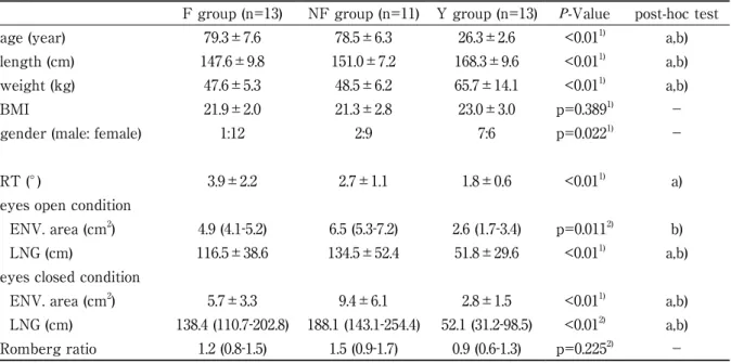
The background data and the RT and standing center of gravity sway data for the three groups are shown in Table 1 (Table 1). In the F group, the thoracic and lumbar vertebrae were fractured in 6 and 8 (includes duplicates) patients, respectively. The number of fractured vertebrae was 1 in 9 cases and ≥2 in 4 cases. In the comparisons among the three groups, significant differences in age, height, and body weight were detected between the F group and Y group (p<0.01), and between the NF group and Y group (p<0.01). BMI did not differ significantly among the three groups. TPS was significantly lower in the F group than in the Y group (p<0.01). The perimeter of the standing center of gravity sway area was significantly greater in the NF group than in the Y group under the eyes open condition (p<0.05). Under the same conditions, the LNG was significantly greater in the F group and NF group than in the Y group (p<0.01). Regarding standing center of gravity sway in the eyes closed condition, the ENV. area and LNG were both significantly greater in the F and NF groups than in the Y group (p<0.01). The area Romberg ratio did not differ significantly among the three groups.
Table 1 The background data and the RT and standing center of gravity sway data for the three groups
F group (n=13) NF group (n=11) Y group (n=13) P-Value post-hoc test
age (year) 79.3±7.6 78.5±6.3 26.3±2.6 <0.011) a,b)
length (cm) 147.6±9.8 151.0±7.2 168.3±9.6 <0.011) a,b)
weight (kg) 47.6±5.3 48.5±6.2 65.7±14.1 <0.011) a,b)
BMI 21.9±2.0 21.3±2.8 23.0±3.0 p=0.3891) −
gender (male: female) 1:12 2:9 7:6 p=0.0221) −
RT (°) 3.9±2.2 2.7±1.1 1.8±0.6 <0.011) a)
eyes open condition
ENV. area (cm2) 4.9 (4.1-5.2) 6.5 (5.3-7.2) 2.6 (1.7-3.4) p=0.0112) b)
LNG (cm) 116.5±38.6 134.5±52.4 51.8±29.6 <0.011) a,b)
eyes closed condition
ENV. area (cm2) 5.7±3.3 9.4±6.1 2.8±1.5 <0.011) a,b)
LNG (cm) 138.4 (110.7-202.8) 188.1 (143.1-254.4) 52.1 (31.2-98.5) <0.012) a,b)
Romberg ratio 1.2 (0.8-1.5) 1.5 (0.9-1.7) 0.9 (0.6-1.3) p=0.2252) −
Mean ± standard deviation, Median (interquartile range)
1) one-way ANOVA, 2) Kruskal-Wallis test
post-hoc test; a) there is a difference between F and Y group. b) there is a difference between NF and Y group. BMI: body mass index, RT: relocation test, ENV. area: environmental area, LNG: total length of COP
4. Discussion
The purpose of this study was to examine TPS in patients with vertebral compression fractures and to perform comparisons between healthy young adults and the elderly. As a result, it was suggested that patients with vertebral compression fractures exhibit reduced TPS compared with healthy young subjects. However, TPS did not differ significantly between the elderly with and without vertebral compression fractures.
Aging and vertebral compression fractures might have been responsible for the reduction in TPS seen in the F group compared with the Y group. Regarding the effects of aging on proprioception, previous studies have reported that the threshold of vibratory sensation increases approximately three times with aging12).
Y group was due to the effects of aging. However, a significant difference in X2 was only observed between the F and Y groups, and no significant difference was seen between the NF and Y groups. This was considered to have been caused by the effects of the vertebral compression fractures themselves in addition to the effects of aging. The following discusses the possibility that the observed reductions in TPS might have occurred due to the effects of the vertebral compression fractures. First, it is important to consider the effects of pain as a cause of vertebral compression fracture-related reductions in TPS. Previous studies have reported that lumbar positional sense was reduced in patients with lower back pain7,13). In addition, it
has been reported that patients with chronic lower back pain exhibited reduced lumbar positional sense, even in the absence of pain14,15). Based on these findings, it is possible that the patients in the N group had
consulted a medical institution complaining of lower back pain, and their TPS decreased due to the effects of the pain.
Next, the effects of increased lumbar muscle fatigue need to be considered as a cause of vertebral compression fracture-related reductions in TPS. Previous studies have reported that increased lumbar muscle fatigue and a concomitant reduction in blood supply adversely affected lumbosacral positional sense15,16). The mechanism responsible for these effects is that ischemia caused by increased muscle fatigue
promotes the accumulation of metabolic substances and adversely affects proprioceptor control17,18). It has
been reported that elderly people with vertebral compression fractures increased muscle fatigue in the trunk muscles19). In the present study, TPS was decreased in the F group, and the abovementioned findings
suggest that the subjects in this group might have increased muscle fatigue in the trunk muscle.
Finally, the reduction in TPS due to vertebral compression fractures might have been due to a reduction in muscle mass in the lumbar region. Previous studies have suggested that the paraspinal muscles, such as the lumbar erector spinae and lumbar multifidus, function as proprioceptors that contribute to TPS20). It
has been reported that the mass of the paraspinal muscles is decreased and the amount of intramuscular fat in the lumbar multifidus is increased in patients with vertebral compression fractures21). Therefore,
it is considered that TPS was decreased in the F group, in which the paraspinal muscles (which act as proprioceptive receptors) might have changed.
In this study, we measured standing body sway in order to evaluate how static posture control is affected by proprioception. As a result, we found that the static posture control abilities of the F and NF groups were lower than that of the Y group. On the other hand, TPS was only lower in the F group than in the Y group. Therefore, it is considered that the main cause of the reductions in static posture control aging was aging rather than proprioception.
The main limitation of this study is that it was limited to patients with vertebral compression fractures who had consulted a medical institution complaining of lower back pain. However, there are many asymptomatic cases of vertebral compression fractures that do not involve pain at consultation3). Therefore,
the results of this study must be interpreted carefully. In addition, the RT only evaluates certain aspects of TPS. In everyday life, positional sense contributes to posture control during dynamic and complex tasks, but does not usually involve conscious decisions22,23). On the other hand, the RT consciously challenges the
subject and only involves a simple exercise. Therefore, when interpreting the results of this research it is important to keep in mind that the RT only evaluates certain aspects of TPS. In addition, the number of subjects in this study was smaller than the planned sample size (20 people per group). The reason was that not enough subjects were recruited.
This study suggests that patients with vertebral compression fractures have lower TPS than healthy adults, but their ability to control static posture is comparable to that of elderly people without vertebral compression fractures.
References
1. Freitas SS, Barrett-Connor E, Ensrud KE, Fink HA, Bauer DC, Cawthon PM, Lambert LC and Orwoll ES : Rate and circumstances of clinical vertebral fractures in older men. Osteoporosis International, 19,
615-623, 2008.
2. Arima K, Abe Y, Nishimura T, Okabe T, Tomita Y, Mizukami S, Kanagae M and Aoyagi K : Assocoation of vertebral compression fractures with physical performance measure among community-dwelling Japanese women aged 40 years and older. BMC Musculoskelet Disord, 18, 176, 2017.
3. O’Neill TW, Felsenberg D, Varlow J, Cooper C, Kanis JA and Silman AJ : The prevalence of vertebral deformity in European men and women: The European Vertebral Osteoporosis Study. Journal of Bone
and Mineral Reseach, 11, 1010-1018, 1996.
4. Greig AM, Bennell KL, Briggs AM, Wark JD and Hodges PW : Balance impairment is related to vertebral fracture rather than thoracic kyphosis in individuals with osteoporosis. Osteoporosis Internationnal, 18, 543-551, 2007.
5. Andersson L, Bolling M, Wirestam R, Holtas S and Stahlberg F : Combined diffusion weighting and CSF suppression in functional MRI. NMR in Biomedicine, 15, 235-240, 2002.
6. Inglis JT, Horak FB, Shupert CL and Rycewicz C : The importance of somatosensory information in triggering and scaling automatic postural responses in humans. Experimental Brain Resesch, 101, 161, 1994. 7. Brumagne S, Cordo P, Lysens R, Verschueren S and Swinnen S : The role of paraspinal muscle spindles
in lumbosacral position sense in individuals with and without low back pain. Spine, 25, 989-994, 2000. 8. Brumagne S, Cordo P and Verschueren S : Proprioceptive weight changes in persons with low back pain
and elderly persons during upright standing. Nuuroscience Letters, 366, 63-66, 2004.
9. Ito T, Sakai Y, Kubo A, Yamazaki K, Ohno Y, Nakamura E, Sato N and Morita Y : Relative proprioceptive weighting ratio of the muscle spindles in old-old lumbar spinal disease patients with a history of falls assessed by local vibratory stimulus. Rigakuryoho Kagaku, 29, 633-638, 2014.
10. Revel M and Deshays C : Cervicocephalic kinesthetic sensitivity in patients with cervical pain. Archives
of Physical Medicine and Rehabilitation, 72, 288-291, 1991.
11. Landis JR and Koch GG : The measurement of observer agreement for categorical data. Biometrics, 33, 159-174, 1977.
12. Kenshalo DR, Han SS and Coons DH eds : Aging effects on cutaneous and kinesthetic sensibilities: Special senses
in aging. University of Michigan, Michigan, 1979.
13. O’Sullivan PB, Burnett A, Floyd AN, Gadsdon K, Logiudice J, Miller D and Quirke H : Lumbar repositioning deficit in a specific low back pain population. Spine, 28, 1074-1079, 2003.
14. Brumagne S, Janssens L, Suuden-Johanson E, Claeys K and Knapen S : Persons with recurrent low back pain exhibit a rigid postural control strategy. European Spine Journal, 17, 1177-1184, 2008.
15. Janssens L, Brumagnne S, Polspoel K, Troosters T and McConnell A : The effect of inspiratory muscles fatigue on postural control in people with and without recurrent lowback pain. Spine, 35, 1088-1094, 2010. 16. Taimela S, Kankaanpaa M and Luoto S : The effect of lumbar fatigue on the ability to sense a change in
lumbar position: a controlled study. Spine, 24, 1322-1327, 1999.
17. Delliaux S and Jammes Y : Effects of hypoxia on muscle response to tendon vibration in humans. Muscle &
Nerve, 34, 754-761, 2006.
18. Jahanson E, Brumagne S, Janssens L, Pijnenburg M, Claeys K and Paasuke M : The effect of acute back muscles fatigue on postural control in individuals with and without recurrent low back pain. European
Spine Journal, 20, 2152-2159, 2011.
19. Shipp KM, Purse JL, Gold DT, Pieper CF, Sloane R, Schenkman M and Lyles KW : Timed loaded standing: A measure of combined trunk and arm endurance suitable for people with vertebral osteoporosis. Osteoporosis International, 11, 914-922, 2000.
20. Cao DY, Khalsa PS and Pickar JG : Dynamic responsiveness of lumbar paraspinal muscle spindles during vertebral movement in the cat. Experimental Brain Resesch, 197, 369-377, 2009.
21. Kim JY, Chae SU, Kim GD and Cha MS : Changes of paraspinal muscles in postmenopausal osteoporotic spinal compression fractures: Magnetic resonance imaging study. Journal of Bone and Mineral Reseach, 20, 75-81, 2013.
22. Cordo PJ and Gurfinkel VS : Motor coordination can be fully understood only by studying complex movements. Progress in Brain Research, 143, 29-38, 2004.
23. Claeys K, Dankaerts W, Janssens L and Brumagne S : Altered preparatory pelvic control during the sit-to-stance-to-sit movement in people with nonspecific low back pain. Journal of Electromyography and