緒 言
著者らはこれまでに,マダイPagrus majorの血液中の好 中 球 に2種 類 の 通 常 型 顆 粒(ordinary chromophobic granule, oϐG; 1型, oϐG-1; 2型, oϐG-2)が存在し1,2),感染症 に罹患したマダイではそれら顆粒の細胞化学的特徴が変化 す る こ と が あ り( こ の 場 合 の 顆 粒 は 異 常 型 顆 粒 extraordinary chromophobic granule(eoϐG)と呼ぶ), また,感染症の種類によっては未感染魚の好中球には観察 さ れ な い 誘 導 型 顆 粒(inducible chromophobic granule, iϐG)が出現することを報告した3-5)。 マダイのエドワジエラ症の原因細菌であるEdwardsiella anguillarumに感染したマダイの好中球には,未感染魚の2 種類の顆粒(oϐG-1とoϐG-2)と同様な特徴を有する顆粒 (oϐG-1EaとoϐG-2Ea)とともにアルカリ性フォスファターゼ (AlP)陽性の誘導型顆粒(iϐGEa)が観察される3)。タイノ エ症の原因寄生虫であるタイノエCeratothoa verrucosaに感 染したマダイでは,oϐG-1とoϐG-2とは異なる細胞化学的特 徴を有する2種類の異常型顆粒(eoϐG-1CvとeoϐG-2Cv)およ びAlP陰性でペルオキシダーゼ(PO)陽性の誘導型顆粒 (iϐGCv)が認められる4)。アミルウージニウム症の原因寄 生虫であるデンプンベンモウチュウAmyloodinium ocellatum の寄生では,マダイ好中球に2種類の顆粒(eoϐG-2Aoと
iϐGAo)が観察される5)。eoϐG-2AoはoϐG-2と類似した構造(顆 粒の中心を取り囲むエオシン好性の層(L0)とその周辺 の難染色性層(L1)からなる)を有するが,oϐG-2では陽 性の酸性フォスファターゼ(AcP)が eoϐG-2Aoでは陰性で あり,oϐG-2では陽性所見が認められないオイルレッドO およびズダンIII染色によってL1が染色される。また, iϐGAoはiϐGEaと同様に難染色性の2層構造(L0とL1)を有す
水産大学校生物生産学科 (Department of Applied Aquabiology, National Fisheries University) †別刷り請求先(corresponding author):[email protected]
体表白濁症に罹患したマダイの好中球顆粒
近藤昌和
†,前川幸平,安本信哉,高橋幸則
Neutrophil Granules of Red Seabream Pagrus major Infected
with Body Surface Cloudiness
Masakazu Kondo
†, Kouhei Maekawa, Shinya Yasumoto and
Yukinori Takahashi
Abstract : Two types of granules were observed in the neutrophils of red seabream Pagrus major infected with
body surface cloudiness (BSC). Both granule types had similar morphologies but different cytochemical characteristics to those of ordinary chromophobic granules (oϐG-1, oϐG-2) from non-infected fish. In this paper, we called the two granule-types from the fish infected with BSC to extraordinary chromophobic granules (type 1, eoϐG-1BSC; type 2, eoϐG-2BSC). The eoϐG-1BSC showed chromophobic, simple morphology (without stratified structure), peroxidase positive, SBB negative and lack of lysozomal enzymes. The eoϐG-2BSC was stratified granule with two-layer structure (inner eosinophilic layer (L0) and outer chromophobic layer (L1)). Lysozomal enzymes (acid phosphatase, ϐ-glucuronidase (ϐ-Glu) and esterases) and peroxidase (PO) were localized in L0 and L1, respectively. Both types of extraordinary granules were Sudan black B negative. Spot formation, a curious phenomenon appeared in PO- stained oϐG-2 (positive L1 and negative L0), was not observed in eoϐG-2BSC. Almost all L0 of eoϐG-2BSC were ϐ-Glu positive. Contrastively, this enzyme activity was detected in a few L0 of oϐG-2.
るが,AlPは検出されず,iϐGEaでは陰性のズダン黒B(SBB), オイルレッドOおよびズダンIII染色によってL0が染色され る。 白 点 病 の 原 因 寄 生 虫 シ オ ミ ズ ハ ク テ ン チ ュ ウ Cryptocaryon irritansの 寄 生 で は,2種 類 の 異 常 型 顆 粒 (eoϐG-1CiとeoϐG-2Ci)が認められ,oϐG-1とoϐG-2では陽性
のSBBが陰性である 体表白濁症は海水中のなんらかの刺激物質が原因と考え られている6)。本研究では非感染性疾病の一つである体表 白濁症に罹患したマダイの好中球について報告する*。
材料および方法
2016年11月に,水産大学校の大型屋外水槽(水量約10 kL)で飼育していた体重約 200 gマダイ(収容尾数:約 200尾)において摂餌量が低下し,ほとんど全ての個体に おいて体表全体に白濁が生じた。飼育水には濾過や殺菌を 行っていない天然海水を掛け流し条件で用いており,発生 時の水温は約17℃であった。病魚の皮膚の病理組織像から, これらのマダイは体表白濁症に罹患したと判断された。本 症を呈するマダイ3尾を実験に供した。各供試魚をキナル ジンで麻酔し,尾柄部血管から採血した。血液塗抹標本の 作製および各種細胞化学染色法は前報2-6)と同様に行った。 体表白濁症が発生した水槽のマダイを発生翌日に別の水槽 へ移動したところ,1週間後に摂餌量が回復し,10日後に は体表の白濁症状は認められなくなった。この間に死亡は 無かった。結果および考察
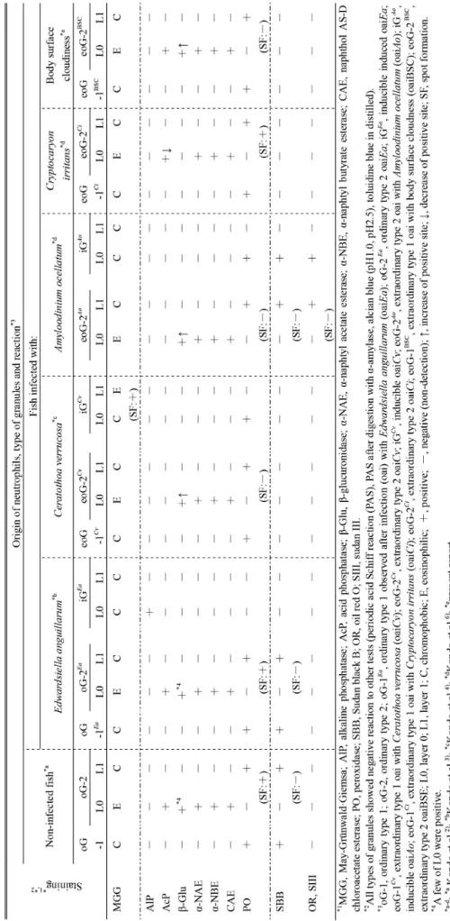
体表白濁症に罹患したマダイ(以後,罹患魚と称す)の 血 液 中 に は 多 数 の 好 中 球 が 観 察 さ れ た。May-Grünwald·Giemsa(MGG)染色では2種類の顆粒,すなわ ち難染色性の顆粒と,エオシン好性のL0とその周囲のL1 からなる顆粒が認められ,未感染魚の好中球に類似してい た(Figs. 1A, 2)。各種細胞化学染色を行ったところ,罹 患魚の好中球はAlP陽性であったが,陽性部位は細胞質基 質であり,陽性顆粒は認められなかった(Fig. 1B)。難染 色性顆粒には成層構造は観察されず,顆粒全体がPO陽性 であり(Fig. 1D),各種リソゾーム酵素は認められなかっ た。また,SBB陰性であり,オイルレッドOおよびズダン III染色にも陽性反応を示さなかった。一方,エオシン好 性 のL0を 有 す る 顆 粒 に は,AcP, ϐ-グ ル ク ロ ニ ダ ー ゼ (ϐ-Glu),α-ナフチルアセテートエステラーゼ,α-ナフチル ブチレートエステラーゼおよびナフトールAS-Dクロロア セテートエステラーゼがL0に局在し,ϐ-Glu陽性のL0が多 数観察された(Fig. 1C)。本顆粒の難染色性領域であるL1 はPO陽性であったが(Fig. 1D),oϐG-2(PO陰性のL0と陽 性のL1からなる)に認められる斑形成(spot formation)は 見られなかった(Table 1)。また,SBB,オイルレッドO およびズダンIII染色のいずれにも陰性であった。罹患魚 好中球の2種類の顆粒は,未感染魚のoϐG-1およびoϐG-2と MGG染色性ならびに構造が類似するが,細胞化学的特徴 が異なることから,異常型顆粒であると言える。罹患魚の 2種類の異常型顆粒をeoϐG-1BSCおよびeoϐG-2BSCと表記す る。eoϐG-1BSCの細胞化学的特徴はタイノエに寄生された マダイの好中球の1型異常型顆粒(eoϐG-1Cv)およびシオ ミズハクテンチュウ寄生マダイにおける1型異常型顆粒 (eoϐG-1Ci)と同じであった(Table 1)。一方,eoϐG-2BSC と同様にエオシン好性のL0と難染色性のL1からなる異常 型顆粒は,タイノエ(eoϐG-2Cv),デンプンベンモウチュ ウ(eoϐG-2Ao)およびシオミズハクテンチュウ(eoϐG-2Ci) の寄生を受けたマダイの好中球で観察されている(Table 1)。 いずれの2型異常型顆粒においてもL0に各種エステラーゼ 活性が検出されているが,AcPはeoϐG-2CvとeoϐG-2Aoでは 認められず,eoϐG-2Ciでは少数のL0に検出されるのに対し て,eoϐG-2BSCでは,未感染魚のoϐG-2と同様に多数のL0に 観察された。また,ϐ-GluはoϐG-2では少数のL0に陽性反応 が 認 め ら,eoϐG-2Ciで は 陰 性 で あ る が,eoϐG-2BSCで は eoϐG-2CvおよびeoϐG-2Aoと同様に多数のL0が陽性である (Table 1)。oϐG-2および4種類のeoϐG-2のいずれも,L0は PO陰性であり,L1にPOが検出されているが,oϐG-2に見 られる斑形成は4種類のうちeoϐG-2Ciのみに観察される。前 述のように,斑形成が認められるoϐG-2とeoϐG-2CiのL0には ϐ-Gluがほとんど検出されず,斑形成が見られないeoϐG-2Cv, eoϐG-2AoおよびeoϐG-2BSCではL0がϐ-Glu陽性である。斑の 形成には,L0内にさらに層が存在する必要が示唆されて いる7)。L0にϐ-Gluが含まれることでL0内の構造が乱れ,斑 が形成されなくなるのかもしれない。oϐG-2のL1はSBB陽 性であり,オイルレッドOおよびズダンIIIには陽性反応を 示さない。一方,4種類のeoϐG-2のうちeoϐG-2Ao以外では * 本研究の一部は,平成29年度日本魚病学会秋季大会(2017年 9月12日)において報告した[318: 近藤昌和, 前川幸平, 安本信哉, 高橋幸則: 体表白濁症に罹患したマダイの好中球顆粒(プログラムおよび講演要旨, 31)]。SBB,オイルレッドOおよびズダンIIIのいずれにも染色さ れない。eoϐG-2AoではL1がいずれの脂肪染色においても陽 性であるが,SBB陽性像はoϐG-2のL1のそれと比べて淡い。 したがって,eoϐG-2AoのL1におけるSBB陽性物質はoϐG-2 のL1の陽性物質とは異なり,oϐG-2のL1に存在するSBB陽 性物質はいずれの種類のeoϐG-2においても含まれないと推 察される。
Fig. 1. Neutrophils from red seabream infected with body surface cloudiness. A,
May-Grünwald・Giemsa; B, alkaline phosphatase; C, ϐ-glucuronidase; D, peroxidase (arrows, eoϐG-1BSC; arrowheads, eoϐG-2BSC). Bars=1 µm.
Fig. 2. Schematic illustration of neutrophil granules from red seabream. A, non-infected fish;
B-E, infected fish (B, Edwardsiella anguillarum; C, Ceratothoa verrucosa; D, Amyloodinium ocellatum; E, Cryptocaryon irritans; F, body surface cloudiness). Open area, chromophobic; closed area, eosinophilic; inner layer, L0; outer layer, L1. Abbreviations as in Table 1.
Table 1.
Comparison of neutrophil granules (chromophobic granule, G) fr
om red seabream
Pagrus major
(modified from Kondo et al.
文 献
1 ) Kondo M, Yasumoto S, Takahashi Y: Two types of granules in neutrophils from red sea-bream Pagrus major. J Nat Fish Univ, 64, 269-271 (2016)
2 ) Kondo M, Yasumoto S, Takahashi Y: Cytochemical characteristics of neutrophil granules from red seabream Pagrus major. J Nat Fish Univ, 65, 141-145 (2017)
3 ) Kondo M, Yasumoto S, Takahashi Y: Inducible granules in neutrophils from red seabream Pagrus major infected with atypical Edwardsiella tarda (=Edwardsiella anguillarum). J Nat Fish Univ, 65, 185-188 (2017)
4 ) 近藤昌和,窪田太貴,前川幸平,安本信哉,高橋幸則: タイノエに寄生されたマダイの好中球顆粒. 水大校研 報, 65, 203-206 (2017) [Kondo M, Kubota T, Maekawa K, Yasumoto S, Takahashi Y: Neutrophil granules of red seabream Pagrus major parasitized with Ceratothoa verrucosa. J Nat Fish Univ, 65, 203-206 (2017) (in Japanese with English abstract)]
5 ) 近藤昌和,前川幸平,窪田太貴,安本信哉,高橋幸則: 原生動物寄生虫(デンプンベンモウチュウ,シオミズ ハクテンチュウ)に寄生されたマダイの好中球顆粒. 水大校研報, 66, 183-187 (2018) [Kondo M, Maekawa K, Kubota T, Yasumoto S, Takahashi Y: Neutrophil granules of red seabream Pagrus major parasitized with protozoean parasits: Amyloodinium ocellatum (Kinetoplastea) and Cryptocaryon irritans (Ciliophora). J Nat Fish Univ, 66, 183-187 (2018) (in Japanese with English abstract)]
6 ) 藤巻由紀夫,富澤 泰,畑井喜司雄,窪田三朗: 体表 の白濁症状を呈するマダイの病理組織学的検索. 魚病 研究, 23, 111-115 (1988) [Fujimaki Y, Tomizawa Y, Hatai K, Kubota SS: A histopathological examination of red sea bream with a symptom of cloudiness on the body surface. Fish Pathol, 23, 111-115 (1988) (in Japanese with English abstract)]
7 ) 近藤昌和,安本信哉,高橋幸則: マダイ好中球の有芯 顆粒の構造: 顆粒における観察光の散乱様現象に基づ く一考察. 水大校研報, 65, 251-253 (2017) [Kondo M, Yasumoto S, Takahashi Y: Structure of neutrophil
pithy granules from red seabream Pagrus major: Possible explanations from light scattering-like phenomenon by the granules under the light microscopic observations. J Nat Fish Univ, 65, 251-253 (2017) (in Japanese with English abstract)]