1.INTRODUCTION
Inorganic phosphate (Pi) retention is a major harmful complication of chronic kidney disease (CKD), leading to secondary hyperparathyroidism and ectopic calcification, and is significant risk for cardiovascular morbidity and mortality (1-3). High-normal serum Pi levels are associated with cardio-vascular events and mortality among individuals
having normal kidney function (4). Therefore, re-stricting dietary Pi is necessary to prevent hyper-phosphatemia followed by a progression of serious complications.
Dietary Pi is the most important factor in body Pi homeostasis (5, 6). Dietary Pi levels control active vitamin D synthesis, parathyroid hormone (PTH) secretion, and renal Pi reabsorption (7-9). Renal Pi reabsorption is a key determinant of serum Pi lev-els in the body (10, 11). A low-Pi diet can lead to al-most 100% renal reabsorption of filtered Pi, whereas a high-Pi diet leads to decreased proximal tubular Pireabsorption (10, 11). The factors controlling the
dietary adaptive system are not known, but do not include PTH, vitamin D, growth hormone, thyroid
ORIGINAL
Effect of dietary components on renal inorganic
phosphate (Pi) excretion induced by a Pi-depleted diet
Ritsuko Ohnishi
1, Hiroko Segawa
1, Tomoyo Ohmoto
1, Shohei Sasaki
1, Ai Hanazaki
1,
Ayaka Mori
1, Kayo Ikuta
1, Junya Furutani
1, Eri Kawakami
1, Sawako Tatsumi
1,
Yasuhiro Hamada
2, and Ken-ichi Miyamoto
1 1Department of Molecular Nutrition and2
Therapeutic Nutrition, Institution of Health Biosciences, the University of Tokushima Graduate School, Tokushima, 770-8503, Japan
Abstract : Dietary inorganic phosphate (Pi) is the most important factor in the regulation of renal Pi excretion. Recent studies suggest the presence of an enteric-renal signaling axis for dietary Pi as well as the existence of a mechanism by which the intestine detects changes in luminal Pi concentrations. The mechanisms of intestinal Pi sensing, however, are unknown. In the present study, we focused on Pi depletion signals and investigated the effects of dietary components on intestinal Pi sensing. After feeding rats experimental diets for 3 days, we investigated urinary Pi excretion and plasma biochemical parameters. Renal Pi excretion was suppressed in rats fed a low-Pi diet (0.02%% Pi). Elimination of die-tary calcium (Ca) completely blocked the suppression of Pi excretion, suggesting that the presence of Ca is essential for the Pi depletion signal. Furthermore, a minimum Ca content of more than 0.02%% was necessary for the Pi depletion signal. Magnesium, lanthanum, and strontium, which are agonists of calcium sensing receptor, instead of Ca, reduced Pi ex-cretion. Therefore, dietary Ca appears to be important for the Pi depletion-sensing mecha-nism in the gastrointestinal tract. In addition, the calcium sensing receptor may be in-volved in the Pi depletion signal. J. Med. Invest. 61 : 162-170, February, 2014
Keywords :dietary phosphate, sensing, calcium, phosphate excretion
Received for publication December 6, 2013 ; accepted January 11, 2014.
Address correspondence and reprint requests to Ken - ichi Miyamoto, PhD, Department of Molecular Nutrition, Institution of Health Biosciences, the University of Tokushima Graduate School, Kuramoto - Cho 3, Tokushima City 770 - 8503 Japan and Fax : +81 - 633 - 7091.
hormone, calcitonin, or fibroblast growth factor-23 (12, 13).
Dietary Pi restriction in humans and animals is associated with enhanced renal tubular Pi reabsorp-tion (14). Urinary excrereabsorp-tion of Pi declines within hours after a reduction in dietary Pi intake, and Pi virtually disappears from the urine within 1 to 2 days. Changes in renal tubular reabsorption of Pi and its urinary excretion may occur before a fall in the plasma concentration of Pi becomes evident (14). This renal response to Pi depletion develops in normal, parathyroidectomized, and vitamin D-de-ficient animals (14). These reports suggest the pres-ence of an enteric-renal signaling axis for Pi, and also imply the existence of a mechanism by which the intestine detects changes in luminal Pi, inde-pendent of known phosphaturic factors (14, 15). It is not known, however, which dietary components, e.g., sucrose, protein, or calcium (Ca), affect intes-tinal dietary Pi sensing. Using a rat model to moni-tor renal Pi excretion induced by a low-Pi diet, we investigated the essential dietary components for Pi depletion signals in the gastrointestinal tract.
2. MATERIALS AND METHODS
2.1. Animal and Diets
Rats were maintained under pathogen-free con-ditions and handled in accordance with the Guide-lines for Animal Experimentation of Tokushima Uni-versity School of Medicine. Male 6-week-old Wistar rats were purchased from Charles River Laborato-ries Japan (Yokohama, Japan). The rats were pro-vided free access to water and standard rat chow (Oriental, Osaka, Japan). Before dietary adaptation, the rats were fed a control diet (CP, 0.6% Pi, 0.6% Ca) based on modified AIN93G for 6 days. There-after the animals were fed test diets (Studies 1-3).
All experimental diets were based on the AIN93G rodent diet. Egg white was used as the protein source instead of casein (16). An AIN93G mineral mixture without Pi and Ca sources was used to adjust the concentration of Ca and Pi for each test diet.
For study 1, the rats were divided into six groups ; a control Pi (CP) diet, and low-Pi (LP) diets 1 through 5. Rats were placed on one of the following diets for 3 days. The CP diet included an AIN93G-based diet without protein, AIN93G mineral mixture without Pi and Ca sources, 20% egg white (protein source), sucrose (a carbohydrate source), KH2PO4,
and CaCO3. Pi levels and Ca levels in the CP diet
were adjusted to 0.6% using KH2PO4 and CaCO3,
respectively. The LP1 diet was the same as the CP diet, except the Pi source (K2HPO4) was eliminated
(final concentration : 0.02% Pi and 0.6% Ca). The LP2 diet was the same as the LP1 diet, except the Pi source (egg white) was eliminated (final concen-tration : 0% Pi and 0.6% Ca). The LP3 diet was the same as the LP2 diet, except the Pi and Ca sources (K2HPO4, CaCO3, and egg white) were eliminated
(final concentration : 0% Pi and 0% Ca). The LP4 diet was the same as the LP3 diet, except the sucrose was removed (final concentration : 0% Pi and 0% Ca). The LP5 diet was the same as the LP4 diet, except Ca was added (final concentration : 0% Pi and 0.6% Ca). The final concentrations of Pi and Ca are shown in Table 1.
For study 2, the rats were divided into six groups (CP) diet, and Ca-modified low-Pi (CaLP) diets 1 through 5. Rats were fed one of the following diets for 3 days. CP is described above. In CaLP1, the Pi source (K2HPO4, and egg white) was eliminated
from the CP diet (final concentration : 0% P and 0.6% Ca). In CaLP2, CaLP3, and CaLP4, the Pi source (K2HPO4, and egg white) was also eliminated from
the CP diet. Ca levels in the CaLP2 diet were ad-justed to 0.3% using CaCO3(final concentration : 0%
Pi and 0.3% Ca). Ca levels in the CaLP3 diet were adjusted to 0.15%. Ca levels in the CaLP4 diet were adjusted to 0.02%. In the CaLP5 diet, both the Pi and Ca sources (K2HPO4, CaCO3, and egg white)
were eliminated from the CP diet (final concentra-tion : 0% Pi and 0% Ca). Final concentraconcentra-tions of Pi and Ca are shown in Table 2.
For study 3, the rats were divided into five groups, each fed a mineral-modified 0% Pi diet. A Ca (0.6% Ca), no Ca (-Ca diet, 0% Ca), magnesium (Mg), lanthanum (La), or strontium (Sr) diet. Mg, La,
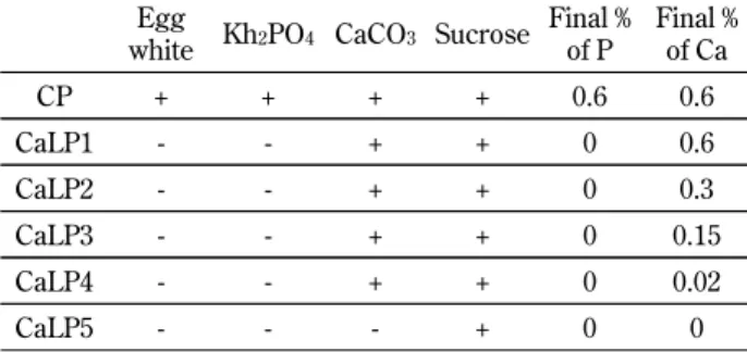
Table 1 Comparison of the experimental diet for study 1 Egg
white KH2PO4 CaCO3 Sucrose Final % of P Final % of Ca CP + + + + 0.6 0.6 LP1 + - + + 0.02 0.6 LP2 - - + + 0 0.6 LP3 - - - + 0 0 LP4 - - - - 0 0 LP5 - - + - 0 0.6
and Sr levels were adjusted to 0.6% using MgCO3,
La2(CO3)3, and SrCO3, respectively. Rats were fed
one of the test diets for 3 days. Final concentra-tions of Pi and Ca in the diets are shown in Table 3.
2.2. Concentrations of plasma Ca, Pi, and PTH
Concentrations of plasma or urinary Pi, and Ca were determined using commercial kits (Wako, Osaka, Japan) (16). Concentrations of plasma PTH were determined using the PTH ELISA kit (Immu-notopics Inc., San Clemente, CA) (16). Tail vein blood collection was performed using a 1-mL syr-inge with a 25-gauge needle at 11 : 00 AM for all studies.
Metabolic cages were used collect urine, and urine pools from rats over a 24-h period were ana-lyzed on the last day of each study (16).
2.3. Statistical Analysis
Data are expressed as means!SE. Statistical analysis was performed using two-factor factorial analysis of variance. A P-value of less than 0.05 was considered to be statistically significant.
3. RESULTS
3.1. Study 1 ; Effect of low-Pi diets on plasma and urinary Pi levels.
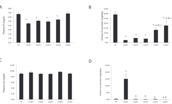
After feeding the rats each diet for 3 days, the bio-chemical parameters were analyzed in the plasma and urine of the animals (Fig. 1). Food intake did not differ among groups (data not shown). The LP1 (0.02% Pi and 0.6% Ca) and LP2 groups (0% Pi and 0.6% Ca) showed hypophosphatemia and a mark-edly reduced urinary Pi excretion compared with the CP group (0.6% Pi and 0.6% Ca ; Fig. 1A and 1B). The LP3 (0% Pi and 0% Ca) and LP4 (deletion of sucrose, 0% Pi and 0% Ca) groups showed nor-mal plasma Pi levels and Pi excretion, as did the CP group. In contrast, the LP5 group (deletion of su-crose, 0% Pi and 0.6% Ca) showed hypophosphate-mia and hypophosphaturia (Fig. 1A and 1B). In the absence of Ca, the low-Pi diet did not affect the lev-els of plasma Pi and urinary Pi excretion (Fig. 1A and 1B). Plasma Ca levels were not significantly dif-ferent among any of the groups (Fig. 1C). The LP1, LP2, and LP5 groups showed hypercalciuria com-pared with the CP group (Fig. 1D). Elimination of dietary sucrose did not affect the plasma Pi and uri-nary Pi levels. In addition, plasma PTH levels were significantly reduced after feeding on the LP1 or LP2 diet (Fig. 1E). Compared with LP1 and LP2, plasma PTH levels were significantly increased in animals fed the LP3 diet. These data suggest that dietary Ca levels are an essential factor for the Pi depletion signal in the diet.
3.2. Study 2 ; Effect of Ca in a low-Pi diet on Pi ex-cretion
To further investigate the effect of Ca concen-tration in a low-Pi diet, we prepared low-Pi diets containing various concentrations (0.6"0%) of Ca (CaLP1-CaLP5 ; Fig. 2). The amounts of food in-take did not differ among any of the groups (data not shown). Animals were fed low-Pi diets with 0.6% (CaLP1), 0.3% (CaLP2), 0.15% (CaLP3), 0.02% (CaLP4), and 0% (CaLP5) Ca for 3 days. Intake amounts of 0.6% (CaLP1), 0.3% (CaLP2), 0.15% (CaLP3), and 0.02% Ca (CaLP4) significantly de-creased plasma Pi levels compared with the CP diet (Fig. 2A). The Pi-free diets containing 0% Ca (CaLP5), however, did not show hypophosphatemia. In contrast, urinary Pi excretion levels were sup-pressed in the CaLP1-5 groups compared with those in the CP group (Fig. 2B). Furthermore, the uri-nary Pi excretion levels were significantly higher
Table 2 Comparison of the experimental diet for study 2 Egg
white Kh2PO4 CaCO3 Sucrose Final % of P Final % of Ca CP + + + + 0.6 0.6 CaLP1 - - + + 0 0.6 CaLP2 - - + + 0 0.3 CaLP3 - - + + 0 0.15 CaLP4 - - + + 0 0.02 CaLP5 - - - + 0 0
Table 3 Comparison of the experimental diet for study 3 Egg
white KH2PO4 CaCO3 Sucrose Final % of P Final % of Ca - Ca - - - + 0 0 Ca - - + + 0 0.6 Mg - - - + 0 0 0.6% of Mg La - - - + 0 0 0.6% of La Sr - - - + 0 0 0.6% of Sr
in the CaLP4 and CaLP5 groups compared with the CaLP1-3 group. Plasma Ca levels were not signifi-cantly different among any of the groups (Fig. 2C). In the CaLP5 group, urinary Ca excretion was sig-nificantly increased compared with the other groups (Fig. 2D). The CaLP4 group had increased urinary Pi excretion, whereas plasma Pi levels were re-duced. This reason for these findings is unclear. Uri-nary Pi excretion is considered an excellent marker of Pi depletion. In this context, we suggest that at least 0.02% Ca content in the diet is essential for the signal of Pi depletion.
3.3. Study 3. Effect of CaR agonist in a low-Pi diet on Pi excretion
To examine the effects of various metal ions as substitutes for Ca, we investigated the effect of Mg, La, and Sr, which are Ca sensing receptor (CaR) agonists, on the Pi depletion signal (17, 18). In this experiment, food intake did not differ among the five
groups (data not shown). The -Ca diet (0% Pi and 0% Ca) did not result in hypophosphatemia or hy-pophosphaturia, as described above (Fig. 1, Fig. 3A, and 3B). The Ca diet (0% Pi and 0.6% Ca) led to hy-pophosphatemia and hypophosphaturia, whereas the Mg diet (0% Pi and 0.6% Mg), the La diet (0% Pi and 0.6% La), and the Sr diet (0% Pi and 0.6% Sr) did not lead to hypophosphatemia (Fig. 3A and 3B). Urinary Pi excretion levels were slightly but signifi-cantly decreased in rats on the Mg diet (Fig. 3B). Similar observations were obtained for the La (Fig. 3A and 3B). The Sr diet did not significantly de-crease the urinary Pi excretion compared with CP diet (Fig. 3B). Among all groups, we observed no significant changes in plasma Ca levels (Fig. 3C). The Ca diet-fed animals exhibited hypercalciuria, but not the other diet groups (Fig. 3D). We then investigated plasma PTH levels after feeding of these diets. In animals fed the Ca diet (0.6% Ca, 0% Pi), plasma PTH levels were markedly decreased
Figure 1 Effect of various diets on plasma and urinary Pi levels
Three days after adaptation to the test diet, concentrations of plasma Pi, Ca, and PTH were determined in tail vein blood. Rats were placed in a metabolic cage to collect urine for 24 h on the last day of the study. (A) Plasma Pi, (B) urinary Pi excretion, (C) plasma Ca, (D) urinary Ca excretion, and (E) plasma PTH levels in rats after feeding with a control (CP), low - Pi (LP)1, LP2, LP3, LP4, or LP5 diet (n = 3 - 4), The study was repeated three times. Values are mean!SE. *p!0.05 versus CP,ap!0.05 versus LP1,bp!0.05 versus LP2, cp!0.05 versus LP3, anddp!0.05 versus LP4.
Figure 2 Effect of Ca concentration in a low - Pi diet on Pi metabolism
Three days after adaptation to test diet, concentrations of plasma Pi and Ca were determined in tail vein blood. Rats were placed in a meta-bolic cage to collect urine for 24 h on the last day of the study. (A) Plasma Pi, (B) urinary Pi excretion, (C) plasma Ca, and (D) urinary Ca excretion levels in rats after feeding with a control (CP), or calcium modified low phosphorus (CaLP) 1 - 5 diet (n = 3 - 4). The study was repeated three times. Values are mean!SE. *p!0.05 versus CP,ap!0.05 versus CaLP1,bp!0.05 versus CaLP2,cp!0.05 versus CaLP3
Figure 3 Effect of divalent ions in a low - Pi diet on Pi metabolism
To examine various metal ions in substitution for Ca, we prepared test diets : magnesium (Mg), lanthanum (La), or strontium (Sr) diets. Three days after adaptation to the test diet, concentrations of plasma Pi, Ca, and PTH were determined in tail vein blood. Rats were placed in a metabolic cage to collect urine for 24 h on the last day of the study. (A) Plasma Pi, (B) urinary Pi excretion, (C) plasma Ca, (D) urinary Ca excretion, and (E) plasma PTH levels in rats after feeding on a - Ca diet (0% Pi and 0% Ca), and phosphorus - depletion diets including Ca (0% Pi and 0.6% Ca), Mg (0% Pi and 0.6% Mg), La (0% Pi and 0.6%La), or Sr (0% Pi and 0.6% Sr) (n = 3 - 4). This study was repeated three times. Values are mean!SE. *p!0.05 versus -CP,#p!0.05 versus Ca,$p!0.05 versus La.
compared with those in animals fed the -Ca diet (Fig. 3E). In animals fed the Mg, La and Sr diet, plasma PTH levels were also significantly lower than those fed the -Ca diet. These data suggest that Mg, La, and Sr partially recover Pi depletion signals in the absence of Ca.
4. DISCUSSION
To clarify the sensing of dietary Pi restriction, we prepared several diets modified for each component (mineral, protein, and carbohydrate), and analyzed renal Pi excretion. In the present study, we prepared five different diets with modified carbohydrate (su-crose), protein, or mineral contents, and found that luminal Ca concentration is an important factor for Pi sensing. Pi restriction diets affect urinary Pi and Ca excretion, plasma PTH, vitamin D, and fibroblast growth factor-23 levels (19). It is not clear whether these measures are true indexes of Pi depletion. Urinary Pi excretion, however, is considered the most important index associated with dietary Pi in-take (20-22).
In the present study, using urinary Pi excretion as an index of Pi depletion, we showed that more than 0.02% Ca is essential for the recognition of a Pi-re-stricted diet. In human studies, dietary Ca is nor-mally plentiful when food is plentiful, so that the soluble Ca ion concentration in the lumen is!5 to 10 mM after a meal (23). In animals fed a diet con-taining 0.6% Ca, the soluble Ca ion concentration in the intestinal lumen is!5 to 10 mM (24). In the Ca-restricted diet (0.02-0.15%), a concentration of less than 1 mM Ca in the lumen was essential for signal-ing Pi restriction.
Although low levels (!1 mM Ca) of luminal Ca might be involved in the Pi depletion signal in the small intestine, it is not clear whether extracellular Ca or transported Ca is involved in sensing in the epithelial cells. It is widely accepted there are two pathways of Ca absorption, one transcellular and the other paracellular (23, 24). Transcellular absorption in a saturable process occurring at a luminal concen-tration up to approximately 5 mM, whereas paracel-lular absorption displays linear or diffusive kinetics between 5 and 200 mM Ca2 +. The transcellular
proc-ess involves three major steps : entry across the brush border, mediated by a TRPV6 ; intracellular diffusion, mediated by calbindin D9k ; and extrusion, mediated largely by PMCA1b (24). This pathway is regulated by 1,25(OH)2D3through the vitamin
D receptor. In a previous study, we investigated the signals of dietary Pi restriction in vitamin D receptor-null mice, but the signals were preserved in vitamin D receptor-null mice (25), suggesting that transcellular Ca transport signals are not likely involved in sensing Pi restriction.
In contrast, nutrients are detected in the lumen of the intestine by chemosensors positioned in both absorptive epithelial cells and enteroendocrine cells where they transduce nutritional information to the body (26). Much has been learned from studies of lingual nutrient detection due to identification of the G protein family, which couples with nutrient-sen-sitive G protein coupled receptors located in the taste buds on the tongue and in the intestine (26, 27). The CaR, a G protein-coupled receptor, is best known and understood for its control of PTH syn-thesis and secretion in response to changes in Ca, Mg, Sr, and La (17, 18). CaRs are distributed along most of the gastrointestinal tract, from the stomach to the large intestine (23, 28). The physiologic role of CaRs is the maintenance of constant blood Ca2 +
levels (1.1-1.3 mM) through continuous adjust-ments of PTH release from the parathyroid chief cells, which are highly sensitive to the slightest changes in extracellular Ca ion concentrations (17, 18). In the present study, we showed that Mg, La, or Sr, which are CaR agonists, can partially substi-tute for dietary Ca. In this context, these data sug-gest that Ca sensing exists and is necessary for recognition of low Pi.
In contrast, for sensing a high-Pi diet, evidence in support of a Pi entero-renal axis also comes from a very recent and convincing study in which Pi or saline were acutely administered in the rat duode-num ; this study was performed in normal (intact) and thyroparathyroidectomized rats and after renal denervation (15). Intestinal, but not gastric, Pi ad-ministration results in a prompt increase in Pi ex-cretion, which is independent of PTH and renal nerve activity. The authors excluded changes in cir-culating fibroblast growth factor-23 or frizzled re-lated protein-4 and demonstrated that homogenates of the duodenal mucosa also shows increased Pi ex-cretion (15). We performed similar studies and con-cluded that the presence of Ca in the high Pi diet was not essential for the dietary Pi signal (data not shown). The existence of Ca is not necessary for recognition of the high Pi diet, but Ca is necessary for recognizing a low-Pi diet. Therefore, it is likely that the supposed sensing mechanism of the Pi de-pletion signal differs from that for sensing a high-Pi
diet (29).
In the present study, we investigated the levels of plasma PTH after feeding of various diets. Martin
et al. reported that dietary Pi acutely controls PTH
release by the parathyroid glands by a mechanism independent of changes in plasma Ca, but that may involve concomitant changes in plasma Pi (30). On the other hand, the ability of phosphonoformate (phosphate analogue) to acutely increase PTH lev-els in the absence of changes in plasma Ca and Pi provides evidence for a signal derived from the gas-trointestinal tract that can trigger PTH release (30). We examined whether PTH-dependent Pi excretion is involved in Pi restriction signals, but the results did not provide a clear answer. Further studies are needed to clarify the mechanisms of PTH secretion by Pi restriction signals.
The characteristics of the Pi sensor molecules in the intestine are still unknown. We suggest that the CaRs play a role in dietary Pi recognition. Indeed, previous studies demonstrated that activation of CaRs in enteroendocrine G-cells and I-cells stimu-lates the secretion of gastrin and cholecystokinin, respectively (31, 32). In addition, CaR detects L-amino acids within the concentration ranges of the diets (33, 34). Depletion of extracellular Ca ion di-minishes the secretion of gluco-insulinotropic pep-tide, glucagon-like peptide-1, and peptide tyrosine tyrosine in response to L-amino acids (34). Thus, the extracellular Ca ion concentration may deter-mine the magnitude of the gut peptide secretion re-sponse. In the present study, we suggest that CaRs are important regulators of Pi sensor activity.
Animal and human studies of CKD suggest that dietary Pi restriction could mitigate nephron loss, curb secondary hyperparathyroidism, and lower blood pressure (35-38). Pi binders are widely used to control hyperphosphatemia in clinical CKD ; however, none of the currently available binders is ideal (39, 40). An alternative approach to the use of binders to limit hyperphosphatemia in CKD is to target the Pi intestinal transporter or sensor di-rectly (39). Identifying the Pi sensor molecules is a potential new target for controlling hyperphos-phatemia.
Finally, using urinary Pi excretion as an index of Pi depletion, we investigated the Pi-sensing mecha-nisms for the Pi restriction diet. Dietary Ca con-centration (0.02% Ca) is a critical parameter for Pi depletion in the gastrointestinal tract. In addition, Ca receptor agonists may improve the Pi-sensing mechanism. Further studies are needed to identify
the target molecule for sensing a Pi restriction sig-nal.
5. REFERENCES
1. Isakova T : Comparison of mineral metabolites as risk factors for adverse clinical outcomes in CKD. Semin Nephrol 33 : 106-117, 2013 2. Kendrick J, Chonchol M : The role of
phos-phorus in the development and progression of vascular calcification. Am J Kidney Dis 58 : 826-834, 2011
3. Turner JM, Bauer C, Abramowitz MK,
Melamed ML, Hostetter TH : Treatment of chronic kidney disease. Kidney Int 81 : 351-362, 2012
4. Kestenbaum B, Sampson JN, Rudser KD,
Patterson DJ, Seliger SL, Young B, Sherrard DJ, Andress DL : Serum phosphate levels and mortality risk among people with chronic kid-ney disease. J Am Soc Nephrol 16 : 520-528, 2005
5. Loghman-Adham M : Adaptation to changes in dietary phosphorus intake in health and in renal failure. J Lab Clin Med 129 : 176-188, 1997 6. Murer H, Lotscher M, Kaissling B, Levi M,
Kempson SA, Biber J : Renal brush border membrane Na/Pi-cotransport : molecular as-pects in PTH-dependent and dietary regulation. Kidney Int 49 : 1769-1773, 1996
7. Miyamoto K, Haito-Sugino S, Kuwahara S, Ohi A, Nomura K, Ito M, Kuwahata M, Kido S, Tatsumi S, Kaneko I, Segawa H : Sodium-de-pendent phosphate cotransporters : lessons from gene knockout and mutation studies. J Pharm Sci 100 : 3719-3730, 2011
8. Miyamoto K, Segawa H, Ito M, Kuwahata
M : Physiological regulation of renal sodium-dependent phosphate cotransporters. Jpn J Physiol 54 : 93-102, 2004
9. Miyamoto KI, Itho M : Transcriptional regula-tion of the NPT2 gene by dietary phosphate. Kidney Int 60 : 412-415, 2001
10. Marks J, Debnam ES, Unwin RJ : Phosphate homeostasis and the renal-gastrointestinal axis. Am J Physiol Renal Physiol 299 : F285-296, 2010
11. Takahashi F, Morita K, Katai K, Segawa H, Fujioka A, Kouda T, Tatsumi S, Nii T, Taketani Y, Haga H, Hisano S, Fukui Y, Miyamoto KI, Takeda E : Effects of dietary Pi on the renal
Na+-dependent Pi transporter NaPi-2 in thy-roparathyroidectomized rats. Biochem J 333 (Pt 1) : 175-181, 1998
12. Farrow EG, White KE : Recent advances in re-nal phosphate handling. Nat Rev Nephrol 6 : 207-217, 2010
13. Kido S, Kaneko I, Tatsumi S, Segawa H, Miyamoto K : Vitamin D and type II sodium-dependent phosphate cotransporters. Contrib Nephrol 180 : 86-97, 2013
14. Kumar R : Phosphate sensing. Curr Opin Nephrol Hypertens 18 : 281-284, 2009
15. Berndt T, Thomas LF, Craig TA, Sommer S, Li X, Bergstralh EJ, Kumar R : Evidence for a signaling axis by which intestinal phosphate rapidly modulates renal phosphate reabsorption. Proc Natl Acad Sci U S A 104 : 11085-11090, 2007
16. Ohi A, Hanabusa E, Ueda O, Segawa H, Horiba N, Kaneko I, Kuwahara S, Mukai T, Sasaki S, Tominaga R, Furutani J, Aranami F, Ohtomo S, Oikawa Y, Kawase Y, Wada NA, Tachibe T, Kakefuda M, Tateishi H, Matsumoto K, Tatsumi S, Kido S, Fukushima N, Jishage K, Miyamoto K : Inorganic phosphate homeostasis in sodium-dependent phosphate cotransporter Npt2b(+)/(-) mice. Am J Physiol Renal Physiol 301 : F1105-1113, 2011
17. McGehee DS, Aldersberg M, Liu KP, Hsuing S, Heath MJ, Tamir H : Mechanism of extra-cellular Ca2+ receptor-stimulated hormone re-lease from sheep thyroid parafollicular cells. J Physiol 502 (Pt 1) : 31-44, 1997
18. Saidak Z, Brazier M, Kamel S, Mentaverri R : Agonists and allosteric modulators of the calci-um-sensing receptor and their therapeutic ap-plications. Mol Pharmacol 76 : 1131-1144, 2009 19. Miyamoto K, Ito M, Tatsumi S, Kuwahata M, Segawa H : New aspect of renal phosphate reab-sorption : the type IIc sodium-dependent phos-phate transporter. Am J Nephrol 27 : 503-515, 2007
20. Steele TH, DeLuca HF : Influence of dietary phosphorus on renal phosphate reabsorption in the parathyroidectomized rat. J Clin Invest 57 : 867-874, 1976
21. Steele TH, Stromberg BA, Underwood JL, Larmore CA : Renal resistance to parathyroid hormone during phosphorus deprivation. J Clin Invest 58 : 1461-1464, 1976
22. Shah SV, Kempson SA, Northrup TE, Dousa TP : Renal adaptation to a low phosphate diet
in rats. J Clin Invest 64 : 955-966, 1979
23. Bronner F : Mechanisms of intestinal calcium absorption. J Cell Biochem 88 : 387-393, 2003 24. Kellett GL : Alternative perspective on intestinal
calcium absorption : proposed complementary actions of Ca(v)1.3 and TRPV6. Nutr Rev 69 : 347-370, 2011
25. Segawa H, Kaneko I, Yamanaka S, Ito M, Kuwahata M, Inoue Y, Kato S, Miyamoto K : Intestinal Na-P(i) cotransporter adaptation to dietary P(i) content in vitamin D receptor null mice. Am J Physiol Renal Physiol 287 : F39-47, 2004
26. Mace OJ, Marshall F : Digestive physiology of the pig symposium : gut chemosensing and the regulation of nutrient absorption and energy supply. J Anim Sci 91 : 1932-1945, 2013 27. Sutherland K, Young RL, Cooper NJ, Horowitz
M, Blackshaw LA : Phenotypic characterization of taste cells of the mouse small intestine. Am J Physiol Gastrointest Liver Physiol 292 : G1420-1428, 2007
28. Chattopadhyay N, Cheng I, Rogers K, Riccardi D, Hall A, Diaz R, Hebert SC, Soybel DI, Brown EM : Identification and localization of extra-cellular Ca(2+)-sensing receptor in rat intes-tine. Am J Physiol 274 : G122-130, 1998 29. Ohnishi R, Segawa H, Kawakami E, Furutani
J, Ito M, Tatsumi S, Kuwahata M, Miyamoto K : Control of phosphate appetite in young rats. J Med Invest 54 : 366-369, 2007
30. Martin DR, Ritter CS, Slatopolsky E, Brown AJ : Acute regulation of parathyroid hormone by dietary phosphate. Am J Physiol Endocrinol Metab 289 : E729-734, 2005
31. Liou AP, Sei Y, Zhao X, Feng J, Lu X, Thomas C, Pechhold S, Raybould HE, Wank SA : The extracellular calcium-sensing receptor is re-quired for cholecystokinin secretion in response to L-phenylalanine in acutely isolated intestinal I cells. Am J Physiol Gastrointest Liver Physiol 300 : G538-546, 2011
32. Feng J, Petersen CD, Coy DH, Jiang JK, Thomas CJ, Pollak MR, Wank SA : Calcium-sensing receptor is a physiologic multimodal chemosensor regulating gastric G-cell growth and gastrin secretion. Proc Natl Acad Sci U S A 107 : 17791-17796, 2010
33. Feldman EJ, Grossman MI : Liver extract and its free amino acids equally stimulate gastric acid secretion. Am J Physiol 239 : G493-496, 1980
34. Mace OJ, Schindler M, Patel S : The regula-tion of K- and L-cell activity by GLUT2 and the calcium-sensing receptor CasR in rat small in-testine. J Physiol 590 : 2917-2936, 2012 35. Gutierrez OM, Wolf M : Dietary phosphorus
restriction in advanced chronic kidney disease : merits, challenges, and emerging strategies. Semin Dial 23 : 401-406, 2010
36. Klahr S, Levey AS, Beck GJ, Caggiula AW, Hunsicker L, Kusek JW, Striker G : The effects of dietary protein restriction and blood-pres-sure control on the progression of chronic renal disease. Modification of Diet in Renal Disease Study Group. N Engl J Med 330 : 877-884, 1994 37. Lumlertgul D, Burke TJ, Gillum DM, Alfrey
AC, Harris DC, Hammond WS, Schrier RW : Phosphate depletion arrests progression of chronic renal failure independent of protein in-take. Kidney Int 29 : 658-666, 1986
38. Alfrey AC : Effect of dietary phosphate restric-tion on renal funcrestric-tion and deteriorarestric-tion. Am J Clin Nutr 47 : 153-156, 1988
39. Weinman EJ, Light PD, Suki WN : Gastroin-testinal Phosphate Handling in CKD and Its Association With Cardiovascular Disease. Am J Kidney Dis 62 : 1006-1011, 2013
40. Hutchison AJ, Smith CP, Brenchley PE : Phar-macology, efficacy and safety of oral phosphate binders. Nat Rev Nephrol 7 : 578-589, 2011