Original
Effects of Low-Intensity Pulsed Ultrasound on Callus Formation:
A Comparative Morphological Study
Masayuki KONNO1,3
, Hidetsugu ASANO1,4
, Yusuke FUJII2
, Yoshiyuki KONISHI5
and Yoshihiro MURAGAKI5
1
Graduate School of Medicine, Tokyo Women s Medical University
2J. Morita-mfg. corp. 3
Yours Dental Parkfield Clinic
4
Pioneer Corporation
5
Faculty of Advanced Techno-Surgery, Institute of Advanced Biomedical Engineering and Science, Tokyo Women s Medical University
(Accepted June 5, 2017)
Ultrasound has a variety of applications as a noninvasive treatment. In particular, it is widely used for its ac-celerating effect on synostosis, through Low Intensity Pulsed Ultra-Sound (LIPUS). In this study, morphological evaluation was carried out of the effect of LIPUS on bone tissue, aimed at identifying the effective initiation tim-ing for the treatment and the bone callus formation period.
A bone defect, 2 mm in both diameter and depth, was formed in the femur of 7-week-old male Sprague-Dawlay rats. 30 rats were assigned randomly into 5 groups: a group with no LIPUS stimulation (0 L); a group with LIPUS stimulation on day 1-3 (1-3 L); a group with LIPUS stimulation on day 1-14 (1-14 L); a group with LIPUS stimulation on day 4-14 (4-14 L); and a group with LIPUS stimulation for day 1-14 without a bone defect (Control). The inflammation period was considered to be 3 days. In order to evaluate the healing acceleration effect three-dimensionally, comparison took place of hard tissue volume, as the number of high CT value voxels, and bone density as the average CT value. LIPUS stimulation during the inflammatory period was found to be of signifi-cant importance: the 1-14 L group s bone volume was signifisignifi-cantly higher at day 7 and day 10 compared to the non-LIPUS stimulated groups in the inflammation period. This study suggests that it is desirable for LIPUS stimulation to be continued from the time of bone injury to the osteogenesis stage, and that LIPUS promotes cal-lus formation, especially by stimulation in the inflammatory phase.
Key Words: LIPUS, ultrasound, cullus formation, CT, bone
Introduction
With the spread of minimally invasive treat-ments, focus has grown on the use of non-invasive ultrasound, which now has applications in a variety of treatments. Among these, there has been
par-ticular attention paid to the application of low-power ultrasonic pulses ( Low Intensity Pulsed Ultra-Sound: LIPUS). Reports based on animal ex-periments have suggested acceleration of both soft tissue1)2)
and hard tissue healing3)∼5)
, with further
ap-:Masayuki KONNO Graduate School of Medicine, Tokyo Women s Medical University, 8―1 Kawada-cho, Shinjuku-ku, Tokyo, 162―8666 Japan
Email: [email protected] doi: 10.24488/jtwmu.87.4_108
Copyright Ⓒ 2017 Society of Tokyo Women s Medical University
! # $
J Tokyo Wom Med Univ
87 (4) 108∼116 (2017)
" # %
plication reported for use on the nervous system6)
. LIPUS treatment has even been reported for use within the osteoporosis model7)8)
. Analysis at a ge-netic level has also identified physical stimulation through LIPUS as promoting cell differentiation of osteoblasts and chondrocytes9)∼11)
. Furthermore, there have also been reports of increased mechani-cal strength12)
and bone mineral content13)
resulting from LIPUS stimulation. The results of LIPUS stimulation on bone union are clear and the applica-tion of LIPUS to aid post-fracture recovery has now become widespread. In addition to its utilization for post-fracture treatment, LIPUS application on im-plants has been reported to shorten the healing pe-riod14)∼18)
. Although there is research regarding the optimal working conditions for LIPUS frequency19)
, currently the only safe and effective conditions for fracture healing are a frequency of 1.5 MHz, a repe-tition frequency of 1.0 kHz, an ultrasound intensity of 30 mW/cm2
, a pulse width of 200μs, and stimula-tion time of 20 minutes/day20)
.
LIPUS has been approved as a medical tool since the 1990s and is used in a variety of applications, such as bone fracture treatment. U.S Clinical trials have acknowledged the effects of LIPUS stimula-tion in the accelerastimula-tion of bone union21)22)
. While there exist studies looking at the optimal period for LIPUS stimulation23)
, review of these studies related to bone union following LIPUS stimulation shows that bone evaluation was predominantly done through assessment of mechanical strength24)
, using two-dimensional imaging25)
. Within these studies there was also a lack of three-dimensional (3D) mor-phological evaluation of bone tissue. The fracture healing phase is made up of the inflammatory phase, the cell growth phase, the callus formation phase and the remodeling phase, with the callus for-mation phase reached between week 1 and week 426)27)
. However, the difference between the accelera-tion effect of LIPUS stimulaaccelera-tion at each of these phases is unclear and there are also no reports on the duration or period of LIPUS stimulation neces-sary for effective bone union. There is, therefore, a possibility that more effective post-bone fracture healing acceleration may be achieved through
LI-PUS stimulation treatment if it is applied over an optimal period and duration. In this study, 3D mor-phological assessment is used on LIPUS stimulation on rats with the bone fracture model. The objective was to clearly show the optimal stimulation period and duration for post-fracture LIPUS treatment.
Materials and Methods
The experiment was done using 7-week-old, male Sprague-Dawlay rats (SD rats, Charles River). The entire bone fracture healing phase is made up of the inflammatory phase, the cell growth phase, the cal-lus formation phase and the remodeling phase26)
. However, in the pilot experiment the period neces-sary for the femur of the SD rats to recover enough strength was approximately two weeks. Therefore, based on this, the experiment duration was set to run over 15 days to reach the callus formation phase.
Preparation of the fracture model rats aimed for stable reproduction of the fracture site and was conducted using a drill ( GC Corp. , Depth Drill 6B341) to create a cavity diameter of 2 mm and a depth of 2 mm at the injury site of the femur. The drill was cooled between procedures in order to prevent burning and was thoroughly flushed with saline to ensure no fragments of bone remained.
In order to investigate the relationship between LIPUS stimulation period and post-bone damage bone union, with reference to Nakajima et al28)
, the inflammatory phase was set at three days and the experiment conducted with the subjects split be-tween the five groups below.
1. Bone damage procedure applied, LIPUS stimu-lation not carried out (0 L group)
2. Bone damage procedure applied, LIPUS stimu-lation carried out from day 1-3 (1-3 L group)
3. Bone damage procedure applied, LIPUS stimu-lation carried out from day 1-14 (1-14 L group)
4. Bone damage procedure applied, LIPUS stimu-lation carried out from day 4-14 (4-14 L group)
5. Control group. Bone damage procedure not ap-plied, LIPUS stimulation carried out from day 1-14 (Control group)
Each of the five groups contained six rats, mak-ing a total of 30 rats. (The day of the bone damage
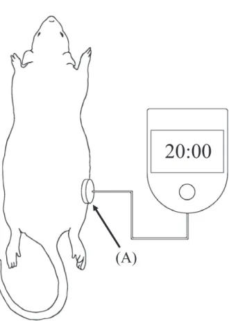
Fig. 1 LIPUS stimulation conditions
(A) is the LIPUS stimulation head unit.
(A)
20:00
procedure was counted as day 0).
LIPUS stimulation of the rats was carried out from day 1or day 4 and was conducted under anes-thesia. During treatment the SAFHS LIPUS stimu-lation equipment was used ( Teijin Pharma Ltd. , SAFHS 4,000 J). Stimulation was carried out with a frequency of 1.5 MHz, a repetition frequency of 1.0 kHz, an ultrasound intensity of 30 mW/cm2
, a pulse width of 200μs and a stimulation time of 20 minutes per day9)
. As shown in Fig. 1, during treatment the ultrasonic wave stimulation unit was affixed to the femur of the anesthetized rat using surgical tape and stimulation was then carried out. Shaving of the target site was conducted before the bone dam-age procedure. Shaving was repeated regularly to enable accurate repeat affixing of the stimulation head. X-ray micro-CT imaging of the rat femurs was performed for all groups on days 1, 3, 7, 10 and 15. The CT imaging was performed using the 3D micro-X-ray system R_mCT2 (manufactured by the Rigaku Corporation). The resolution of the CT de-vice was 59-μm and the scan was carried out with
an FOV diameter of 30 mm, a height of 30 mm, and an exposure time of two minutes.
The experiment received the permission of the animal experimentation ethics committee of Tokyo Women s Medical University (15-23) and was car-ried out in compliance with its standards.
Evaluation was carried out based on the CT val-ues. A CT value of 200HU was set as the threshold for soft tissue; values below this threshold were ex-cluded from the evaluation. Bone volume was calcu-lated as the number of voxels. In addition, bone den-sity was calculated as the sum of the CT values di-vided by the number of voxels, that is, the average CT value. It should be noted that due to the proper-ties of ultrasound, LIPUS stimulation can be re-flected and refracted depending on the tissue boundary surface. This is particularly the case for hard tissue boundaries, meaning the actual site stimulated by ultrasound is limited to the part fac-ing the stimulation unit29)30)
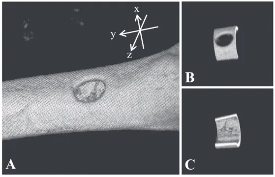
. In consideration of this, in the analysis of this study, the femur was divided into halves on the LIPUS stimulation surface and non-stimulation surface, and the volume of each bone and the density of bone were measured. Spe-cifically, as shown in A in Fig. 2, the central axis of the hole was set as the x axis, the central axis of the femur was set as the y axis, and the z axis formed the right hand coordinate system defined by the x axis and the y axis. At this time, it was cut out into a cylindrical shape with a diameter of 9.4 mm and a height of 2.4 mm with the y axis as the central axis and the xz plane as the object plane, and it was di-vided into two on the yz plane, LIPUS stimulation side (Fig. 2B) and non-stimulation side (Fig. 2C).
Analysis of variance (ANOVA) and multiple com-parison tests (Tukey-Kramer method) were applied on the measurement results from each group: re-sults from the LIPUS stimulation side and non-stimulation side together and results solely from the LIPUS stimulation side (10 groups in total).
Results
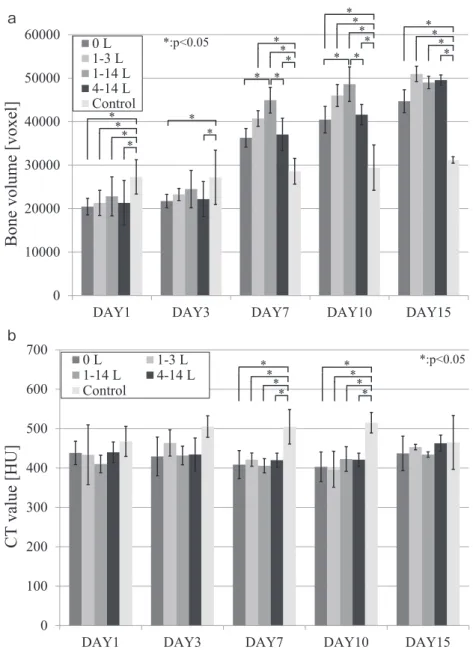
Fig. 3(a) shows the bone volume results for the LIPUS stimulation side.
Over the LIPUS stimulation period no clear change in bone volume of the control group was
ob-Fig. 2 An image obtained by Micro CT
A is a 3D reconstructed bone image. B shows the analyzed area including the bone defect. C is the other side of B.
B
C
x
z
y
A
served. Within the other groups, an increase of bone volume for bone union was confirmed regard-less of the presence or absence of LIPUS stimula-tion or the period of the stimulastimula-tion.
Comparing the results at day 1, the bone volume of the control group was significantly higher than that of the bone-damaged groups ( p < 0.05 ) , but there was no clear difference among the bone-damaged groups. The results for day 3 were the same as for day 1; however the differences between the bone-damaged groups and the control group had become smaller. By day 7, the bone volume of the bone-damaged groups had become higher than that of the control group. A trend was seen in the bone-damaged groups that underwent LIPUS stimulation during the inflammatory period (1-3 L group, 1-14 L group), with higher bone volume com-pared to the other bone-damaged groups ( 0 L group, 4-14 L group). In particular, the bone volume of the 1-14 L group (44,933 ± 2,958 voxels) was sig-nificantly higher (p < 0.05) in comparison to the 0 L group ( 36,288 ± 4,490 voxels ) and 4-14 L group (37,035 ± 3,962 voxels). The same trend seen at day 7 was also apparent at day 10: the bone volume of the 1-14 L group (48,613 ± 3,738 voxels) was
signifi-cantly higher (p < 0.05) than the 0 L group (40,461 ± 5,157 voxels ) and the 4-14 L group ( 41,599 ± 2,354 voxels ) . The differences between the bone-damaged groups and the control group had also in-creased. By day 15, the difference in bone volume among the bone-damaged groups and the control group was significantly smaller (p < 0.05); however there was no significant difference between any combination of the bone-damaged groups.
Fig. 3(b) shows the transition result of the bone density for the LIPUS stimulation side.
At day 1 no differences in bone density were ob-served between each pair of groups. By day 3 dif-ferences in bone density between the control group and the bone-damaged groups had begun to appear, the bone-damaged groups having lower values than the control group and being relatively soft. At day 7, the average bone density of the control group was 505 ± 8, significantly different (p < 0.05 ) to those of the bone-damaged groups: the 0 L group (403 ± 29 HU), the 3 L group (397 ± 46 HU), the 1-14 L group (423 ± 20 HU), and the 4-1-14 L group (421 ± 18 HU). The results at day 10 were the same as for day 7 and a significant difference was again ob-served (p < 0.05): the average bone density of the
Fig. 3 Time series graph showing bone volume (a) and bone CT value (b) (LIPUS stimu-lated side). 0 10000 20000 30000 40000 50000 60000
DAY1 DAY3 DAY7 DAY10 DAY15
0 L 1-3 L 1-14 L 4-14 L Control
Bone volume [voxel]
*:p<0.05 * * * * * * * * * * * * * * * * * * ** * 0 100 200 300 400 500 600 700
DAY1 DAY3 DAY7 DAY10 DAY15
0 L 1-3 L 1-14 L 4-14 L Control
CT
value [HU]
*:p<0.05 * * * * * * * * a a b bcontrol group was 515 ± 22, while the average den-sity of the remaining bone-damaged groups showed no substantial change. By day 15, difference in aver-age bone density among the groups was almost no longer apparent.
It should be noted that no clear change was ob-served in bone volume or bone density of the non-LIPUS stimulated side.
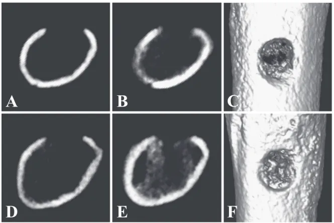
Fig. 4 shows a typical example of the micro-CT images from the 0 L and 1-14 L groups. A and B show the micro-CT images of the 0 L group on day 1 and day 7 respectively. D and E are the CT im-ages of the 1-14 L group on day 1 and day 7. Imim-ages C and F are 3D diagrams of the CT images from the
0 L group and the 1-14 L group at day 7. Comparing B and E, it can be seen that by day 7 both cancel-lous bone impermeability and hard bone volume of the 1-14 L group had increased. It is also apparent from the 3D images that compared to C (0 L group) bone formation can be clearly confirmed at the bone injury site in F (1-14 L group).
Discussion
The experimental results show that at day 1 and day 3, although bone volume for the non-bone-damaged control group was significantly higher, no difference was observed among the bone-damaged groups. This difference in comparison to the control group is attributed to being due to the volume of
Fig. 4 Comparison between an LIPUS non-stimulated rat and a stimulated rat A and B
are CT images from an LIPUS non-stimulated rat (0 L). A is a cross-sectional image on day 1. B is a cross-sectional image on day 7. C is a 3D reconstructed image of B. D-F show the LIPUS stimulated rat data (1-14 L).
A
B
C
D
E
F
bone removed as part of the bone damage treat-ment. By day 7 and day 10 the bone volume of the 1-14 L group, which underwent continued LIPUS stimulation, had greatly increased. In particular, a significant difference in bone volume was seen when compared to groups that had not undergone LIPUS stimulation during the inflammatory phase (0 L group, 4-14 L group). Based on this difference, LIPUS stimulation during the inflammatory phase is thought to be an important factor in accelerating bone recovery. The 1-3 L group, which had LIPUS stimulation only during the inflammatory phase, showed a tendency towards higher bone volume compared to the 0 L group; however, a significant difference was not seen. In regards to bone volume, even if LIPUS stimulation is only applied during the inflammatory phase, there may be a small healing acceleration effect. At day 15, significant differences in bone volume was observed between the bone-damaged groups and the control group: however no difference was found among the bone-damaged
groups themselves. From these results it can be considered that application of LIPUS immediately after bone damage accelerates bone volume in-crease during the early period; however, after cal-lus formation bone volume becomes roughly equal regardless of the presence, duration or period of LI-PUS stimulation.
On the other hand, the bone density results show that at day 7 and day 10, the bone-damaged groups had a lower average bone density compared to the control group, suggesting the formation of imma-ture bone as part of the bone healing process. How-ever, clear differences among the bone-damaged groups themselves were not observed. At day 15 there was no longer a difference in average bone density among the groups, suggesting LIPUS stimulation does not affect bone density after heal-ing.
The results above suggest that after bone injury, LIPUS stimulation in the inflammatory phase and the cell growth phase accelerate the formation of
immature bone, speeding up bone recovery in the early period. Furthermore, LIPUS stimulation does not affect bone volume or bone density at the injury site after healing and has little effect on normal bone. Therefore LIPUS should be used only during the healing process. Initiating LIPUS stimulation in the early period following bone injury, during the inflammatory phase and cell growth phase before the callus formation phase, is expected to accelerate bone union effectively. Also, while Azuma et al23)
re-ported an increase in mechanical strength resulting from LIPUS, the results from this experiment sug-gest an increase in mechanical strength could occur due to an increase in bone volume, without a change in bone density.
It also has been reported by Jingusi et al15)
and Iwabuchi et al16)
that as LIPUS stimulation is damp-ened by soft tissue the position of the stimulation head directly affects the therapeutic effect. In this experiment using rats, the comparison results from the LIPUS stimulation side and non-stimulation side suggest that healing acceleration is limited to the LIPUS stimulation side. Sasaki et al19)
reported that the LIPUS stimulation accelerates capillary vessel formation in the LIPUS stimulation side. Accelera-tion of capillary vessel formaAccelera-tion is limited to the LI-PUS stimulation side, suggesting that there is a re-lationship with our study. In terms of human appli-cation, for example in the case of a complete frac-ture, there is a possibility that further healing accel-eration effects may be obtained through LIPUS stimulation of the entire fracture site.
LIPUS treatment has been reported as affecting osteoblasts, osteoclasts, cartilage cells and mesen-chymal cells31)
, but the specific mechanisms, stimula-tion times and durastimula-tion involved remain unclear. This experiment suggests the optimal stimulation time as being the period before callus formation.
In the future, similar verification of reports that LIPUS stimulation is different on mature rats25)
is necessary to seek a more effective method of treat-ment. In addition to this, 3D morphological evalu-ation of bone tissue and implant treatment after tooth extraction is required to investigate if a simi-lar bone regeneration effect can also be expected.
Conclusion
This study evaluated the callus formation accel-eration effect of LIPUS stimulation, from the point of bone damage to callus formation, using CT imag-ing for 3D morphological evaluation, in order to ver-ify the effective LIPUS stimulation period and dura-tion. The group that underwent continuous stimula-tion directly following bone damage showed an in-creased bone volume in the early period compared to the other groups. This suggests that continuous LIPUS stimulation after bone injury occurs is effec-tive in the acceleration of callus formation.
Acknowledgments
We would like to thank Yoshiharu Kato, Professor of Tokyo Women s Medical University, for his valuable ad-vice, including regarding the experimental methods, as well as Masanori Maeda and Manabu Tamura, Assis-tant Professor of Tokyo Women s Medical University Institute of Advanced Biomedical Engineering and Sci-ence for their support in creating this paper.
During the LIPUS stimulation experiment, free loan of the SAFES 4000J machine was provided by the Tei-jin Pharma Corporation.
References
1)Maeda T, Masaki C, Kanao M et al: Low-intensity pulsed ultrasound enhances palatal mucosa wound healing in rats. J Prosthodont Res 57: 93―98, 2013 2)Nishimura S, Tatsumi J, Hayashi K et al: Effect of
low-intensity pulsed ultrasound on wound healing after periodontal surgery. The Journal of Meikai Dental Medicine 42: 98―109, 2013 (in Japanese) 3)Duarte LR: The stimulation of bone growth by
ul-trasound. Arch Orthop Trauma Surg 101: 153―159, 1983
4)Pilla AA, Mont MA, Nasser PR et al: Non-invasive low-intensity pulsed ultrasound acceler-ates bone healing in the rabbit. J Orthop Trauma 4: 246―253, 1990
5)Wang F, Li Y, Yang Z et al: Effect of low-intensity pulsed ultrasound on a rat model of dentin-dental pulp injury and repair. Ultrasound Med Biol 43 : 163―175, 2017
6)Sato M, Motoyoshi M, Shinoda M et al: Low-intensity pulsed ultrasound accelerates nerve re-generation following inferior alveolar nerve tran-section in rats. Eur J Oral Sci 124: 246―250, 2016 7)Wu S, Kawahara Y, Manabe T et al:
Low-intensity pulsed ultrasound accelerates osteoblast differentiation and promotes bone formation in an
osteoporosis rat model. Pathobiology 76 : 99 ― 107, 2009
8)Cheung WH, Chin WC, Qin L et al: Low intensity pulsed ultrasound enhances fracture healing in both ovariectomy-induced osteoporotic and age-matched normal bones. J Orthop Res 30: 129―136, 2012
9)Mukai S, Ito H, Nakagawa Y et al: Transforming growth factor-beta 1 mediates the effects of low-intensity pulsed ultrasound in chondrocytes. Ultra-sound Med Biol 31: 1713―1721, 2005
10)Kidokoro T, Takeuchi K, Murakami H et al: Ef-fects of low-intensity pulsed ultrasound on os-teoblastic cells derived from rat bone marrow. J Jpn Soc Oral Implant 20: 450―458, 2007 (in Japanese) 11)Suzuki A, Takayama T, Suzuki N et al: Daily low-intensity pulsed ultrasound-mediated osteogenic differentiation in rat osteoblasts. Acta Biochim et Biophys Sin 41: 108―115, 2009
12)Jingusi S: Effects of low-power ultrasonic pulse ir-radiation on femur fracture healing in rats. J Jpn Soc Fracture Repair 21: 655―658, 1999 (in Japanese) 13)Mayr E, Laule A, Suger G et al: Radiographic
re-sults of callus distraction aided by pulsed low-intensity ultrasound. J Orthop Trauma 15: 407―414, 2001
14)Ganzorig K, Kuroda S, Maeda Y et al: Low-intensity pulsed ultrasound enhances bone forma-tion around miniscrew implants. Arch Oral Biol 60: 902―910, 2015
15)Liu Q, Liu X, Liu B et al: The effect of low-intensity pulsed ultrasound on the osseointegration of titanium dental implants. Br J Oral Maxillofac Surg 50: 244―250, 2012
16)Nakanishi Y, Wang P-L, Ochi M et al: Low-intensity pulsed ultrasound stimulation signifi-cantly enhances the promotion of bone formation around dental implants. J Hard Tissue Biol 20: 139― 145, 2011
17)Zhou H, Hou Y, Zhu Z et al: Effect of low-intensity pulsed ultrasound on implant osseointegration in ovariectomized rats. J Ultrasound Med 35: 747―754, 2016
18)Naka T, Yokose S: Effect of low-intensity pulsed ultrasound on osseointegration in a rat model. J Jpn Soc Oral Implant 25: 31―39, 2012 (in Japanese) 19)Sasaki H, Monden K, Yoshinari M et al:
Compari-son of angiogenesis in bone defect healing process
due to the difference in the frequency of low-intensity pulsed ultrasound (LIPUS). J Hard Tissue Biol 25: 157―164, 2016
20)Jinguji S, Matsushita T: Basic and Clinical Applica-tion of Low-intensity Pulse Ultrasound Treatment for Fractures, medical review, Osaka (2008) 21)Heckman JD, Ryaby JP, McCabe J et al:
Accel-eration of tibial fracture-healing by non-invasive, low-intensity pulsed ultrasound. J Bone Joint Surg Am 76: 26―34, 1994
22)Kristiansen TK, Ryaby JP, McCabe J et al: Accel-erated healing of distal radial fractures with the use of specific, low-intensity ultrasound. A multicenter, prospective, randomized, double-blind, placebo-controlled study. J Bone Joint Surg Am 79: 961―973, 1997
23)Azuma Y, Ito M, Harada Y et al: Low-intensity pulsed ultrasound accelerates rat femoral fracture healing by acting on the various cellular reactions in the fracture callus. J Bone Miner Res 16: 671―680, 2001
24)Warden SJ, Fuchs RK, Kessler CK et al: Ultra-sound produced by a conventional therapeutic ul-trasound unit accelerates fracture repair. Phys Ther 86: 1118―1127, 2006
25)Yoshida A, Sasaki H, Furuya Y et al: Effect of low-intensity pulsed ultrasound on bone-healing process in murine low-turnover osteoporosis model. J Hard Tissue Biol 22: 301―309, 2013
26)Bone Biology, ( 2nd ed) . (Suda R, Ozawa H, Taka-hashi H eds), Ishiyaku pub, Tokyo (2016)
27)Matsumoto Dental University Graduate School
of hard tissue research group : Hard Tissue
Re-search Handbook, Matsumoto Dental University Press, Nagano (2008)
28)Nakajima A, Yamazaki M, Takahashi K: Recent progress in fractured healing research through mo-lecular and cellular biology. Chiba Medical Journal
86: 83―91, 2010 (in Japanese)
29)Jinguji S: Basic and clinical fracture treatment us-ing low-power ultrasonic pulse. Orthopedics Ortho-pedic Surgery 51: 1471―1477, 2000 (in Japanese) 30)Iwabuchi T: Medical ultrasound analysis of
ultra-sonic wave transmission of the femoral intramedul-lary fixed model. Ultrasound Techno 18: 32―36, 2006 31)Claes L, Willie B: The enhancement of bone regen-eration by ultrasound. Prog Biophys Mol Biol 93 : 384―398, 2007
低出力超音波パルス照射における仮骨形成への影響の形態的比較に関する研究 1東京女子医科大学大学院医学系研究科 2株式会社モリタ 3ユアーズ歯科パークフィールドクリニック 4パイオニア株式会社 5東京女子医科大学先端生命医科学研究所先端工学外科学分野(指導:村垣善浩教授) コ ン ノ マサユキ ア サ ノ ヒデツグ フ ジ イ ユウスケ コ ニ シ ヨシユキ ムラガキ ヨシヒロ 今野 雅之1,3 ・浅野 秀胤1,4 ・藤井 優輔2 ・小西 良幸5 ・村垣 善浩5 超音波はその非侵襲性に注目が集まり,様々な治療に用いられるようになってきた.中でも低出力超音波パルス (Low Intensity Pulsed Ultra-Sound:LIPUS)による骨癒合促進効果が広く利用されている.しかしながら LIPUS 照射期間と時期の仮骨形成に関する研究では,骨の機械的強度や X 線画像による評価が主であり,骨組織の 3 次元的な形態評価は少ない.本研究は LIPUS 照射による仮骨形成を 3 次元的に形態評価し,骨癒合に最適な照射 時期および期間を明らかにする.7 週齢の雄 SD ラットを使用し実験を行った.大腿骨にドリルを用いて直径 2 mm,深さ 2 mm の骨損傷を形成した.炎症期が 3 日であることを考慮し,5 群に分け実験を行った.骨損傷処置 を行い LIPUS 照射を行わなかった群(0 L 群),骨損傷処置を行い Day 1∼3 に LIPUS 照射した群(1-3 L 群),骨 損傷処置を行い Day 1∼14 に LIPUS 照射した群(1-14 L 群),骨損傷処置を行い Day 4∼14 に LIPUS 照射した群 (4-14 L 群),コントロールとして骨損傷処置を行わずに Day 1∼14 に LIPUS 照射した群(Cont 群),各群 6 匹の 計 30 匹で,実験日数は 15 日とした.LIPUS 照射は周波数 1.5 MHz,繰り返し周波数 1.0 kHz,超音波強度 30 mW/ cm2
,パルス幅 200μs,照射時間は 1 日あたり 20 分間とし,全群に対して Day 1,3,7,10,15 にラット大腿骨の X 線マイクロ CT 撮影を行った.骨の癒合促進効果を 3 次元的に評価するために,CT 値を元に軟組織を除く硬組織 のボクセルデータの数を体積とみなした骨体積と平均 CT 値による骨の密度を比較した.Day 7,Day 10 では LI-PUS を当て続けた 1-14 L 群の骨体積が大きく,特に炎症期における LILI-PUS 照射のない群との比較では有意に骨 体積が大きいことから,炎症期の LIPUS 照射が特に重要であることがわかった.本研究より LIPUS 照射は,骨損 傷直後から仮骨形成期までの間に照射し続けることが望ましく,LIPUS は特に炎症期に照射することで仮骨形成 を促進させることが示唆された.