Expansion of Genetic Testing in the Division of Functional Genomics, Research
Center for Bioscience and Technology, Tottori University from 2000 to 2013
Kaori Adachi*†
*Division of Functional Genomics, Research Center for Bioscience and Technology, Tottori University, Yonago 683-8503, Japan and †Division of Child Neurology, Department of Brain and Neuroscience, School of Medicine, Tottori University Faculty of Medicine, Yonago 683-8503, Japan
ABSTRACT
Background At the Division of Functional Genomics, Research Center for Bioscience and Technology, Tottori University, we have been making an effort to establish a genetic testing facility that can provide the same screen-ing procedures conducted worldwide.
Methods Direct sequencing of PCR products is the main method to detect point mutations, small deletions and insertions. Multiplex ligation-dependent probe am-plification (MLPA) was used to detect large deletions or insertions. Expansion of the repeat was analyzed for triplet repeat diseases. Original primers were con-structed for 41 diseases when the reported primers failed to amplify the gene. Prediction of functional effect of human nsSNPs (PolyPhen) was used for evaluation of novel mutations.
Results From January 2000 to September 2013, a total of 1,006 DNA samples were subjected to genetic testing in the Division of Functional Genomics, Research Cen-ter for Bioscience and Technology, Tottori University. The hospitals that requested genetic testing were located in 43 prefectures in Japan and in 11 foreign countries. The genetic testing covered 62 diseases, and mutations were detected in 287 out of 1,006 with an average muta-tion detecmuta-tion rate of 28.5%. There were 77 samples for prenatal diagnosis. The number of samples has rapidly increased since 2010.
Conclusion In 2013, the next-generation sequencers were introduced in our facility and are expected to pro-vide more comprehensive genetic testing in the near fu-ture. Nowadays, genetic testing is a popular and powerful tool for diagnosis of many genetic diseases. Our genetic testing should be further expanded in the future.
Key words genetic testing; prenatal diagnosis; germ-line mutation
The development of new technologies such as microar-ray analysis and next-generation sequencing has enabled more comprehensive screening for mutations that cause inherited diseases.1, 2 According to GeneTests (http://
www.genetests.org/), screening methods covering more than 2,000 genes are currently available.
Corresponding author: Kaori Adachi adachika@med.tottori-u.ac.jp Received 2013 December 5 Accepted 2013 December 19
Abbreviations: DHPLC, denaturing high performance liquid chromatography; MLPA, multiplex ligation-dependent probe amplification; PolyPhen, prediction of functional effect of human nsSNPs; SSCP, single strand conformation polymorphism
In Japan, however, genetic testing has not yet be-come a common practice in the medical field, since public insurance covers only screenings for mutations in 36 genes. Mutation hunting on several genes is con-ducted on a research basis, which as expected depends on the research project and is not ensured to be provided in the future.
In the Division of Functional Genomics, Research Center for Bioscience and Technology, Tottori Univer-sity, we have established a genetic testing facility that can provide the same screening procedures conducted worldwide. This system started as a service for screening mutations responsible for several types of inherited dis-orders of children, and now has expanded to cover more than 60 inherited disorders. Human DNA samples have been provided not only from Tottori University Hospital but also from other medical facilities all over Japan and foreign countries. Here the development of the system from January 2000 to September 2013 is reported. SUBJECTS AND METHODS
Subjects
From January 2000 to September 2013, a total of 1,006 DNA samples were subjected to genetic testing in our facility. They were from 13 departments at Tottori Uni-versity Hospital, 123 hospitals in Japan and 11 in foreign countries (Table 1). Most of the DNA was extracted from peripheral blood. Exceptions include 24 from cul-tured fibroblasts and 3 from tissue. For prenatal diagno-sis, 40 DNA samples were from chorionic villi, 36 from cultured amnionic cells and 1 from umbilical blood. The DNA was subjected to genetic testing for 62 inherited diseases (Table 2).
The samples were collected according to “Guide-line for Genetic Testing” (August 2005) approved by the Association of Genetic Medicine (10 societies and
38
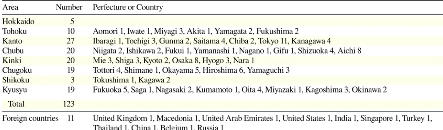
Table 1. The number of medical facilities that requested for genetic testing in our laboratory*
Area Number Perfecture or Country Hokkaido 5
Tohoku 10 Aomori 1, Iwate 1, Miyagi 3, Akita 1, Yamagata 2, Fukushima 2
Kanto 27 Ibaragi 1, Tochigi 3, Gunma 2, Saitama 4, Chiba 2, Tokyo 11, Kanagawa 4 Chubu 20 Niigata 2, Ishikawa 2, Fukui 1, Yamanashi 1, Nagano 1, Gifu 1, Shizuoka 4, Aichi 8 Kinki 20 Mie 3, Shiga 3, Kyoto 2, Osaka 8, Hyogo 3, Nara 1
Chugoku 19 Tottori 4, Shimane 1, Okayama 5, Hiroshima 6, Yamaguchi 3 Shikoku 3 Tokushima 1, Kagawa 2
Kyusyu 19 Fukuoka 5, Saga 1, Nagasaki 2, Kumamoto 1, Oita 4, Miyazaki 1, Kagoshima 3, Okinawa 2 Total 123
Foreign countries 11 United Kingdom 1, Macedonia 1, United Arab Emirates 1, United States 1, India 1, Singapore 1, Turkey 1, Thailand 1, China 1, Belgium 1, Russia 1
*Tottori University Hospital excluded.
1 committee), and also according to the “Guidelines for Genetic Tests and Diagnoses in Medical Practice” (February 2011) approved by the Japanese Association of Medical Sciences. Prenatal diagnosis was approved
by the Ethical Committee of Tottori University.
Methods
Direct Sequencing of the PCR product is the main method to detect point mutations, small deletions and insertions. In the past, single strand conformation poly-morphism (SSCP) and denaturing high performance liquid chromatography (DHPLC) were used in mutation screenings for porphyria, holoprosencephaly and Joubert Syndrome. Mutations in the aberrant PCR product de-tected by SSCP or DHPLC were confirmed by direct sequencing. SSCP and DHPLC were used from Janu-ary 2000 to October 2001, and from July 2004 to April 2010, respectively.
The length of the triplet repeat was determined in the diagnosis of myotonic dystrophy, fragile X syndrome and dentatorubral-pallidoluysian atrophy. Multiplex ligation-dependent probe amplification (MLPA) was used for diagnosis of Fabry disease, Rett syndrome, Pelizaeus-Merzbacher disease, Duchenne muscular dys-trophy, von Hippel-Lindau disease and spinal muscular atrophy, because these diseases are often caused by large deletions and insertions. Real-time PCR was specially used to detect deletions or insertions in the genes that cause Pelizaeus-Merzbacher disease, Gaucher disease, Menkes disease and metachromatic leukodystorophy. Disease and gene information
Information on diseases and causative genes was ob-tained from Online Mendelian Inheritance in Man and Gene database provided by the National Center for Bio-technology Information.
PCR
Before 2009, the primers were designed according to pub-lished data with variable rates of successful amplification. From 2010, almost all primers were designed originally in our laboratory using Primer3 software utilizing the gene information from Entrez Gene and RefSeq, resulting in successful amplification in almost all cases. The coding sequence with 70 bp of the flanking area of each exon was amplified by PCR. The annealing temperature was set relatively high (around 63 ˚C) (Table 2). The sequences of the primers are available on request.
AmpliTaq Gold (Life Technologies, Carlsbad, CA) was routinely used except for the amplification of GC rich sequences (more than 70%). For GC rich sequences, TaKaRa LA Taq (Takara Bio, Otsu, Japan) was used. The reaction mixture contained in a 10 µL reaction vol-ume 10 to 100 ng of template DNA, 2.5 U AmpliTaq Gold, 250 µM or 400 µM dNTP, 1 µM of each primer (forward and reverse) and was run at the following con-ditions: initial denaturation at 95 ˚C for 5 min, followed by 30 cycles with denaturaion at 95 ˚C for 1 min, anneal-ing at 60 ˚C for 1 min, extension at 72 ˚C for 1 min and final extension at 72 ˚C for 5 min. For GC-rich or rela-tively long sequences, 0.5 U TaKaRa LA Taq was used instead of AmpliTaq Gold. The reaction contained 400 µM dNTP and was run as follows: initial denaturation at 94 ˚C for 1 min, followed by 30 cycles with denaturaion at 94 ˚C for 30 s, annealing at 60 ˚C for 30 s, extension at 72 ˚C for 2 min and final extension at 72 ˚C for 5 min.
For diagnosis of incontinentia pigmenti and Fukuyama-type congenital muscular dystrophy, long insertion or deletion was detected according to the meth-ods described by Bardaro et al.3 and Kato et al.4,
respec-tively.
Direct sequencing
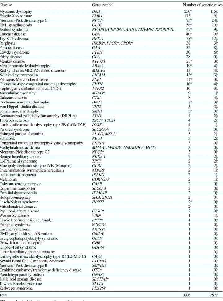
electro-Table 2. List of inherited diseases surveyed for genetic mutations in our facility
Disease Gene symbol Number of genetic cases
Myotonic dystrophy DM1 250* 115‡
Fragile X syndrome FMR1 173 19‡
Niemann-Pick disease type C NPC1† 73* 24‡
GM1 gangliosidosis GLB1 56* 20‡
Joubert syndrome NPHP1†, CEP290†, AHI1†, TMEM67, RPGRIP1L 42* 9‡
Gaucher disease GBA 40* 9‡
Tay-Sachs disease HEXA 38* 12‡
Porphyria HMBS†, PPOX†, CPOX† 38 3‡
Pompe disease GAA 32 8‡
Cowden syndrome PTEN 30 6‡
Fabry disease GLA 28 5‡
Menkes disease ATP7A† 23* 3‡
Metachromatic leukodystrophy ARSA† 19* 4‡
Rett syndrome/MECP2-related disorders MECP2 13 4‡
X-linked hydrocephalus L1CAM 13* 3‡
Pelizaeus-Merzbacher disease PLP1 11* 3‡
Fukuyama-type congenital muscular dystrophy FKTN 10* 4‡
Nephrogenic diabetes insipidus (NDI) AVPR2 10 3‡
Myotubular myopathy MTM1† 9 1‡
Galactosialidosis CTSA 8 4‡
Duchenne muscular dystrophy DMD 7* 3‡
Von Hippel-Lindau disease VHL† 5 3‡
Spinal muscular atrophy SMN1 5* 0‡
Dentatorubral-pallidoluysian atrophy (DRPLA) ATN1 4 2‡
Tuberous sclerosis TSC1†, TSC2† 4 2‡
Limb-girdle muscular dystrophy type 2B (LGMD2B) DYSF† 4 1‡
Pendred syndrome SLC26A4† 3 3‡
Enlarged parietal foramina ALX4†, MSX2† 3 2‡
Sialidosis NEU1 3 2‡
Congenital muscular dystrophy-dystroglycanopathy FKRP† 3 0‡
Methylmalonic acidemia MMAA†, MMAB†, MMADHC†, MUT† 3 0‡
Niemann-Pick disease type C2 NPC2† 3 0‡
Benign hereditary chorea NKX2-1 2 2‡
Li-Fraumeni syndrome TP53 2 2‡
Mucopolysaccharidosis type IVB (Morquio) GLB1 2 2‡
Dyschromatosis symmetrica hereditaria ADAR† 2 1‡
Incontinentia pigmenti IKBKG 2 1‡
Melanoma CDKN2A† 2 1‡
Calcium-sensing receptor CASR 2 0‡
Dopamine transporter SLC6A3 2 0‡
Familial dysautonomia IKBKAP 2 0‡
Holoprosencephaly SHH, ZIC2† 2 0‡
Lesch-Nyhan syndrome HPRT1 2* 0‡
Mitochondrial diseases – 2 0‡
Papillon-Lefevre disease CTSC† 2 0‡
Werner Syndrome WRN† 1 1‡
Ceroid lipofuscinosis, neuronal, 1 PPT1† 1 0‡
Feingold syndrome MYCN† 1 0‡
Gardner syndrome AXIN1† 1 0‡
GM2-gangliosidosis, AB variant GM2A† 1 0‡
Greig cephalopolydactyly syndrome GLI3† 1 0‡
Growth hormone receptor GHR 1 0‡
Klippel-Feil syndrome GDF6† 1 0‡
Leber hereditary optic neuropathy – 1 0‡
Limb-girdle muscular dystrophy type 1C (LGMD1C) CAV3 1 0‡
Nevoid Basal Cell Carcinoma syndrome PTCH1† 1 0‡
Niemann-Pick disease type B SMPD1† 1 0‡
Ornithine carbamoyltransferase deficiency disease OTC† 1 0‡
Pseudohypoparathyroidism GNAS† 1 0‡
Sialic acid storage disease SLC17A5† 1 0‡
Townes-Brocks syndrome SALL1 1 0‡
Zellweger syndrome PEX26† 1 0‡
Total 1006 287‡
*The number includes the cases of prenatal diagnosis †The PCR primers were originally designed in our laboratory
40
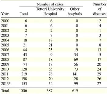
Table 3. The number of cases subjected to genetic testing from January 2000 to September 2013
Number of cases Number Tottori University Other of Year Total Hospital hospitals diseases
2000 6 6 0 2 2001 6 6 0 4 2002 2 2 0 1 2003 7 7 0 3 2004 18 18 0 7 2005 21 21 0 8 2006 44 25 19 13 2007 43 9 34 5 2008 87 18 69 17 2009 74 18 56 20 2010 128 55 73 24 2011 219 78 141 29 2012 198 70 128 26 2013* 153 54 99 27 Total 1006 387 619 * January to September 2013.
phoresis and were purified by Ultrafree-DA centrifugal filter unit (Millipore, Billerica, MA). The sequence was determined by using BigDye Terminator v3.1 Cycle Se-quencing Kit and capillary sequencer 3130xL Genetic Analyzer (Life Technologies).
SSCP
As described previously, PCR products were separated on 12% polyacrylamide gel at room temperature or 4 ˚C, and were visualized by silver staining.5
DHPLC
A WAVE-MD Mutation Detection System (Transgenomic, Omaha, NE) based on the DHPLC method was used to detect heteroduplex states according to the manufac-turer’s instructions.
Fragment analysis for triplet repeat diseases PCR in a total volume of 10 µL contained 100 ng of DNA, 2.5 U AmpliTaq Gold DNA Polymerase, 250 µM dNTP, 10% DMSO, 1 µM each primer (forward and reverse), 0.1 µM of the fluorescent labeled probe (FAM, HEX or NED) and was run as follows: initial denatur-ation at 94 ˚C for 5 min, followed by 30 cycles with denaturation at 95 ˚C for 1 min, annealing at 55 ˚C for 1 min, extension at 72 ˚C for 1 min, and final extension at 72 ˚C for 5 min. The PCR products were mixed with GeneScan 500 ROX Size Standard (Life Technologies). After incubation at 95 ˚C for 5 min, the samples were immediately cooled and electrophoresed by 3130xL Ge-netic Analyzer. Before 2005, ALFred DNA Sequencer (Amersham Pharmacia Biotech, Piscataway, NJ) was used.
MLPA
An MLPA kit (MRC-Holland, Amsterdam, the Nether-lands) was used according to the manufacturer’s instruc-tions. In brief, 80 mg of DNA in 6% of glycerol was in-cubated at 98 ˚C for 15 min for initial denaturation. The final reaction mixture was electrophoresed by a 3130xL Genetic Analyzer using a 50 cm-long capillary POP-7. Real-time PCR
When there was any inconsistency in the results of mu-tations in family members, real-time PCR was used to confirm the presence of deletions or duplications around the mutations.
Data analysis
Sequence data was analyzed by Sequence Scanner (Life Technologies) and Genetyx Software (Genetyx, Shibuya, Japan). Variant Reporter Software (Life Technologies) was used for detection of mutations from 2011. The
mu-tations were confirmed using the human genome muta-tion database (BIOBASE, Halchtersche, Germany) and SNP database. Prediction of functional effect of human nsSNPs (PolyPhen) was used for evaluation of novel mu-tations.6
Fragment analysis data was analyzed by Peak Scan-ner Software v1.0 (Life Technologies) for the repeat size. MLPA data was analyzed by Coffalyzer Software (MRC-Holland).
The personale and running processes of the facility After October 2010, the working force to run the facility consists of 1 senior medical genetist, 1 assistant
profes-Fig. 1. The number of DNA samples subjected to genetic testing from January 2000 to September 2013. Vertical bars show the number of cases. Light gray shows the number from other hospi-tals, dark gray shows the number from Tottori University Hospital. Dots show the number of diseases.
0 5 10 15 20 25 30 35 0 50 100 150 200 250 2000 2001 2002 2003 2004 2005 2006 2007 2008 2009 2010 2011 2012 2013 * Number of diseases Number of cases Year
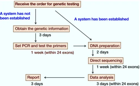
Fig. 2. A flow chart of the genetic diagnosis using direct sequencing of PCR products. When the system has not been established, it is necessary to obtain the genetic information and design new PCR primers. In this case, it takes at least 3 weeks to detect mutations. When the system has already been established, it takes 2 weeks.
sor and 6 part-time staff. The diagnostic procedures are separated into steps such as DNA preparation, PCR, purification, etc., each of which takes approximately 3 working hours, and the samples were processed sequen-tially by the part-time staff. The senior medical genetist is responsible for genetic counseling to the patients and family members, as well as conformation of mutations and preparation of the final report.
RESULTS
Distribution of the hospitals that requested for ge-netic testing
The hospitals that requested for genetic testing were lo-cated 43 in prefectures of Japan and in 11 foreign coun-tries (Table 1).
The number of DNA samples subjected to genetic testing
A total of 1,006 genetic tests covering 62 diseases were performed from January 2000 to September 2013 (Table 2). The numbers of samples exceeded 50 in myotonic dystrophy, fragile X syndrome, Niemann-Pick disease type C and GM1-gangliosidois. Original primers were constructed for 41 diseases. The number of genetic tests gradually increased year by year and did so rapidly af-ter we introduced a new service system in 2010 (Table 3, Fig. 1). All samples were analyzed individually, and mutations were detected in 287 out of 1,006 with an average mutation detection rate of 28.5%. Prenatal di-agnosis covered 13 diseases and 77 DNA samples were analyzed.
Flow chart of genetic testing
After 2010, it started to take at least 2 weeks for each ge-netic test to be completed. An additional one and a half weeks is required if it is necessary to settle new screen-ing procedures (Fig. 2). PolyPhen software was used to predict whether any mutation detected was disease-causing or not.
DISCUSSION
We have received a number of requests for genetic test-ing from all over Japan. Institutes that provide genetic testing are rare, and our institute provides a unique func-tion for the Japanese scientific and medical fields. Actu-ally, genetic testing has contributed to several scientific reports.7–18 The number of cases of myotonic dystrophy
was particularly high partly because the genetic testing was approved by Ministry of Health, Labour and Wel-fare of Japan as “highly advanced medical treatment”. It may also be because our facility is the only one ap-proved for conducting prenatal diagnosis of this disease in Japan.19 The numbers of cases of fragile X syndrome,
Niemann-Pick disease type C, GM1-gangliosidosis and other lysosomal storage diseases are also high because these diseases were the subjects of active research at Tottori University.20–22
Direct sequencing by a capillary sequencer is the main method of genetic testing and the size of the repeat was analyzed for triplet repeat diseases. MLPA was used for deletions or insertions of exons. For Joubert syndrome, 19 or more disease-causing genes were re-ported. Direct sequencing of every exon of all genes is impractical. In such cases, SSCP or DHPLC was used
42 for initial screening. These methods are prone to give false-negative results and it will be better to use next-generation sequencers,23–25 which were introduced to
Tottori University in 2013.
The efficiency of genetic testing has improved and new lines of genetic testing have been enabled since 2010 by the improvement of running processes. Genetic testing is often still conducted based on research, and the expense is usually paid for by research funds. Because the expense is limited and genetic testing is necessary for clinical diagnosis of many inherited diseases, the costs should be included in medical expenses. At Tottori University Hospital, we have settled on basic original rules that enable us to refer the cost to the patient or cli-ent.
Genetic testing performed for research lacks the high quality required in clinics. In the United States and Europe, the quality of genetic testing is secured by Clinical Laboratory Improvement Amendments.26
Un-fortunately there is no guideline for quality control in Japan. At our facility, testing is performed according to the Standard Operating Procedures and the information is shared by each member. Genetic testing is now a pop-ular and powerful tool for diagnosis of many inherited diseases. We should promote it further in the future.
Acknowledgments: I would like to thank: Professor Eiji Nanba and all technical staff of the Division of Functional Genomics, Research Center for Bioscience and Technology for building our system of genetic testing and helpful discussion; Dr. Kousaku Ohno, Emeritus Professor of Tottori University and the staff of the Division of Child Neurology, Department of Brain and Neurosci-ence, Faculty of Medicine for collection of the samples and help-ful discussion; and Professors Tatsuo Watanabe, Ichiro Hisatome and Haruaki Ninomiya for helpful discussion. I would also like to acknowledge all the doctors at our hospital and other hospitals who provided samples. Finally, thanks to the Center for Promoting Next-Generation Highly Advanced Medicine at Tottori University Hospital, which supported the acceptance of samples and sending reports.
This work was supported by a JSPS Grant-in-Aid for Sci-entific Research (B) 21659257, 22390207 and 25293230, Health Labour Sciences Research Grant (Research on Measures for In-tractable Diseases) H21-InIn-tractable-public-022, H22-InIn-tractable- H22-Intractable-public-002, H22-Intractable-public-014 and H22-Intractable- public-126.
The authors declare no conflict of interest. REFERENCES
1 Jacob HJ. Next-generation sequencing for clinical diagnostics. N Engl J Med. 2013;369:1557-8. PMID: 24088040.
2 Veltman JA, Brunner HG. De novo mutations in human genet-ic disease. Nat Rev Genet. 2012;13:565-75. PMID: 22805709. 3 Bardaro T, Falco G, Sparago A, Mercadante V, Gean Molins E,
Tarantino E, et al. Two cases of misinterpretation of molecular results in incontinentia pigmenti, and a PCR-based method to discriminate NEMO/IKKgamma dene deletion. Hum Mutat.
2003;21:8-11. PMID: 12497627.
4 Kato R, Kawamura J, Sugawara H, Niikawa N, Matsumoto N. A rapid diagnostic method for a retrotransposal insertional mutation into the FCMD gene in Japanese patients with Fukuyama congenital muscular dystrophy. Am J Med Genet A. 2004;127A:54-7. PMID: 15103718.
5 Zhang H, Nanba E, Yamamoto T, Ninomiya H, Ohno K, Mizuguchi M, et al. Mutational analysis of TSC1 and TSC2 genes in Japanese patients with tuberous sclerosis complex. J Hum Genet. 1999;44:391-6. PMID: 10570911.
6 Wu J, Jiang R. Prediction of deleterious nonsynonymous single-nucleotide polymorphism for human diseases. Scienti-ficWorldJournal. 2013:675851. PMID: 23431257.
7 Fujimoto S, Manabe Y, Fujii D, Kozai Y, Matsuzono K, Takahashi Y, et al. A novel mutation of the GAA gene in a pa-tient with adult-onset Pompe disease lacking a disease-specific pathology. Intern Med. 2013;52:2461-4. PMID: 24190153. 8 Chiba Y, Komori H, Takei S, Hasegawa-Ishii S, Kawamura N,
Adachi K, et al. Niemann-Pick disease type C1 predominantly involving the frontotemporal region, with cortical and brain-stem Lewy bodies: an autopsy case. Neuropathology. 2013. PMID: 23711246.
9 Sekijima Y, Nakamura K, Kishida D, Narita A, Adachi K, Ohno K, et al. Clinical and serial MRI findings of a sialidosis type I patient with a novel missense mutation in the NEU1 gene. Intern Med. 2013;52:119-24. PMID: 23291686.
10 Xiong H, Higaki K, Wei CJ, Bao XH, Zhang YH, Fu N, et al. Genotype/phenotype of 6 Chinese cases with Niemann-Pick disease type C. Gene. 2012;498:332-5. PMID: 22326530. 11 Muraoka T, Murao K, Imachi H, Kikuchi F, Yoshimoto T,
Iwama H, et al. Novel mutations in the gene encoding acid -1,4-glucosidase in a patient with late-onset glycogen storage disease type II (Pompe disease) with impaired intelligence. Intern Med. 2011;50:2987-91. PMID: 22185990.
12 Higaki K, Li L, Bahrudin U, Okuzawa S, Takamuram A, Yamamoto K, et al. Chemical chaperone therapy: chaper-one effect on mutant enzyme and cellular pathophysiology in -galactosidase deficiency. Hum Mutat. 2011;32:843-52. PMID: 21520340.
13 Watanabe T, Yoshida Y, Adachi K, Nanba E, Yamamoto O. Multiple subcutaneous hard nodules. Clin Exp Dermatol. 2010;35:681-2. PMID: 20642802.
14 Ishii K, Hosaka A, Adachi K, Nanba E, Tamaoka A. A Japa-nese case of fragile X associated tremor/ataxia syndrome (FXTAS). Intern Med. 2010;49:1205-8. PMID: 20558944. 15 Kinoshita T, Hanaki K, Nagaishi J, Kawashima Y, Adachi K,
Nanba E, et al. Variation analysis of beta3-adrenergic receptor and melanocortin-4 receptor genes in childhood obesity. Pedi-atr Int. 2007;49:133-7. PMID: 17445027.
16 Maeda N, Horie Y, Sasaki Y, Adachi K, Nanba E, Nishida K, et al. Three novel mutations in the protoporphyrinogen oxi-dase gene in Japanese patients with variegate porphyria. Clin Biochem. 2000;33:495-500. PMID: 11074242.
17 Maeda N, Horie Y, Adachi K, Nanba E, Kawasaki H, Daimon M, et al. Two deletion mutations in the hydroxymethylbilane synthase gene in two unrelated Japanese patients with acute intermittent porphyria. J Hum Genet. 2000;45:263-8. PMID: 10944860.
18 Kotani K, Shimomura T, Murakami F, Ikawa S, Kanaoka Y, Ohgi S, et al. Allele frequency of human endothelial nitric oxide synthase gene polymorphism in abdominal aortic aneu-rysm. Intern Med. 2000;39:537-9. PMID: 10888208.
Yamamoto T, et al. Prenatal diagnosis of congenital myotonic dystrophy in two Japanese families: direct mutation analysis by a non-radioisotope PCR method and haplotype analysis with flanking DNA markers. Brain Dev. 1996;18:122-6. PMID: 8733903.
20 Nanba E, Kohno Y, Matsuda A, Yano M, Sato C, Hashimoto K, et al. Non-radioactive DNA diagnosis for the fragile X syndrome in mentally retarded Japanese males. Brain Dev. 1995;17:317-21. PMID: 8579216.
21 Yamamoto T, Nanba E, Ninomiya H, Higaki K, Taniguchi M, Zhang H, et al. NPC1 gene mutations in Japanese patients with Niemann-Pick disease type C. Hum Genet. 1999;105:10-6. PMID: 10480349.
22 Nishimoto J, Nanba E, Inui K, Okada S, Suzuki K. GM1-gan-gliosidosis (genetic beta-galactosidase deficiency): identifica-tion of four mutaidentifica-tions in different clinical phenotypes among Japanese patients. Am J Hum Genet. 1991;49:566-74. PMID: 1909089.
23 Tsuji S. Genetics of neurodegenerative diseases: insights from high-throughput resequencing. Hum Mol Genet. 2010;19:R65-70. PMID: 20413655.
24 Najmabadi H, Hu H, Garshasbi M, Zemojtel T, Abedini SS, Chen W, et al. Deep sequencing reveals 50 novel genes for recessive cognitive disorders. Nature. 2011;478:57-63. PMID: 21937992.
25 Koshimizu E, Miyatake S, Okamoto N, Nakashima M, Tsurusaki Y, Miyake N, et al. Performance comparison of bench-top next generation sequencers using microdroplet PCR-based enrichment for targeted sequencing in patients with autism spectrum disorder. PLoS One. 2013;8:e74167. PMID: 24066114.
26 Tsuji S, Matsubara Y, Okuyama T, Goto Y, Saito K, Miyachi H, et al. [General working report for a study on the effective enforcement system for the diagnosis by genetic techniques: from the second quarter of 2010 to the first quarter of 2012. Research on measures for intractable diseases; Japan Health and Labour Sciences Research Grant]. MHLW Grant System [Internet]. Tokyo: National Institute of Public Health (Japan); 1997 - . [cited 2013 Jun 10]. 118 p. Available from: http:// mhlw-grants.niph.go.jp/niph/NIDD00:201231014B. Japanese.