総 説
プリオン病
―本邦の特徴と診断のポイント―
三條 伸夫
*水澤 英洋
要旨:プリオン病は正常プリオン蛋白が伝播性を有する異常プリオン蛋白に変化し蓄積することにより発症す る.孤発性,遺伝性,獲得性の 3 種類があり,本邦では 1999 年からの約 10 年間のサーベイランス調査で 1,320 名の患者が確認され,硬膜移植 CJD 例が多いことや遺伝性 CJD で本邦に特異的な変異例が多いなどの特徴があ る.孤発性古典型では急速進行性の認知症,四肢のミオクローヌス,MRI 拡散強調画像で大脳皮質と基底核の高信 号,脳波で PSD などの特徴的な所見をみとめるが,非典型例も少なくない.臨床症状,髄液検査,遺伝子検索によ り的確な診断をくだすことが,病態の解明,二次感染予防,心理サポートにおいて重要である. (臨床神経 2010;50:287-300) Key words:プリオン病,クロイツフェルト・ヤコブ病,伝達性海綿状脳症,ゲルストマン・ストロイスラー・シャイン カー病,致死性家族性不眠症 はじめに プリオン病は正常プリオン蛋白が何らかの理由で伝播性を 有する異常プリオン蛋白に変化し,主に中枢神経内に蓄積す ることにより神経細胞変性をおこすまれな致死性疾患であ る.プリオン病の代表的なタイプである孤発性クロイツフェ ルト・ヤコブ病(CJD)は 1 年間に 100 万人に 1 人程度の割合 で発症することが知られている1).我が国では 1999 年から臨 床調査個人票や感染症法の登録制度をもちいたサーベイラン ス調査が続けられており,2009 年 12 月までに 2,400 名以上の 患者の調査がおこなわれ,1,320 名がプリオン病として登録さ れている.未だに有効な治療法は無く,プリオン病の約 80% を占める孤発性古典型 CJD は症状の進行が早く,発症から 3∼7 カ月で確実に無動性無言になり2)3),多くは肺炎などの合 併症で死亡する.異常プリオン蛋白は伝播性を有する感染因 子そのものと考えられており,蛋白性感染粒子という意味で 作られた言葉がプリオンである.プリオン病は人畜共通感染 症であり,伝達性海綿状脳症(transmissible spongiform en-cephalopathy;TSE)としてヒト以外では羊や山羊のスクレ ピー(Scrapie),牛の牛海綿状脳症(bovine spongiform en-cephalopathy;BSE),鹿の慢性消耗病(chronic wasted dis-ease;CWD)などが知られている.我が国では 2003 年から五 類感染症に分類されており,医師は診断後 7 日以内に保健所 へ報告することが義務づけられている. プリオン蛋白 正常プリオン蛋白の遺伝子は第 20 番染色体に存在し,ヒト のプリオン蛋白は 253 個のアミノ酸で構成されており(Fig. 1),アミノ基末端側(N 末側)に小胞体移行シグナルやオクタ ペプチドリピート配列(8 アミノ酸配列の 5 回くりかえし)が あり,カルボキシル基末端側(C 末側)の 181 番目と 197 番目 のアスパラギンがゴルジ体で糖鎖修飾を受ける(後述).また, 179 番目と 214 番目のシステインはジスルフィド結合を受 け,231 番 目 の セ リ ン は 細 胞 膜 上 に 係 留 さ れ る た め の glycosyl-phosphatidyl-inositol(GPI)アンカーが付加する4). マウスではほぼすべての臓器での発現が確認されているが, 脳での発現量がもっとも多い.脳では海馬,尾状核,視床の神 経細胞や周辺の神経網(ニューロピル)で多く発現している. 正常プリオン蛋白の機能は未だ明らかにされておらず,細胞 レベルでの抗ストレス作用5)や,アポトーシス誘発促進作用6) などの報告があり,ノックアウト・マウス個体においては神 経系をふくめ発達段階での異常をみとめず7),異常プリオン蛋 白感染に対し抵抗性であることなどが確認されている8).異常 プリオン蛋白は正常プリオン蛋白とアミノ酸配列は同じであ るが高次構造がことなっており(Fig. 2),正常プリオン蛋白で は 3% 以下 で あ るβ シート構造が異常プリオン蛋白では 40% 以上になっている9).異常プリオン蛋白は,正常プリオン 蛋白とことなり難溶性で凝集しやすく,蛋白分解酵素 prote-inase K により切断はされても分解されない性質を持ってい * Corresponding author: 東京医科歯科大学大学院脳神経病態学〔〒113―8519 東京都文京区湯島 1―5―45〕 東京医科歯科大学大学院脳神経病態学 (受付日:2010 年 3 月 3 日)Fig. 1 Constitution ofprion protein and frequency ofcodon 129 polymorphism.Prion protein is254 amino acidsin length and a highly conserved protein among the mammalian species.GPIanchoris attached to the C-terminuspostcleavage ofthe signalsequence,and addition oftwo N-linked glyc o-sylationsoccursatAsn 181 and 197.A disulfide bridge isformed between Cys179 and 214. To-ward the N-terminusisthe octapeptide repeatregion which hasHisresiduessuggested to play a role in metalbinding.Atthe end ofN-terminus,endoplasmicreticulum transition signalsequence iscleaved atthe Golgiapparatus.Two polymorphismicsitesare known atcodon 129 and 219.Al -though 129MM form isthe mosttype in Japan,129MV the mostcommon in UK.
codon 129 polymorphism MM MV VV (%)
Japan 92 8 0
UK 37 51 12
NH3 COOH
51 91 131 179 214
GPI anchor binding site Codon 219 polymorphism codon 129 polymorphism octa-peptide repeat PHGGGWGQ/−/−/−/PHGGGWGQ glycosylation site 253 22 ER transition signal S-S bond 181 197 231
Fig. 2 Conformationalchange ofprion proteins.In prion disease,endogenousnormalprion protein convertsfrom the alpha-helix-rich form (left)into the mainly beta-sheetcontaining form (right), which leadsto the accumulation ofamyloid fibrilsin the centralnervoussystem (CNS)and to neurodegeneration.(文献(40)より改変)
[Huang, et al. 1995] る(Fig. 3)10).プリオン蛋白がプリオン病における病因物質で あると同時に,感染因子であるとする「プリオン仮説」は 1982 年に Stanley Prusiner により提唱され11),その後正常型プリ オン蛋白不在状態における不感染性8),in vitro における正常 型から異常型への変換12),リコンビナントプリオン蛋白の感 染性13),in vitro 産生の異常型様プリオン蛋白の感染性14)など が報告され,プリオン仮説は広く支持されるようになった.プ リオン蛋白異常化の機序に関しては,異常プリオン蛋白 1 分
Table 1 Classification ofhuman prion diseases.
IdiopathicPrion disase SporadicCJD
Classicform orHeidenhain variant;MM1/MV1 Ataxicform;VV2,MV2 (Kuru-plaquesvariant)
Thalamicform (fatalsporadicinsomnia;FSI,MM2-thalamicform);MM2 Corticalform;MM2 (MM2-corticalform),VV1
Environmentally acquired Prion disease Kuru
IatrogenicCJD (dura mater,neurosurgery,depth electrode,cornealtransplant,human growth hormon,human gonadtrophin) variantCJD
Genetic(hereditary)Prion disease FamilialCJD
Gerstmann-Sträussler-Scheinkerdisease (GSS) Fetalfamilialinsomnia (FFI)
others
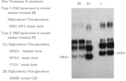
Fig. 3 Western blotting patternsofprion proteinsofsporadicCJD digested by protease K.Prot e-inase K-resistantprion protein fragmentsfrom brain extractsof3 subjectswith sporadicCJD and 2 subjectswith variantCJD representative ofthe three groups.The fragmentsmigrate asthree majorisoforms,unglycosylated,monoglycosylated and diglycosylated forms.Two typesofthe fragmentsin size and glycosylation were detected in sporadicCJD (1 and 2A),and anothertype wasdetected in variantCJD (2B).The top ofthe three bandsin each lane isdiglycosylated form and the bottom isunglycosylated form.Strongerbandsindicate higheramountofproteinsthan weakerbands,which isa classificationalindex.
After Proteinase K treatment, Type 1; 21kD (processed at around
aminno terminal 82) Diglycoform<Non-glycoform
MM1, MV1: classic form Type 2; 19kD (processed at around
aminno terminal 97) 2A; Diglycoform<Non-glycoform MM2A:thalamic form MV2A:ataxic form VV2A:ataxic form 2B; Diglycoform>Non-glycoform MM2B: variant CJD 19KD 2B 2A 1 21KD [遅発性ウイルス感染に関する調査研究班報告書,2002] 子と正常プリオン蛋白 1 分子が会合してヘテロダイマーとな ることで正常プリオン蛋白が異常プリオン蛋白に変換すると する「ヘテロダイマー説」15)と異常プリオン蛋白が複数結合し て核(seed)を形成し,この seed が正常プリオン蛋白を取り 込むことにより異常プリオン蛋白化し重合・増大してゆくと する「核依存性重合モデル」16)の 2 通りの説がある.さらに, 異常プリオン蛋白による神経細胞変性のメカニズムもまだ明 らかにされていないが,感染動物の脳では接種後に神経樹状 突起の変性に先立って Notch-1 の発現量が増加することが報 告されており17),関連性が示唆されている. プリオン蛋白遺伝子には正常多型があり(Fig. 1),1 つは 129 番目のアミノ酸(codon 129)がメチオニン(M),または バリン(V)かでそれぞれ MM,MV,VV の 3 種類に分類さ れる.日本人では MM 型がほとんどであるのに対し,白人で は MM 型は約半数程度である(Fig. 1).もう 1 つは codon 219 の多型でグルタミン酸(E)とリジン(K)の組み合わせで EE, EK,KK の 3 種類である.codon 129 のバリンや codon 219 のリジンの多型はプリオン病感染に対し抵抗性が指摘されて いる18)∼20).
Fig. 4 Frequency ofprion diseasesin Japan. Idiopathic (77.2%) sporadic CJD: 1,019 cases Hereditary (16.7%) Familial CJD: 172 cases (13.0%)* GSS: 45 cases (3.4%) FFI: 3 cases (0.2%) Environmentaly aquired (6.1%) Variant CJD: 1 case (0.1%)
Dura-graft associated CJD: 80 cases (6.0%)
*including 6 cases without confirmation of PrP gene mutation 2 cases were unclassified (0.2%)
1,305 cases during Apr, 1999-Sep, 2009 M : F ratio 554 : 751 (42 : 58%) Age at Onset 65.5±12.6 (15-94) yo Duration 18.5±22.3 (1-226) mo
Fig. 5 Periodicsynchronousdischarge (PSD)ofEEG detected in sporadicclassicCJD patient.In the majority ofcasesgeneralized bi-ortriphasicperiodicsharp wave complexesappearwith a fr e-quency ofaround 1-2 persecond (left).In an appropriate clinicalcontext,the appearance ofthis EEG pattern isstrongly supportive ofa diagnosisofsporadicCJD.In laterstage ofmostcases,this periodicdischarge gradually disappearand slow wavesbecome dominant.
At onset 5 month later
Fp F aT mT pT C P O ヒトのプリオン病 ヒトのプリオン病は病因により,原因不明の特発性(孤発性 CJD;sporadic CJD(sCJD)),プリオン蛋白遺伝子変異によ る 遺 伝 性(家 族 性 CJD;Gerstman-Sträussler-Scheinker 病 (GSS);致 死 性 家 族 性 不 眠 症(fatal familial insomnia: FFI)),他のプリオン病からの感染による獲得性(environ-mentally aquired)(ク ー ル ー,医 原 性,変 異 型(variant: vCJD))の 3 種類に分類される(Table 1). 異常プリオン蛋白は proteinase K 処理により N 末側が断 片化され,ウエスタンブロッティングにより糖鎖修飾の違い による 3 本のバンドが検出される(Fig. 3).181 番目と 197 番目の両方のアスパラギンに糖鎖が付加した diglycoform, いずれか一方のみの monoglycoform,糖鎖付加のない non-glycoform があり,non-non-glycoform が 21kDa のものを 1 型,19 kDa のものを 2 型とし,このバンドのパターン(1 と 2)とプ リオン蛋白遺伝子の codon 129 の多型によるアミノ酸の組み 合わせ(MM と MV と VV)により,sCJD の分類がなされて いる(Parchi 分類;Table 1 孤発性 CJD の欄)2) .最近,prote-inase K で分解される異常プリオン蛋白が蓄積する病型も報 告された21). 本邦では 1996 年に vCJD の発症を受けてプリオン病に関 する緊急全国調査に始まり,1997 年から 1999 年まで CJD および類縁疾患調査がおこなわれ,1999 年から今日までは特 定疾患の調査研究班の中にサーベイランス委員会を組織して
Fig. 6 MRIimagesofsporadicclassicCJD.Asymmetricalhyperintensity ofbasalganglia and corti calribbon isseen in MRIofpatients(upperpanelsofDWI).In laterstage,hyperintensity ofwhite matterisdetected in some patients.
T1WI DWI
Fig. 7 Pathology ofsporadicclassicCJD.(a)Microscopicappearance ofHE staining ofthe brain r e-vealed spongiform change.(b)Synaptic-type deposition ofprion protein wasdetected in atypical sCJD cases(Provided by prof.Masahito Yamada ofKanazawa University Graduate SchoolofMedi -calScience).
a
b
調査が続けられている.2009 年 9 月までに 1,305 名がプリオ ン病と認定されており,男女比は 42:58,発症年齢は平均 65.5,罹病期間は平均 18.5 カ月であり,孤発性 CJD が 77.2%, 遺伝性が 16.7%,獲得性が 6.1% であった(Fig. 4).感染性プ リオン病は 1 例の変異型 CJD 以外すべて硬膜移植後 CJD で あり,新規発症の頻度は減少しつつあるが,最長の潜伏期はFig. 8 SPECT of MM2 thalamic form of sporadic CJD. Cerebralblood flow (CBF)study using SPECT showed re duction ofthe CBF in the thalamusaswellasthe cerebral cortex (provided by Dr.YuseiShiga ofAoba Neurosur gery Hospital). 30 年であり,今後も発症があるものと思われる.遺伝性プリ オン病は遺伝子検索の普及により近年その頻度が増加し,現 在は 30 種以上の変異が知られている.本邦の遺伝性プリオン 病では家族歴のないタイプの方がむしろ多いので注意が必要 である. 孤発性 CJD22)23) (A)孤発性古典型 CJD 急速に進行する認知症症状とミオクローヌスを特徴として おり,Parchi 分類の MM1 と MV1 に相当する.罹患率は 100 万人に 1 人で,平均年齢が 67.1 歳であり,臨床病期は以下の 3 期に分類されている. 第 1 期はおおよそ 1∼2 カ月間で,食欲低下,不安感などの 不定愁訴,歩行障害,視覚異常を呈する.第 2 期は急速に認知 症が進行し,数カ月以内に高度の認知症を呈し,会話や歩行が 不可能になる.錐体路症状,錐体外路症状,小脳失調,ミオク ローヌスなども出現し,典型例では脳波で周期性同期生放電 (periodic synchronous discharge:PSD)をみとめ(Fig. 5), 脳 MRI では拡散強調画像や FLAIR 画像で大脳皮質や基底 核に非対称性の高信号(Fig. 6)をみとめる.髄液検査では 14-3-3 蛋白やタウ蛋白が高値となる.3∼7 カ月で第 3 期となり, 無動無言状態に陥り,除皮質硬直や屈曲拘縮を呈する.経口摂 取が不可能となるため,経鼻経管栄養や胃瘻造設が施され,褥 瘡や肺炎により死亡する.神経病理学的に海綿状脳症をみと め,シナプス型のプリオン蛋白の沈着をみとめる(Fig. 7). (B)孤発性視床型 CJD(MM2-th)
孤発性致死性不眠症(sporadic fatal insomnia:SFI)や視床
変性症と呼ばれることもあり,病変が視床にとくに強くみと められる.致死性家族性不眠症(FFI)のような症状を呈する 例もあるが,認知症,運動失調,自律神経症状など初発症状は 様々で,発症年齢は平均 52.3 歳(36 歳∼71 歳)である.経過 は 8∼24 カ月で平均 15.6 カ月と比較的緩徐である.脳波では PSD をみとめず,MRI 拡散強調画像でも信号変化はみとめな い.髄液 14-3-3 やタウ蛋白も正常であることが多い.脳血流シ ンチ(SPECT)で両側視床の血流低下をみとめることが早期 診断に有用であることが報告されている(Fig. 8)24).神経病理 学的には視床と下オリーブ核に変性をみとめ,大脳皮質には 軽度の海綿状変化をみとめることがある.Parchi 分類では MM2A に相当する. その他の孤発性 CJD の特徴を Table 2 に,診断基準を Ta-ble 3 にまとめた. 遺伝性プリオン病 我が国のサーベイランスデータによると,1999 年 4 月から 2009 年 9 月までの約 10 年間でプリオン病と判定(possible 以上)された 1,305 例のうち 215 例(16.5%)が遺伝性プリオ ン病であった.一方,EUROCJD プロジェクト参加国(オース トラリア,オーストリア,カナダ,フランス,ドイツ,イタリ ア,オランダ,スロバキア,スペイン,スイス,イギリス)の 1993 年から 2002 年までの 10 年間でプリオン病(probable 以上)の判定を受けた 4,441 例のうち 455 例(10.2%)が遺伝 性 プ リ オ ン 病 で あ っ た25)26).各 国 と の 比 較 に お い て も EUROCJD 参加国における遺伝性プリオン病の数はもっとも 多いイタリアで 115 名と我が国の約半数にすぎず,我が国に おける遺伝性プリオン病の頻度は高い可能性がある.また,遺 伝性プリオン病のタイプ別統計(Fig. 9)をみると,我が国に ほぼ特有な V180I,M232R,P105L の 3 型の合計で 125 例と半 数以上を占めており,欧米の比率とはことなっている.現在ま でに 30 種類以上の遺伝子変異と 15 種類の欠失・挿入が報告 されている(Fig. 10). (A)V180I 変異 CJD プ リ オ ン 蛋 白 遺 伝 子 コ ド ン 180 の Valine(V)か ら Iso-leucine(I)への変異による家族性 CJD で,本邦では遺伝性プ リオン病全体の 40% ともっとも多くみとめられているが,欧 米では 1 例の報告のみである25).発症年齢は 44∼93 歳で,平 均約 76 歳である.初発症状は記銘力障害,または失語や失行 などの高次脳機能障害であり,緩徐に進行するため,アルツハ イマー病などと誤診されている例もある.神経学的には小脳 失調や視覚障害は示さず,ミオクローヌスの出現も多くはな い.全経過の平均は約 1.9 年であり,数年にわたるばあいもあ る.末期には寝たきりから無動無言状態となり,感染症などで 死亡する.これまで,V180I で家族内発症が確認された報告は 無く,一見孤発性の発症様式であるが,非典型的な症状で診断 がつきにくいため,診断にはプリオン病遺伝子検索が必須で ある.脳波では約 10% で PSD をみとめる.ほぼ全例で脳 MRI の拡散強調画像で後頭葉と中心溝前後を除いたほぼ全
Table 2 Clinicalclassification and PrPsctypesofsporadicCJD. others PrPSC MRI-DWI hyper intensity tau 14-3-3 PSD myoclonus progression Age at onset Clinical form synapticform + + + + + subacute 60s classic MM1 synapticform + + + + + subacute 60s classic MV1 SPECT (synapticform) - - - - - slow 50s thlamic MM2th (synapticform) + (+ ) (+ ) rare + subacute 60s cortical MM2c plaque form +th (+ ) rare rare + slow 60s ataxic MV2 plaque form +th (+ ) (+ ) rare rare slow 60s ataxic VV2 synapticform + (+ ) (+ ) - - slow 40s cortical VV1 ataxia smallplaque - ? - - - 20 (10 - 60) 62 (48 - 71) cortical PSPr*
*PSPr:Protease-sensitive prionpathy (Gambettietal,Ann Neurol2008;63:697 ― 708) **th:thalamus
Table 3 Diagnosticcriteria ofsporadicCJD.
Possiblecase
Progressive dementia;and EEG atypicalornotcarried out;and duration < 2 years;and
atleasttwo outofthe following clinicalfeatures:myoclonus,visualorcerebellardisturbance,pyramidal,extrapyramidaldysfunction,akinetic mutism.
Probablecase(in theabsenceofan alternativediagnosisfrom routineinvestigation)
Progressive dementia;
atleasttwo ofthe following fourclinicalfeatures:myoclonus,visualorcerebellardisturbance,pyramidal/extrapyramidaldysfunction,aki -neticmutism,with
a typicalEEG (generalized triphasicperiodiccomplexesatapproximately one persecond),whateverthe clinicalduration ofthe disease, and/or
a positive 14-3-3 assay in CSF and a clinicalduration leading to death in < 2 years.
Definitecase
Neuropathologicalconfirmation (see end ofCJD case classification below);and/or
confirmation ofprotease-resistantprion protein (PrP)(immunocytochemistry orWestern Blot);and/or presence ofscrapie-associated fibrils.
Fig. 9 Prevalence ofgeneticprion diseasesin Japan and EUROCJD countries.V180Imutation is the mostcommon in Japan,while E200K isthe mostin EUROCJD countries.Prevalence ofother mutationsisalso differentbetween Japan and EUROCJD countries.
Japan (215 patients) V180I P102L E200K M232R P105L D178N-FFI Miscellaneous E200K V210I D178N-FFI P102L D178N-CJD A117V 120bp insertion Miscellaneous
EUROCJD project countries (455 patients)
域に大脳皮質のリボン状の高信号と基底核領域の高信号をみ とめ,大脳皮質全体が浮腫状に腫脹する(Fig. 11).髄液検査 では 14-3-3 蛋白とタウ蛋白が陽性にならない例も多い. (B)P102L 変異 GSS プリオン蛋白遺伝子コドン 102 の Proline(P)から Leucine (L)への変異による GSS(GSS102)は GSS のうちでもっとも 頻度の高いものであり,遺伝性プリオン病全体の中では約
Fig. 10 Normalpolymorphism and pathologicalmutationsofPRNP gene.Pathologicalmutations highlighted by bold lettersare common in Japan.
NH3 COOH E219K M129V Insertions (2-24 repeats) Deletion (2 repeats) P102L P105L P105T G114V A117V S132I A133V Y145Stop R148H Y163Stop D167G N171S D178N V180I T183A M187R E196K E200K V203I R208H V210I E211Q Q212P Q217R M232R Normal polymorphism Pathogenic mutation
Fig. 11 Brain MRIoffamilialCJD (V180I)patient.DWIofMRIdemonstrated remarkable high-i n-tensity with swelling in the cerebralcortex exceptforthe medialregions,posteriorto the pariet o-occipitalsulciand nearthe centralsulci.
DWI FLAIR 16% を占める.発症年齢は 40∼60 歳代で,平均約 53.7 歳であ る.初発症状は歩行障害であり,その後に認知症をともなって 両者が緩徐に進行する.神経学的には四肢の小脳失調,眼振, 構音障害,下肢異常感覚,腱反射の低下,病的反射,認知症が みとめられる.ミオクローヌスの出現はまれである.平均罹病 期間は 4.5 年である.末期には寝たきりから無動無言状態と なり,感染症などで死亡する.比較的急速に進行する亜型が存 在する.脳波上約 20% に PSD をみとめる.髄液検査では NSE や 14-3-3 蛋白の上昇は通常みとめない.脳 MRI の拡散 強調画像または FLAIR 画像にて大脳皮質と大脳基底核や視 床の高信号がみとめられることがある. (C)E200K 変異 CJD プリオン蛋白遺伝子コドン 200 の Glutamate(E)から Ly-sine(K)への変異による家族性 CJD である.遺伝性プリオン 病のうちでは我が国では 2 番目,欧米ではもっとも頻度が高 い(2009 年 12 月現在).浸透率はほぼ 100% であるが,家族 内発症例のない例も報告されている.わが国のサーベイラン スのデータによると発症平均年齢は 58.4 歳で EUROCJD の データとほぼ同じである.症状は孤発性古典型に類似し,急速 進行の認知症,全身のミオクローヌスを呈し,数カ月以内に無 動 性 無 言 に な る.わ が 国 で の 平 均 罹 病 期 間 は 1.1 年 で, EUROCJD のデータよりも長期生存傾向がある.特定の地域
Table 4 Duration from dura matertransplantation yearto disease onset.
Duration from transplantation to disease onset(year)
total 30 29 28 27 26 25 24 23 22 21 20 19 18 17 16 15 14 13 12 11 1-10 Year 1 1 1975 76 77 2 1 1 78 2 1 1 79 2 1 1 1980 3 1 1 1 81 8 1 1 2 4 82 14 1 1 5 1 6 83 25 1 1 2 1 1 1 4 2 12 84 23 2 1 2 2 2 1 1 12 85 29 1 2 4 4 2 3 2 1 10 86 19 2 2 1 3 1 2 8 87 3 1 1 1 88 2 2 89 1990 1 1 91 92 1 1 93 135 1 1 4 3 3 4 4 9 9 12 17 4 6 58 合計
(CJD surveillance committee in Japan,Feb.2009)
に偏る傾向があることが指摘されている.脳波では PSD をみ とめ,脳 MRI では孤発性古典型と同様の大脳皮質・基底核の 信号変化を拡散強調画像でみとめる.髄液検査でも 14-3-3 蛋 白,タウ蛋白が陽性となる. (D)その他の遺伝性プリオン病 M232R 変異 CJD はわが国にほぼ特異的で,孤発性古典型 CJD と同様の臨床経過,検査所見を呈する例が多いが,一部 緩徐進行性の非典型例も報告されている.家族内発症例は報 告されていない.V180I との鑑別にはプリオン病遺伝子検索 が必須である.平均発症年齢が 65.0 歳,平均罹病期間は 1.7 年である. P105 変異 GSS も我が国に特徴的であり,2009 年 12 月まで に 6 家系の報告があり, すべて Leucine(L)への変異である. 海外では 2 家系の報告があるが,Serine(S)と Threonine (T)の家系であり27)28),P105L は本 邦 の み で あ る.40∼50 歳の若年で発症し非常に緩徐に進行する例が多い.失調症状 と錐体路症状が主症状で約半数で痙性対麻痺をみとめる.不 随意運動を主体とする例もみとめられ,同一家系内での症状 のバリエーションがある29). 獲得性(感染性)プリオン病 欧米でもちいられている Environmentally-acquired prion desases の日本語訳として,近年では感染性ではなく獲得性 プリオン病という名称がもちいられている.大きく分けて クールー,医原性 CJD,変異型 CJD の 3 種類がある. (A)クールー(Kuru)
パプアニューギニア(Papua New Guinea)の東部高地のオ カパ(Okapa)地域のフォーレ(Fore)族(集落)のカニバリ ズムが原因で感染が蔓延したが,1959 年より順次各地域に対 しカニバリズムの禁止が徹底されていった結果,1959 年以降 に生まれた子供からはクールーの発症は報告されていない. 最近では,潜伏期間が最長 50 年位であることが指摘されてい る30). (B)医原性 CJD 医原性 CJD の感染源として報告されているものには,ヒト 屍体乾燥硬膜,CJD 患者由来の角膜,深部脳波電極,脳外科 手術の際の手術器具,vCJD 患者の献血由来の輸血などがあ る. (1)硬膜移植による CJD(硬膜 CJD,dCJD) 脳外科手術時のヒト由来乾燥硬膜の移植により CJD が感 染したと考えられる例で,アルカリ処理をしていないドイツ 製のヒト屍体由来の乾燥硬膜(商品名 Lyodura)を使用してい たことが証明されている.サーベイランスでは調査した 129 例のうち 117 例で使用硬膜が確認され(2008 年 11 月時点), すべて Lyodura であった.Lyodura の使用を中止してから患 者の発生は減少しているが,依然として新規症例の報告があ り,孤発例との鑑別に注意を要する(Table 4).潜伏期は 1∼ 30 年(平均 12)年で,発症年齢は手術時期によるが平均約 57 歳と,孤発性 CJD とくらべると若い.初発症状は小脳失調が 多く,眼球運動障害,視覚異常の出現頻度が高い傾向がある. その他の臨床症状に孤発性古典型 CJD と大差なく,PSD や
Table 5 Diagnosticcriteria ofvariantCJD.
Possible
A patientwith the itemsunder(I)above and atleast4 itemsunder(II) EEG doesnotshow the typicalappearance ofsporadicCJD
Probable
A patientwith the itemsunder(I)and atleastfouritemsunder(II) Bilateralpulvinarhigh signalon MRIbrain scan
EEG doesnotshow the typicalappearance ofsporadicCJD although generalized periodiccomplexesmay occasionally be seen atthe laterstagesofthe disease.
OR
A patientwith itemsunder(I)and a positive tonsilbiopsy.
Definite
A patientwith the itemsunder(I)above Neuropathologicalconfirmation ofvCJD. (I)
Progressive psychiatricdisorder Clinicalduration > 6 months
Routine investigationsdo notsuggestan alternative diagnosis No history ofpotentialiatrogenicexposure
No evidence ofa familialform ofTSE (transmissible spongiform encephalopathies). (II)
Early psychiatricsymptoms(depression,anxiety,apathy,withdrawal,delusions) Persistentpainfulsensory symptoms(pain and/ordysaesthesia)
Ataxia
Chorea/dystonia ormyoclonus Dementia.
(III)
EEG unknown ordoesnotshow the typicalappearance ofsporadicCJD (generalized triphasicperiodicc om-plexesatapproximately one persecond)
Bilateralsymmetricalpulvinarhigh signalon MRIbrain scan (relative to otherdeep gray-matternuclei). (IV)
Positive tonsilbiopsy.
Fig. 12 Brain MRIofvariantCJD.MRIofthe brain (FLAIR and DWI)showshigh signalin the me dialand posteriorthalamus(hokey stick sign)in the Japanese case (provided by doctorsin charge ofthe case). T2 FLAIR DWI ミオクローヌスが出現し,罹病期間も 1∼2 年である.硬膜移 植による CJD の約 30% の患者は発症 1 年後にも簡単な応答 が可能であるような緩徐進行性の症状を呈する非古典型(プ ラーク型とも呼ばれている)である.このばあいミオクローヌ スや PSD はみられないか,みられても出現が遅いことが多 い31). (C)変異型 CJD BSE 罹患牛由来の食品の経口摂取によって牛からヒトに 伝播したと考えられている.1994 年よりイギリスを中心に発 生しており,2009 年 9 月現在,累積患者数は 210 名を越えて
Table 6 Tissue infectivity in sporadicand variantCJD. VariantCJD SporadicCJD Tissue High High
Brain,spinalcord,cranialand spinalganglia,dura mater
High High
Opticnerve and retina
Medium Medium
Othereye tissues,olfactory epithelium
Medium Low Appendix Medium Low Tonsil Medium Low Spleen Medium Low
Otherlymphoid tissues
Low Low
Blood *
Low Low
Othertissues,including dentalpulp and gingivaltissue High:> =107ID50/g;Medium 104-107ID50/g;Low < 104 ID50/g
* Even low infectivity levelscould be importantbecause large quantitiesofblood and plasma derivatives
are used to treatindividualpatients.
These quantitiesgreatly exceed the trace amountofprotein remaining on surgicalinstrumentsafter decontamination.
Managementofpossible exposure to CJD through medicalprocedures2005,CIP
いる.イギリス以外では,フランス,アイルランド,イタリア, 香港,アメリカ,カナダ,オランダおよび日本で報告がある. vCJD の全例でプリオン蛋白遺伝子 Codon 129 多型は MM 型である.発症年齢は 12∼74 歳であるが,平均 29 歳と若年で ある.初期には抑鬱,焦燥,不安,自閉,無関心,不眠,強迫 観念,錯乱,興奮,異常な情動,性格変化,異常行動,記憶障 害などの精神症状が中心である.進行すると認知症が徐々に 顕著となり,また全例に失調症状をみとめるようになる.顔・ 四肢の痛み,異常感覚,感覚障害も高頻度にみとめられる.ミ オクローヌスはみとめられるが,古典型 CJD にみられる程に は明瞭でなく,出現期間,頻度ともに少ない.経過は緩徐進行 性で罹病期間は平均 18 カ月である.末期には約半数が無動無 言状態となる.脳波では通常 PSD をみとめず,髄液検査では 約半数で 14-3-3 蛋白が陽性となる.脳 MRI では視床枕に T2 強調画像で高信号領域がみとめられる(視床枕徴候:pulvi-nar sign)(Fig. 12).大脳基底核も高信号領域を呈することが あるが,vCJD では視床の病変の方が大脳基底核よりも明瞭 であり,大脳皮質のリボン状の高信号領域はみとめられない. 本邦の vCJD 例は長期経過後,PSD や拡散強調画像で大脳皮 質や基底核の高信号を呈しており,注意が必要である.視床内 側も同時に高信号領域を呈することがある(ホッケー杖徴 候:hockey stick sign).Table 5 に診断基準を示した.
プリオン病の感染予防 プリオン病は発症後のみならず潜伏期間においても患者に 対して使用した器具や,患者から提供された臓器などを介し て32),さらに近年では vCJD において血液を介して伝播する 可 能 性 が 指 摘 さ れ て い る33).英 国 CJD incident panel で は CJD 患者における各組織の感染性を Table 6 のように分類し ている. 本邦では脳外科手術後に CJD であることが判明し, 同一器具による手術を受けた事例が報告された34)ことなどか ら, 2008 年に感染予防のためのガイドラインが改訂された. 現在推奨されている消毒・滅菌方法は,①焼却可能な器具,用 具はすべて焼却,②器具に付着した組織片をできるかぎり取 り除いた後,3%SDS 溶液にて 3∼4 分間 100℃ 煮沸し,手作 業またはウォッシャーディスインフェクターによる洗浄後に プレバキューム方式のオートクレーブで 134℃10 分処置,③ 軟性内視鏡などの加圧・加熱処理ができない手術器具に関し ては適切な洗浄剤による充分な洗浄後に過酸化水素低温ガス プラズマ滅菌による洗浄・不活化処理,④病理標本に関して は 90% 蟻酸で 1 時間処理とされている.詳細は「プリオン病 感染予防ガイドライン(2008 年版)」を参照していただきたい (http:!!prion.umin.jp!guideline!cjd_2008all.pdf). プリオン病の遺伝カウンセリングと心理ケア プリオン病ではプリオン蛋白遺伝子の多型が病態にかかわ ることと,家族歴のない遺伝性プリオン病例が多数みとめら れることより,サーベイランス調査において遺伝子検索が積 極的に勧められている.更に,発症前診断は原則としておこな わないことより浸透率に関する情報が少ないことで,血縁者 には心理的な負担や不安を抱えている方も少なくない.その ような患者,血縁者に対し心理カウンセラーによる心理カウ ンセリングをおこない,情報提供と理解の支援,心理的社会的 支援などを厚生労働省研究班の事業としておこなっている (http:!!prion.umin.jp!prion!counseling.html). 拡散強調画像標準化作業 我が国の CJD の診断基準にはふくまれていないが,プリオ ン病における MRI 拡散強調画像(DWI)における大脳皮質と 基底核の高信号はプリオン病,とくに CJD に特有の所見であ ることが広く知られるようになった35)36).しかし,DWI におけ
る画像処理・表示条件は各施設・装置・担当者によりこと なっているため,軽微な早期病変の見逃しや正常構造と病変 の誤認がおこっている可能性がある.そこで,厚生労働省プリ オン病および遅発性ウイルス感染症に関する調査研究班プリ オン病画像小委員会(湯浅龍彦委員長)では DWI 画像におけ る大脳皮質,および基底核の高信号をより特異的に表出する た め の 手 法 と し て,ASIST-Japan(Acute Stroke Imaging Standardization Group Japan)が考案した DWI 表示条件標準 化手法を CJD に応用する研究をおこなっている.この方法で は,DWI と同時に取得される b0 画像の正常脳実質(主に視 床)の信号強度を計測しその値を window 幅(WW)とし,そ の 1!2 を window レベル(WL)とする手法であり,その有効 性は読影実験により検証されている37)∼39). プリオン病診断のポイント プリオン病の診断のポイントは,まず大部分を占める孤発 性の古典型 CJD の特徴を熟知し,非典型例が存在することに 留意することが重要である.脳波(PSD),MRI:DWI 高信号, 髄 液:14-3-3!タ ウ 蛋 白 高 値,遺 伝 子 多 型・変 異,SPECT (PET):視床の血流低下をチェックすることでほとんどの ものは診断可能である.プリオン病および遅発性ウイルス感 染症に関する調査研究班では様々な支援体制を整えており, 診療上の疑問や支援に関して活用していただければ幸甚であ る(http:!!prion.umin.jp!). 文 献
1)Dalsgaard NJ. Prion diseases. An overview. APMIS 2002; 110:3-13.
2)Parchi P, Castellani R, Capellari S, et al. Molecular basis of phenotypic variability in sporadic Creutzfeldt-Jakob dis-ease. Ann Neurol 1996;39:767-778.
3)Parchi P, Giese A, Capellari S, et al. Classification of spo-radic Creutzfeldt-Jakob disease based on molecular and phenotypic analysis of 300 subjects. Ann Neurol 1999;46: 224-233.
4)Hachiya NS, Sakasegawa Y, Kaneko K. [Unfolding chap-erone as a prion protein relating molecule ] . Rinsho Shinkeigaku 2003;43:817-819.
5)Brown DR, Schulz-Schaeffer WJ, Schmidt B, et al. Prion protein-deficient cells show altered response to oxidative stress due to decreased SOD-1 activity. Exp Neurol 1997; 146:104-112.
6)Paitel E, Fahraeus R, Checler F. Cellular prion protein sensitizes neurons to apoptotic stimuli through Mdm 2-regulated and p53-dependent caspase 3-like activation. J Biol Chem 2003;278:10061-10066.
7)Bueler H, Fischer M, Lang Y, et al. Normal development and behaviour of mice lacking the neuronal cell-surface PrP protein. Nature 1992;356:577-582.
8)Brandner S, Isenmann S, Raeber A, et al. Normal host prion protein necessary for scrapie-induced neurotoxic-ity. Nature 1996;379:339-343.
9)Riesner D. Biochemistry and structure of PrP (C) and PrP (Sc). Br Med Bull 2003;66:21-33.
10)Oesch B, Westaway D, Walchli M, et al. A cellular gene encodes scrapie PrP 27-30 protein. Cell 1985;40:735-746. 11)Prusiner SB. Novel proteinaceous infectious particles
cause scrapie. Science 1982;216:136-144.
12)Kocisko DA, Come JH, Priola SA, et al. Cell-free formation of protease-resistant prion protein. Nature 1994;370:471-474.
13)Legname G, Baskakov IV, Nguyen HO, et al. Synthetic mammalian prions. Science 2004;305:673-676.
14)Castilla J, Saa P, Hetz C, et al. In vitro generation of infec-tious scrapie prions. Cell 2005;121:195-206.
15)Cohen FE, Pan KM, Huang Z, et al. Structural clues to prion replication. Science 1994;264:530-531.
16)Lansbury PT. Mechanism of scrapie replication. Science 1994;265:1510.
17)Ishikura N, Clever JL, Bouzamondo-Bernstein E, et al. Notch-1 activation and dendritic atrophy in prion disease. Proc Natl Acad Sci U S A 2005;102:886-891.
18)Shibuya S, Higuchi J, Shin RW, et al. Codon 219 Lys allele of PRNP is not found in sporadic Creutzfeldt-Jakob dis-ease. Ann Neurol 1998;43:826-828.
19)Bishop MT, Hart P, Aitchison L, et al. Predicting suscepti-bility and incubation time of human-to-human transmis-sion of vCJD. Lancet Neurol 2006;5:393-398.
20)Ironside JW, Bishop MT, Connolly K, et al. Variant Creutzfeldt-Jakob disease: prion protein genotype analy-sis of positive appendix tissue samples from a retrospec-tive prevalence study. Bmj 2006;332:1186-1188.
21)Gambetti P, Dong Z, Yuan J, et al. A novel human disease with abnormal prion protein sensitive to protease. Ann Neurol 2008;63:697-708. 22)山内一也, 立石 潤. スローウイルス感染とプリオン. 東 京: 近代出版; 1995. 23)厚生労働省特定疾患・遅発性ウイルス感染調査研究班. ク ロイツフェルトヤコブ病診療マニュアル(改訂版).東京: 厚生労働省; 2002.
24)Hamaguchi T, Kitamoto T, Sato T, et al. Clinical diagnosis of MM2-type sporadic Creutzfeldt-Jakob disease. Neurol-ogy 2005;64:643-648.
25)Kovacs GG, Puopolo M, Ladogana A, et al. Genetic prion disease: the EUROCJD experience. Hum Genet 2005;118: 166-174.
26)Ladogana A, Puopolo M, Croes EA, et al. Mortality from Creutzfeldt-Jakob disease and related disorders in
Europe, Australia, and Canada. Neurology 2005;64:1586-1591.
27)Rogaeva E, Zadikoff C, Ponesse J, et al. Childhood onset in familial prion disease with a novel mutation in the PRNP gene. Arch Neurol 2006;63:1016-1021.
28)Tunnell E, Wollman R, Mallik S, et al. A novel PRNP-P 105 S mutation associated with atypical prion disease and a rare PrPSc conformation. Neurology 2008;71:1431-1438.
29)Yamada M, Itoh Y, Inaba A, et al. An inherited prion dis-ease with a PrP P105L mutation: clinicopathologic and PrP heterogeneity. Neurology 1999;53:181-188.
30)Collinge J, Whitfield J, McKintosh E, et al. Kuru in the 21 st century ― an acquired human prion disease with very long incubation periods. Lancet 2006;367:2068-2074. 31)Noguchi-Shinohara M, Hamaguchi T, Kitamoto T, et al.
Clinical features and diagnosis of dura mater graft associ-ated Creutzfeldt Jakob disease. Neurology 2007 ; 69 : 360-367.
32)Will RG. Acquired prion disease: iatrogenic CJD, variant CJD, kuru. Br Med Bull 2003;66:255-265.
33)Hewitt PE, Llewelyn CA, Mackenzie J, et al. Creutzfeldt-Jakob disease and blood transfusion: results of the UK Transfusion Medicine Epidemiological Review study. Vox Sang 2006;91:221-230.
34)三條伸夫, 佐々木真理, 水澤英洋ら. 脳外科手術による CJD リスク保因可能性者事例の経過と注意すべきプリオン病 画像診断のポイント. CI 研究 2009;印刷中.
35)Macfarlane RG, Wroe SJ, Collinge J, et al. Neuroimaging findings in human prion disease. J Neurol Neurosurg Psy-chiatry 2007;78:664-670.
36)Shiga Y, Miyazawa K, Sato S, et al. Diffusion-weighted MRI abnormalities as an early diagnostic marker for Creutzfeldt-Jakob disease. Neurology 2004;63:443-449. 37)Sasaki M, Ida M, Yamada K, et al. Standardizing display
conditions of diffusion-weighted images using concurrent b0 images: a multi-vendor multi-institutional study. Magn Reson Med Sci 2007;6:133-137.
38)湯浅龍彦, 佐々木真理. プリオン病における MRI 診断の ピットフォールとその対策. 厚生科学研究費補助金 プリ オン病及び遅発性ウイルスに関する調査研究.平成 18 年度分担研究報告書. 2007. 39)湯浅龍彦, 佐々木真理. プリオン病早期診断における標準 化拡散強調画像の精度検証. 厚生科学研究費補助金 プリ オン病及び遅発性ウイルスに関する調査研究.平成 19 年度分担研究報告書. 2008.
40)Huang Z, Prusiner SB, Cohen FE. Scrapie prions: a three-dimensional model of an infectious fragment. Fold Des 1995;1:13-19.
Abstract Prion disease
―The characteristics and diagnostic points in Japan― Nobuo Sanjo, M.D., Ph.D. and Hidehiro Mizusawa, M.D., Ph.D.
Department of Neurology and Neurological Science, Graduate School of Medical and Dental Science, Tokyo Medical and Dental University
Prion disease develops when normal prion proteins change into transmissible abnormal prion proteins and the converted proteins accumulate in the brain. The Japanese Creutzfeldt-Jakob Disease (CJD) Surveillance Com-mittee has identified 1,320 patients with prion diseases in the 10 years since 1999 (classified into 3 types: sporadic, 77.2%; hereditary, 16.7%; and environmentally acquired, 6.1%). Compared with patients in other countries, a rela-tively larger number of Japanese patients characteristically have dura mater graft-associated CJD and hereditary prion diseases. All the environmentally acquired cases, except 1 case of variant CJD, were acquired from dura grafts. Although most patients were diagnosed with a classical subtype of sporadic CJD (sCJD), whose features in-clude rapidly progressing dementia, myoclonus, hyperintensity in the cerebral cortex and basal ganglia in diffusion-weighted magnetic resonance imaging, and periodic synchronous discharge in electroencephalography, the number of cases with atypical symptoms, such as MM2 (0.8%), MV2 (0.2%), VV1 (0%), and VV2 (0.2%) sub-types of sCJD cases, was not negligible. Appropriate diagnosis should be made based on clinical features, neurora-diological findings, CSF findings (14-3-3 and total tau proteins), and genetic analysis of polymorphisms. Hereditary prion diseases are classified into 3 major phenotypes: familial CJD (fCJD); Gerstmann-Straeussler-Scheinker dis-ease (GSS), which mainly presents as spinocerebellar ataxia; and fatal familial insomnia. Many mutations of the prion protein gene have been identified, but V180I (fCJD), P102L (GSS), and E200K (fCJD) mutations were the most common among the fCJD cases in Japan. Without a family history, genetic testing is necessary to distinguish even seemingly sporadic CJD from fCJD. Accurate diagnosis is important for clarification of the pathological process, prevention of secondary infection, and also psychological support.
(Clin Neurol 2010;50:287-300) Key words: Prion disease, Creutzfeldt-Jakob disease, transmissible spongiform encephalopathy,