CANCER RESEARCH INSTITUTE
事務部長Director 課 長 Chief
研究企画係 Research Planning Affairs 研究協力係
Research Cooperative Affairs 研 究 部 門
Research Department
セ ン タ ー Center
がん研究所
Cancer Research Institute
(所 長)
(Director)
共 同 利 用 施 設 Central Facilities
が ん 病 態 制 御 研 究 部 門 Department of Cancer Biomedicine がん分子細胞制御研究部門 Department of Molecular Cancer Cell Biology
が ん 幹 細 胞 研 究 セ ン タ ー Center for Cancer and Stem Cell Resarch
分 子 標 的 が ん 医 療 研 究 開 発 セ ン タ ー Molecular and Celluar Targeting Translational Oncology Center
細 胞 機 能 統 御 研 究 分 野 Division of Molecular Virology and Oncology 分 子 生 体 応 答 研 究 分 野 Division of Molecular Bioregulation 免 疫 炎 症 制 御 研 究 分 野 Division of Immunology and Molecular Biology
中 央 研 究 室 Central Research Facilities 図 書 室 Library
細 胞 情 報 調 節 研 究 分 野 Division of Cell Biology
ゲ ノ ム 分 子 病 態 研 究 分 野 Division of Molecular Pathology シ グ ナ ル 伝 達 研 究 分 野 Division of Molecular Cell Signaling
遺 伝 子 ・ 染 色 体 構 築 研 究 分 野 Division of Molecular Genetics 腫 瘍 遺 伝 学 研 究 分 野 Division of Genetics
幹 細 胞 医 学 研 究 分 野 Division of Stem Cell Medicine
腫 瘍 制 御 研 究 分 野 Division of Translational and Clinical Oncology 機 能 ゲ ノ ミ ク ス 研 究 分 野 Division of Functional Genomics 腫 瘍 動 態 制 御 研 究 分 野 Division of Tumor Dynamics and Regulation 腫 瘍 内 科 研 究 分 野 Division of Medical Oncology 腫 瘍 外 科 研 究 分 野 Division of Surgical Oncology 金沢大学がん研究所概要目次,機構 Organization,職員数 Number of Staff
はじめに Preface………1 歴代所長・附属センター長 Successive Directors・Directors of Center ……2
沿革 Historical Chart………3
研 究 活 動 Research Activities
がん分子細胞制御研究部門 Department of Molecular Cancer Cell Biology…4 細 胞 情 報 調 節 研 究 分 野 Division of Cell Biology………5 ゲ ノ ム 分 子 病 態 研 究 分 野 Division of Molecular Pathology ………6 シ グ ナ ル 伝 達 研 究 分 野 Division of Molecular Cell Signaling…………7 がん病態制御研究部門 Department of Cancer Biomedicine ………8 細 胞 機 能 統 御 研 究 分 野 Division of Molecular Virology and Oncology …9 分 子 生 体 応 答 研 究 分 野 Division of Molecular Bioregulation ………10 免 疫 炎 症 制 御 研 究 分 野 Division of Immunology and Molecular Biology…11 がん幹細胞研究センター Center for Cancer and Stem Cell Research ………12 遺伝子・染色体構築研究分野 Division of Molecular Genetics………13 腫 瘍 遺 伝 学 研 究 分 野 Division of Genetics………14
分子標的がん医療研究開発センター Molecular and Celluar Targeting Translational Oncology Center 16〜17 腫 瘍 制 御 研 究 分 野 Division of Translational and Clinical Oncology…18 機 能 ゲ ノ ミ ク ス 研 究 分 野 Division of Functional Genomics …………19 腫 瘍 動 態 制 御 研 究 分 野 Division of Tumor Dynamics and Regulation 20 腫 瘍 内 科 研 究 分 野 Division of Medical Oncology ………21 腫 瘍 外 科 研 究 分 野 Division of Surgical Oncology ………22
共 通 施 設 Central Facilities
自動セルソーター等 Automated Cell Sorter ………23〜24 各種シンポジウム開催状況 Research Activities………25
基 礎 統 計 Foundation Statistics
決算額(運営費交付金)等………26〜27 Settlement of accounts for Each Year(Subsidy from the National Government)
教 育 活 動 Educational Activities
大学院生・研究生数 Graduate Students and Research Students………27 交流協定校 Partner Universities and Faculties………27
所 在 地 Campus Addresses
金沢大学がん研究所概要目次
Cancer Research Institute Contents
機 構
Organization
職 員 数
Number of Staff
講 師 助 教 計准教授
教 授
平成21年7月1日現在 副課長Vice-Chief
金沢大学がん研究所は,文部科学省唯一のがん研究所と して,昭和42年に臨床研究部門を含む8研究部門制で設 立され,その後順次整備を行い,10部門制となりました。
平成9年度に3大部門制に拡大改組するとともに,新規抗 がん治療法などの開発を目指す 分子標的薬剤開発セン ター を設置しました。この間,がん研究所では,がん転 移に関わるタンパク分解酵素の発見,がんの病態成立に密 接に関与しているケモカイン・アポトーシス・血管新生因 子の機能解明を始めとした基礎研究に大きな成果を挙げる とともに,新規の抗がん剤の開発にも力を注いできました。
平成16年4月に国立大学は独立法人化されるとともに,
がん研究所を取り巻く研究環境も大きく変化しました。さ らには,がん研究そのものについても,基礎研究の成果を 診断・治療法の開発に結びつける努力が,近年一層求めら れるに至っています。がん研究所におきましては,このよ うな社会的要請に応えるべく,今日のがん診療において未 解決な点が多い, 転移 再発 薬剤耐性 の克服を目指す 研究の国際的なレベルの拠点となるべく,平成18年度に 3大部門1センターから2大部門2センターへと改組いた しました。2センターの一つである「がん幹細胞研究セン ター」においては,転移・再発・薬剤耐性に密接に関与し ている「がん幹細胞」の実態解明を目指すとともに,もう一 つの「分子標的がん医療開発センター」においては「がん幹 細胞研究センター」ならびに基盤研究部門と連携しつつ,
先進的ながんの診断・治療法の開発を目指す体制を構築い たしました。
がん研究所では,これまでにも臨床医から理工系出身者 までの幅広い分野の研究者を結集して,転移・再発・薬剤 耐性の克服を目指す研究を推進してまいりました。本年度 末の角間キャンパスへの移転を契機に,さらに一層幅広い 分野の研究者の参加のもと,がんの克服を目指した研究を 推進することを目指しています。
平成21年度の金沢大学がん研究所概要を刊行するにあ たり,一層のご理解・ご支援をお願い申し上げます。
金沢大学がん研究所長
向 田 直 史
Kanazawa University Cancer Research Institute was found- ed as the only cancer research institute of the Ministry of Education in 1967. Cancer Research Institute started with 8 departments including clinical section of Surgical Oncology and Medical Oncology departments, and was later expanded to 10 departments. In 1997 the former departmental structure was abolished, and replaced with a super-department structure.
Center for the Development of Molecular-targeted Drugs was also established at the same time. Cancer Research Institute has been producing epoch-making achievements in a wide variety of fields, such as the discovery of proteinases, identifications of function of chemokine, apoptosis, and angiogenic factors, and development of novel anti-cancer drugs.
National universities became Independent Academic Bodies in 2004, and far-reaching changes have occurred around the institute. Simultaneously, in cancer research, more endeavors are required to translate the achievements in basic research to clin- ics. Thus, in order to become a center of excellence to overcome unsolved clinical situations in cancer, especially metastasis, recurrence, and drug resistance, Cancer Research Institute was reorganized to establish 2 fundamental departments and 2 cen- ters in 2006. Center for Cancer Stem Cell Research is investigat- ing cancer stem cells, which are presumed to be involved in metastasis, recurrence, and drug resistance, while Molecular and Cellular Targeting Translational Oncology Center is trying to develop a new cancer diagnosis and treatment in collaboration with Center for Cancer Stem Cell Research and 2 fundamental departments.
In Cancer Research Institute, researchers from a variety of fields including natural science, engineering, and clinical medi- cine have assembled to establish a cutting-edge research locus, to conquer metastasis, recurrence, and drug resistance. With the movement to Kakuma Campus at the end of this fiscal year, we are trying to collaborate with researchers in more wide varieties of fields, to overcome stubborn cancer.
With the publication of the 2009 Kanazawa University Cancer Research Institute Outline, I would like to request your continuous support and understanding.
Naofumi MUKAIDA, M.D., Ph.D.
Director,
Cancer Research Institute
はじめに
Preface
歴代所長・歴代附属病院長・附属センター長
Successive Directors・Successive Directors of the Institute Hospital・Directors of Center
■歴代研究所長・研究施設長 Successive Directors 1942.14.18〜1954. 3. 31 石 坂 伸 吉 結核研究所長 1954. 4. 1〜1954. 6. 30 戸 田 正 三 結核研究所長事務取扱 1954. 7. 1〜1958. 6. 30 岡 本 肇 結核研究所長 1958. 7. 1〜1961. 6. 30 柿 下 正 道 〃 1961. 7. 1〜1962. 6. 30 斎 藤 幸一郎 〃 1962. 7. 1〜1966. 6. 30 石 崎 有 信 〃 1966. 7. 1〜1967. 5. 31 伊 藤 亮 〃 1961. 4. 1〜1967. 5. 31 岡 本 肇 癌研究施設長 1967. 6. 1〜1967. 8. 14 岡 本 肇 がん研究所長事務取扱 1967. 8. 15〜1968. 3. 31 岡 本 肇 がん研究所長 1968. 4. 1〜1971. 3. 31 石 川 太 刀 雄 丸 〃
1971. 4. 1〜1975. 1. 30 伊 藤 亮 がん研究所長事務取扱 1975. 1. 31〜1978. 4. 1 伊 藤 亮 がん研究所長 1978. 4. 2〜1982. 4. 1 越 村 三 郎 〃 1982. 4. 2〜1984. 4. 1 倉 田 自 章 〃 1984. 4. 2〜1988. 3. 31 波田野 基 一 〃 1988. 4. 1〜1990. 3. 31 右 田 俊 介 〃 1990. 4. 1〜1993. 3. 31 亀 山 忠 典 〃 1993. 4. 1〜1997. 3. 31 高 橋 守 信 〃 1997. 4. 1〜2001. 3. 31 磨 伊 正 義 〃 2001. 4. 1〜2005. 3. 31 山 本 健 一 〃 2005. 4. 1〜2009. 3. 31 佐 藤 博 〃 2009. 4. 1〜 向 田 直 史 〃
■歴代附属病院長 Successive Directors of the Institute Hospital 1964. 4. 1〜1965. 7. 31 水 上 哲 次 結核研究所附属病院長 1965. 8. 1〜1966. 2. 1 石 崎 有 信 〃
1966. 2. 1〜1967. 6. 1 倉 金 丘 一 〃
1967. 6. 1〜1982. 4. 20 倉 金 丘 一 がん研究所附属病院長 1982. 4. 20〜1983. 1. 31 磨 伊 正 義 がん研究所附属病院長事務取扱 1983. 2. 1〜1991. 1. 31 磨 伊 正 義 がん研究所附属病院長 1991. 2. 1〜1993. 1. 31 澤 武 紀 雄 〃
1993. 2. 1〜1997. 1. 31 磨 伊 正 義 〃 1997. 2. 1〜2001. 3. 31 澤 武 紀 雄 〃
2001. 4. 1〜2001. 9. 30 澤 武 紀 雄 〃 附属病院長を命ずる
■附属がん幹細胞研究センター長 Center for Cancer and Stem Cell Research 2006. 4. 1〜2009. 3. 31 向 田 直 史
2009. 4. 1〜 平 尾 敦
■附属分子標的がん医療研究開発センター長 Molecular and Cellular Targeting Translational Oncology Center 2006. 4. 1〜 源 利 成
ISHIZAKA, Shinkichi Director of Tuberculosis Research Institute TODA, Shozo Acting Director of Tuberculosis Research Institute OKAMOTO, Hajime Director of Tuberculosis Research Institute KAKISHITA, Masamichi 〃
SAITO, Koichiro 〃
ISHIZAKI, Arinobu 〃
ITOU, Ryo 〃
OKAMOTO, Hajime Director of Cancer Research Institute OKAMOTO, Hajime Acting Director of Cancer Research Institute OKAMOTO, Hajime Director of Cancer Research Institute ISHIKAWA, Tachiomaru 〃
ITOU, Ryo Acting Director of Cancer Research Institute ITOU, Ryo Director of Cancer Research Institute KOSHIMURA, Saburo 〃
KURATA, Yoriaki 〃
HATANO, Motoichi 〃
MIGITA, Shunsuke 〃
KAMEYAMA, Tadanori 〃 TAKAHASHI, Morinobu 〃
MAI, Masayoshi 〃
YAMAMOTO, Ken-ichi 〃
SATO, Hiroshi 〃
MUKAIDA, Naofumi 〃
MIZUKAMI, Tetsuji Director of Tuberculosis Research Institute Hospital ISHIZAKI, Arinobu 〃
KURAKANE, Kyuichi 〃
KURAKANE, Kyuichi Director of Cancer Research Institute Hospital MAI, Masayoshi Acting Director of Cancer Research Institute Hospital MAI, Masayoshi Director of Cancer Research Institute Hospital
SAWABU, Norio 〃
MAI, Masayoshi 〃
SAWABU, Norio 〃
SAWABU, Norio 〃 Appoint a Hospital Director
MUKAIDA, Naofumi HIRAO, Atsushi
MINAMOTO, Toshinari
沿 革
Historical Chart
Tuberculosis Research Facility was established in School of Medicire for "the study of chemotherapy of tuberculosis".
Tuberculosis Research Institute was established by expanding the Facility. Three departments, Department of Pharmaceutics, Department of Microbial Immunology and Department of Chemistry, opened for "the basic and applied research for the prevention and treatment of tuberculosis".
Department of Medical Examination and Treatment opened in Izumi-honmachi, kanazawa.
The Tuberculosis Research Institute was attached Kanazawa University.
Two departments were renamed ; Department of Pharmaceutics to Department of Pharmacology, Department of Medical Examination and Treatment to Department of Clinic.
Department of Pathophysiology opened.
Clinical facility of the Department of Clinic renamed as Tuberculosis Research Institute Hospital.
The Department of Clinic and the Tuberculosis Research Institute Hospital moved to Yoneizumi-machi, Kanazawa.
Cancer Research Facility was established in School of Medicine for "the basic biological study of cancer". Department of Biochemistry opened.
Department of Virology opened.
Department of Molecular Immunology opened.
Cancer Research Institute was established combining the Tuberculosis Research Institute and the Cancer Research Facility. The institute started with eight departments ; Molecular Biology, Virology, Molecular Immunology, Immunology, Pathophysiology, Pharmacology, Experimental Therapeutics and Clinic.
Tuberculosis Research Institute Hospital was renamed as Cancer Research Institute Hospital.
Department of Biophysics opened.
A new building for basic research departments was built and opened at present site in Takaramachi, Kanazawa.
Department of Surgery opened. Department of Clinic was renamed as Department of Internal Medicine.
An office building was built for the Cancer Research Institute Hospital.
Ten departments were reorganized to be three departments (consist 14 divisions) and one center.
Department of Molecular Oncology, Department of Molecular and Cellular Biology, Department of Basic and Clinical Oncology and Center for the Development of Molecular Target Drugs opened.
The Hospital was merged with the University Hospital.
Three departments (consist 14 divisions) and one center were reorganized to be two departments and two center. Department of Molecular Cancer Cell Biology, Department of Cancer Biomedicine, Center for Cancer
■結核研究所 Tuberculosis Research Institute 1940.12. 6
金沢医科大学に「結核の化学療法に関する研究」のため結核研究施設が設 置された。
1942. 3. 20
金沢医科大学附属結核研究所となり「結核の予防及び治療に関する学理 並びにその応用研究」を目的とし,薬理製剤,細菌免疫及び化学の3研 究部門に増設された。
1947. 7. 3
金沢市泉本町に診療部門が増設された。
1949. 5. 31
金沢大学附置の結核研究所となった。
1963. 3. 18
薬理製剤部門が薬理部門に,診療部門が臨床部門に研究部門名が変更された。
1963. 4. 1
病態生理部門が増設された。
1964. 4. 1
臨床部門の診療施設が結核研究所附属病院に改称された。
1967. 3.
臨床部門及び附属病院が金沢市米泉町に新築移転された。
■医学部附属癌研究施設 Cancer Research Facility, School of Medicine 1961. 4. 1
医学部に「癌の基礎生物学的研究」のため附属癌研究施設が新設され,
研究部門は生化学部門が設置された。
1964. 4. 1
ウイルス部門が増設された。
1966. 4. 5
分子免疫部門が増設された。
■がん研究所 Cancer Research Institute 1967. 6. 1
「がんに関する学理及びその応用の研究」を目的に,結核研究所と医学部 附属癌研究施設が統合され金沢大学がん研究所となり,分子生物,ウイ ルス,分子免疫,免疫生物,病態生理,薬理,化学療法及び臨床の8研 究部門が設置された。
結核研究所附属病院は,がん研究所附属病院に改称された。
1968. 6. 1
生物物理部門が増設された。
1969. 4. 3
基礎研究系の研究棟が金沢市宝町の同構内現在地に新築移転された。
1977. 4. 18
外科部門が増設され,臨床部門が内科部門に研究部門名が変更された。
1983. 3. 30
附属病院に管理棟(軽量鉄骨)及び渡り廊下が増築された。。
1997. 4. 1
10部門を3大部門(14研究分野)1センターに改組し,腫瘍分子科学,
細胞制御,腫瘍制御の3大部門及び分子標的薬剤開発センターを置く。
2001. 4. 1
附属病院は医学部附属病院と統合された。
2006. 4. 1
3大部門(14研究分野)1センターを2大部門2センターに改組し,が ん分子細胞制御研究部門,がん病態制御研究部門の2大部門及びがん幹
がん分子細胞制御研究部門
Department of Molecular Cancer Cell Biology
■
ゲノム分子病態研究分野 Division of Molecular Pathology
教授山本 健一
Professor YAMAMOTO, Ken-ichi
助教清水 弘子
Assistant Professor SHIMIZU, Hiroko
助教林 直之
Assistant Professor HAYASHI, Naoyuki
助教小林 昌彦
Assistant Professor KOBAYASHI, Masahiko
■
シグナル伝達研究分野 Division of Molecular Cell Signaling
教授善岡 克次
Professor YOSHIOKA, Katsuji
助教佐藤 時春
Assistant Professor SATO, Tokiharu
■
細胞情報調節研究分野 Division of Cell Biology
准教授黒木 和之
Associate Professor KUROKI, Kazuyuki
助教木戸 敬治
Assistant Professor KIDO, Yukiharu
助教天野 重豊
Assistant Professor AMANO, Shigetoyo
Division of Cell Biology
細胞情報調節研究分野
遺伝情報発現の課程には多くのタンパク質,RNA,及 びその複合体が関与しており,細胞の増殖,維持のための 機能の発現ばかりでなく,細胞のがん化にも関わっている。
本研究分野では,RNAの生成機構及び機能の解析を中心 とした遺伝子発現調節機構の研究を進めるとともに,B型 肝炎ウイルスの分子生物学的研究を行っている。主な研究 課題は次の通りである。
1)低分子RNAの構造と機能及び発現機構の解析 2)B型肝炎ウイルスの感染機構の解明
Various RNAs, proteins and their complexes participate in gene expression and concern with not only maintenance and proliferation but also transformation of the cells. We are focusing on revealing transcription and maturation mechanisms and relationship between structure and function of RNAs. We are also studying molecular biology of hepadna viruses. Main projects of this division are as follows.
1) Structure and function of low molecular weight RNAs.
2) Infection mechanism of hepatitis B viruses.
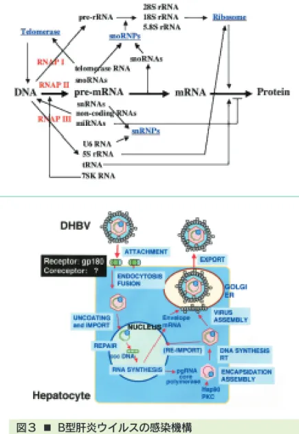
図1 ■ 遺伝情報発現に関わる低分子RNA
細胞内には多種類の低分子RNAが存在し,遺伝情報発現の様々な課程で 機能し,細胞の増殖,維持に関わっている。我々は遺伝子ノックアウト法 を用いてmiRNA及びsnoRNAの機能解析を行っている。
Fig. 1■ Low molecular weight RNAs are concerned in gene expression
Various low molecular weight RNAs function at many stages in gene expression and are concerned with maintenance and proliferation of the cells.
We are working on the functional characterization of miRNAs and snoRNAs.
図2 ■ U13 snoRNAによる18SリボソームRNAの修飾 U13 snoRNAの機能を明らかにするため,U13 snoRNA遺伝子を欠失さ せたトリ細胞を作製した。野性株とノックアウト細胞株を比較することに より,U13 snoRNAは18SリボソームRNAの3'-末端付近のCの修飾に関 与することが明らかになった。
Fig. 2■ U13 snoRNA guides modification of 18S rRNA By the using of targeted disruption, we could obtain chicken cell lines those are deficient in the expression of U13 snoRNA. In these cells, the modifications of a cytidine residue near the 3'-end of 18S rRNA was inhibited completely.
図3 ■ B型肝炎ウイルスの感染機構
B型肝炎ウイルスの感染機構を知るため,ダックB型肝炎ウイルス(DHBV)
をモデルに,DHBV蛋白質と結合する宿主蛋白質を探索している。その結 果,このウイルスのレセプターである新規カルボキシペプチダーゼ gp180を発見したが,感染成立にはさらに第二の宿主因子が必要である ことがわかってきた。
Fig. 3■ Infection mechanism of hepatitis B viruses
To understand the nature of the uptake pathway for hepadnaviruses, we have begun the search for the host proteins that interacts to envelope proteins of the duck hepatitis B virus (DHBV) as a model of these viruses. After our finding of novel carboxypeptidase gp180, which is now regarded as a host receptor,
Division of Molecular Pathology
ゲノム分子病態研究分野
ゲノム分子病態分野は,発ガン剤や抗ガン剤を含む様々 なDNA損傷ストレスにたいする細胞の応答機構,特に細 胞のアポトーシス,とその異常の病態像を分子レベルで解 明し,ガンの病因の解明と予防・診断・治療への応用を図 ろうとしている。具体的には,
1)DNA損傷ストレスに対する細胞応答において司令塔 的な役割を果たしているataxia telangiectasia原因 遺伝子(ATM)ファミリーの機能の解明と,その異常に よって起こる免疫不全・小脳失調・発ガンの病態解析 2)様々なDNA損傷ストレスがどのような因子によって 認識され,ATMファミリーが活性化されるのかの解明 3)DNA損傷ストレスによって活性化されてストレス応 答に重要な役割を果たしていると考えられているc- Ablファミリー, BRCA1, Chk2等ののストレス応答 に果たす役割,特にDNA修復における役割の解明,
等の研究が現在進行している。
DNA damage is a constant threat to eukaryotic cells and defective response to this threat increases genetic instability, ultimately leading to cancer. The goal of our research is to clarify how cells recognize DNA damage and transduce signals to cell cycle checkpoint control, DNA repair and apoptosis machineries.
To achieve this goal, we are currently studying the activation and functions of ATM (a gene mutated in ataxia telangiectasia) family in cellular response to DNA damage, using knockout cells. We are also studying how c-Abl family, BRCA1 and Chk2 are activated and what roles these factors play in the response.
Division of Molecular Cell Signaling
シグナル伝達研究分野
細胞内シグナル伝達経路の異常な活性化は細胞の癌化を 誘導することが知られている。私たちは,主要な細胞内シ グナル伝達経路の一つであるMAPキナーゼ(MAPK)カス ケードに注目し,
・MAPKカスケードの in vivo における機能の解明
・MAPKカスケードの特異性を保持する分子機構の解明 を目指して研究を進めている。
Abnormal activation of intracellular signaling pathways often leads to tumors. The goal of our project is to elucidate the functions of MAP kinase (MAPK) cascades in vivo, which are major intracellular signaling pathways, and the molecular mechanisms of how the specificity of MAPK cascades is maintained.
図1 ■ MAPKカスケードの in vivo における役割,及び足 場タンパク質によるキナーゼ複合体の形成
MAPKカスケードは細胞の増殖,分化,及びアポトーシスにおいて重要な 役割を担っている。足場タンパク質は,MAPKカスケードの主要な構成成 分であるMAPK,MAPKキナーゼ(MAPKK),及びMAPKKキナーゼ
(MAPKKK)と複合体を形成することによりMAPKカスケードの特異性を保 持すると考えられる。
Fig. 1■ Function of MAPK cascade in vivo, and Formation of Multikinase Complex by Scaffold Protein
Recent studies indicate that MAPK cascades, in which major components are MAPK, MAPK kinase (MAPKK), and MAPKK kinase (MAPKKK), play important roles in cell proliferation, differentiation, and apoptosis. Scaffold proteins could contribute to the specificity determination of MAPK cascades.
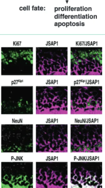
図2 ■ 発達期小脳における足場タンパク質JSAP1と活性 型 JNK MAPK の発現
免疫組織化学法による解析を行い,足場タンパク質JSAP1および活性型 JNK MAPKは発達期マウス小脳の外顆粒層を形成する顆粒前駆細胞
(GCP),特に増殖を停止したGCPで強く発現していることを見出した。
この結果は,JSAP1-JNKシグナル伝達系がGCPの増殖阻害,および分化 促進に関わることを強く示唆している。またJSAP1-JNK系は,髄芽腫発 生に関与していると考えられる。JSAP1(JNK MAPK経路の足場タンパク 質),P-JNK(活性型 JNK MAPK),K i 67(細胞増殖マーカー),p27Kip1(細 胞増殖停止マーカー),NeuN(細胞分化マーカー)。
Fig. 2■ Expression of the scaffold protein JSAP1 and active JNK in developing mouse cerebellum
During the development of the cerebellum, massive clonal expansion of granule cell precursors (GCPs) occurs in the outer part of the external granular layer (EGL). We have provided evidence that the scaffold protein JSAP1 and active JNK were expressed preferentially in the post-mitotic inner EGL progenitors in the developing cerebellum. These results suggest that JSAP1 promotes the cell-cycle exit and differentiation of GCPs by modulating JNK activity in cerebellar development. It is conceivable that JSAP1-JNK signaling would be involved in the development of medulloblastoma. JSAP1, a scaffold protein for JNK MAPK cascades; P-JNK, phosphorylated (activated) JNK;
Ki67, a proliferation marker; p27Kip1, a negative regulator of the GCP cell cycle; NeuN, a neural differentiation marker.
がん病態制御研究部門
Department of Cancer Biomedicine
■
細胞機能統御研究分野 Division of Molecular Virology and Oncology
教授佐藤 博
Professor SATO, Hiroshi
准教授滝野 隆久
Associate Professor TAKINO, Takahisa
准教授遠藤 良夫
Associate Professor ENDO, Yoshio
准教授久野 耕嗣
Associate Professor KUNO, Kouji
■
分子生体応答研究分野 Division of Molecular Bioregulation
教授向田 直史
Professor MUKAIDA, Naofumi
助教馬場 智久
Assistant Professor BABA, Tomohisa
■
免疫炎症制御研究分野 Division of Immunology and Molecular Biology
教授須田 貴司
Professor SUDA, Takashi
助教今村 龍
Assistant Professor IMAMURA, Ryu
助教木下 健
Assistant Professor KINOSHITA, Takeshi
Division of Molecular Virology and Oncology
細胞機能統御研究分野
目的と研究課題
正常細胞においてがん遺伝子,がん抑制遺伝子の変異が 蓄積した結果としてがんが発生し,悪性化する。悪性化し たがんは組織内へ浸潤し,遠隔臓器へ転移する。我々はが ん化,悪性化そして転移性獲得の過程を分子レベルで明ら かにすると共にその成果を診断・治療法へと応用すること を目指している。
がんの組織内への浸潤には組織・基底膜の破壊を伴う。
我々は1994年にがん転移の鍵を握るタンパク分解酵素を 発見しMT1-MMPと命名した(Nature, 1994)。MT1- MMPは細胞浸潤のみならず増殖・運動などの調節にも重 要な役割を果たしているとのデーターが蓄積しつつある。
Aim and Projects on going
Accumulation of mutation in ocogenes and tumor suppressor genes in normal cells results in malignant tumors. Malignant tumors invade into tissues and finally metastasize to distant organs. The goal of our project is to elucidate the molecular mechanism of tumor metastasis and develop diagnostic and therapeutic application.
Tumor invasion into tissue requires degradation of tissue basement membrane. We discovered a protease which is the key enzyme for tumor metastasis, and named it as MT1-MMP (Nature, 1994). Accumulating evidences indicate that MT1-MMP plays important roles in not only tumor invasion but also regulation of tumor growth and migration.
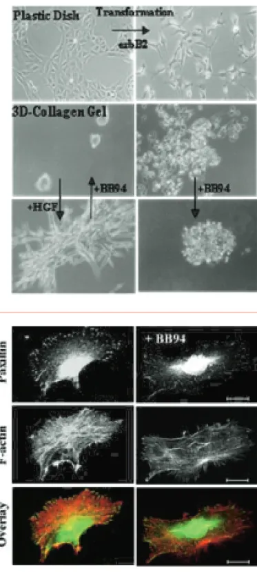
図1 ■ 上皮細胞のがん化に伴うMT1-MMPの発現と浸潤
正常上皮細胞株MDCKはがん遺伝子(erbB2)によりトランスフォームし,がん細 胞の形態を示すとともにMT1-MMPを発現する。コラーゲンゲル内での培養では 正常細胞は凝集して増殖するのに対してMT1-MMPを発現するがん化した細胞は 浸潤性の増殖をする。MMP阻害剤BB94の添加によりコラーゲンゲル内での浸潤 は抑制される。また,正常MDCK細胞はHGF添加によりコラーゲンゲル内で管空 を形成する。この管空形成もMT1-MMPを阻害することにより完全に抑制される。
Fig. 1■ Induction of MT1-MMP and Invasive Growth by Ocnogenic Transformation of Normal Epithelial Cells
Normal epithelial MDCK cells were transformed with oncogne (erbB2), and showed tumor phenotype including MT1-MMP expression. Normal cells grow to form cysts in collagen gel, but transformed cells which express MT1-MMP show invasive growth.
Tumor invasive growth is suppressed by the addition of MMP inhibitor BB94. Normal MDCK cells form branching tubules upon addition of HGF, which is also suppressed by BB94
図2 ■ 細胞運動とMT1-MMP
MT1-MMPを発現するHT1080細胞をコラーゲン上で培養するとパキシリンで可 視化された細胞接着斑とアクチンの走行により細胞運動の状態が見える。BB94 の添加によりMT1-MMPを阻害すると細胞接着班の局在が変化し,極性を喪失し て細胞は静止状態となる。MT1-MMPは細胞接着斑のターンオーバーを促進する ことにより運動シグナルを増強している。
Fig. 2■ Cell Migration and MT1-MMP
HT1080 cells were cultured on collagen, which express MT1-MMP, and were stained for paxillin to visualize focal adhesion and actin. Addition of MT1-MMP inhibitor BB94 altered the localization of focal adhesion, reduced cell polarity and suppressed cell migration. MT1-MMP enhances motility signal by stimulating turnover of focal adhesion.
Division of Molecular Bioregulation
分子生体応答研究分野
目的,研究課題,最近の主要な成果
組織障害に対して,生体は炎症反応を行い,組織障害を 軽 減 す る よ う に 働 く 。 し か し , 過 剰 な 炎 症 反 応 は , Helicobacter pyloriiの慢性感染で見られるように,組織 障害を進行させ,時にがんを発症させる。
固形がん組織中の線維芽細胞・血管内皮細胞などのいわ ゆるストローマ細胞と白血球は,がん細胞との相互作用を 通して,ケモカインを始めとする炎症性サイトカインを始 めとする生理活性物質を産生する。産生された因子は,が んの進展・転移に重要な役割を果たしている。本研究分野 では,
1)ケモカイン関連遺伝子欠損マウスを用いた解析から,
ケモカインががんの発症・進展に,種々の面から関与 していることを明らかにしつつある。
2)セリン/スレオニン・キナーゼ活性を保有する,原が ん遺伝子Pim-3の発現が,肝臓・膵臓におけるがん病 変で亢進していて,好アポトーシス分子Badの不活性 化を通して,がん細胞のアポトーシスを抑制し,がん の進展に寄与している可能性を明らかにした。このこ とは,Pim-3を分子標的とした新たな抗がん療法の可 能性を示唆している。
Aims, Ongoing Projects, and Recent Achievements
Inflammatory responses occur upon tissue injuries, to reduce tissue damage. If inflammatory responses are exaggerated and prolonged as observed in chronic infection with Helicobacter pylorii, tissue injuries continue, leading sometimes to carcinogenesis.
By interacting with tumor cells, stroma cells and leukocytes can produce various bioactive substances including chemokines.
The produced molecules can affect tumor progression and metastasis. We are elucidating the interaction between tumor cells and stroma cells and obtained the following results recently.
1) By using mice deficient in chemokine-related genes, we are showing that chemokines can contribute to tumor development and progression by exerting various activities.
2) We revealed that the expression of a serine/threonine kinase, Pim-3, was aberrantly enhanced in malignant lesions of liver and pancreas. Moreover, aberrantly expressed Pim-3 can inactivate a proapoptotic molecule, Bad by phosphorylating its serine residue, and eventually prevent apoptosis of tumor cells.
Thus, Pim-3 may be a good molecular target for cancer treatment.
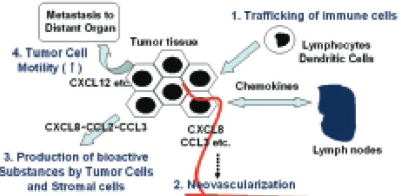
図1 ■ ケモカインのがん病態における役割
ケモカインは,①免疫担当細胞のがん病巣から所属リンパ節への移動過程 の調節,②腫瘍血管新生の誘導,③がん細胞の運動性亢進による転移能の 亢進以外に,がん細胞・ストローマ細胞からの種々の生理活性物質の産生 を誘導し,がん病態の形成に関与している。
Fig. 1■ Roles of chemokines in tumor progression and metastasis processes
Various chemokines contributes to progression and metastasis through the following functions.
a . Regulation of immune cell trafficking b. Induction of neovascularization c . Enhancement of tumor cell motility
d. Induction of production of bioactive substances by tumor and stromal cells
図2 ■ がん病変で発現亢進するセリン/スレオニン・キ ナーゼPim-3
がん病変で発現亢進するPim-3は,好アポトーシス分子,Badをリン酸化 し,不活性化することによって,がん細胞のアポトーシスを抑制している。
Fig. 2■ Aberrant expression of a serine / threonine kinase, Pim-3 in malignant lesions
Pim-3, aberrantly expressed in various malignant lesions, inactivates a pro-
Division of Immunology and Molecular Biology
免疫炎症制御研究分野
私たちの体を構成している一つ一つの細胞は,必要に応 じて自殺する能力を備えている。アポトーシス(枯死)とは,
この様な機能的,能動的細胞死の典型である。放射線など で遺伝子に多くの傷がついた時も,細胞はがん細胞になる 前に自殺することで,がんの出現を防いでいる。
一方,我々は,アポトーシス誘導蛋白Fasリガンドに対 する中和抗体が,肝炎などの炎症性疾患の動物モデルで治 療効果を示すことを明らかにした。さらに慢性肝炎から肝 癌を発症する動物モデルで,この抗体を用いて肝炎の治療 を行うと,肝癌の発症も抑制された。これらの結果から,
我々はFasリガンドのシグナル伝達経路が炎症性疾患の治 療薬,慢性炎症に伴う発がんの予防薬の標的になりうると 考え,研究を進めている。
最近の研究から,Fasリガンドに限らず,アポトーシス と炎症の双方に関わる蛋白が多数発見されている。すなわ ち,アポトーシスと炎症(の一部)の機構は共通の祖先的生 体防御システムから,機能的にも密接に関連しながら進化 したものと考えられる。この様な視点から,我々はアポ トーシスと炎症の双方に関連する蛋白因子を同定し,その 生体防御における役割とがんとの関わりを解明することを 目指している。
Each cell composing our body has an ability to kill itself when necessary. Apoptosis is a common type of such functional and active cell death. To prevent oncogenesis, cells often die by apoptosis when their genes were severely damaged.
On the other hand, we have demonstrated that a neutralizing antibody against Fas ligand (FasL), an apoptosis-inducing protein, has therapeutic potential in animal models of inflammatory diseases including hepatitis. Furthermore, using this antibody, we successfully prevented hepatic cancer development in an animal model of chronic hepatitis. Currently, we are exploring the signal transduction pathway of FasL, which is a potential target of drugs therapeutic for inflammatory diseases and/or preventive for cancer associating with chronic inflammation.
Recent studies have revealed that besides FasL, many other proteins have roles in both apoptosis and inflammation. We are exploring the function of such proteins, which could be important players in biodefense and cancer.
図1 ■ 慢性肝炎モデルにおける抗Fasリガンド抗 体の治療効果
肝臓にB型肝炎ウイルス抗原を発現するマウスに同抗原で免疫し たマウスのリンパ球を移植すると,慢性肝炎を発症し,一年以上 後にほぼ100%肝癌を発症する。このモデルで,抗Fasリガンド 抗体をマウスに投与すると,肝炎ばかりでなく肝癌も抑制された。
Fig. 1■ Therapeutic effect of an anti-FasL antibody in an animal model of chronic hepatitis Transplantation of HBs antigen-primed lymphocytes into transgenic mice expressing HBs antigen in the liver caused chronic hepatitis, and after one year or more, led to hepatic cancer. Administration of an anti-FasL antibody not only ameliorated hepatitis, but also prevented cancer development.
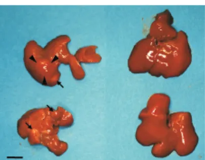
図2 ■ 抗Fasリガンド抗体を投与しなかった場合(左)と投与 した場合(右)の慢性肝炎モデルマウスの肝臓
感作リンパ球移植後15ヶ月。抗体非投与マウスの肝臓(左)は萎縮し,大小の 腫瘤(矢頭および矢印)が出来ている。これらの腫瘤が肝癌であることは組織学 的に確認した。これに対し,抗体投与マウスの肝臓(右)は大きさも組織学的に もほぼ正常である。
Fig. 2■ Livers from mice treated (right) or untreated (left) with an anti-FasL antibody
Fifteen months after the lymphocyte transplantation. Untreated livers shrunk and carried multiple tumors (arrow heads and arrows). Histological analyses revealed that these tumors were hepatic cancer. On the other hand, treated livers were almost normal in size and histology.
がん幹細胞研究センター
Center for Cancer and Stem Cell Research
■
遺伝子・染色体構築研究分野 Division of Molecular Genetics
教授平尾 敦
Professor HIRAO, Atsushi
助教仲 一仁
Assistant Professor NAKA, Kazuhito
助教田所 優子
Assistant Professor TADOKORO, Yuko
■
腫瘍遺伝学研究分野 Division of Genetics
教授大島 正伸
Professor OSHIMA, Masanobu
助教大島 浩子
Assistant Professor OSHIMA, Hiroko
Division of Molecular Genetics
遺伝子・染色体構築研究分野
幹細胞とは,各組織あるいは細胞の源となる細胞であり,
多系統の細胞に分化する 多分化能 と幹細胞を再び作る 自己複製能 を持つ細胞と定義される細胞である。幹細 胞プールが個体の生涯に亘って維持され続けるためには,
自己複製能を適切に制御する必要がある。我々は,これま でDNA損傷応答やPI3K-AKT経路に関与する分子が幹細 胞の自己複製に重要な役割を果たしていることを明らかに してきた。このことは,老化や発がんのメカニズムと幹細 胞制御の共通性を示唆するものである。
近年,がん組織中に,幹細胞的役割を持つ がん幹細胞 の存在が示され,がん治療の真の標的細胞として注目され ている。正常幹細胞とがん幹細胞の共通および相違点を見 極めることによって,がんの根治を目指した新たな治療法 の開発に寄与できると考えられる。
Stem cells are defined as cells that have the ability to perpetuate through self-renewal, and develop into mature cells of a particular tissue through differentiation. Appropriate controls of stem cell functions are critical for maintaining tissue homeostasis.
We have revealed that genes that are involved in DNA damage responses or PI3K-AKT signaling contribute to the maintenance of stem cell self-renewal capacity. Thus, signaling pathways for control of tumorigenesis or senescence may be involved in stem cell regulation.
Recent evidence has demonstrated that in tumors only a minority of cancer cells has the capacity to proliferate extensively and form new tumors. These tumor-initiating cells, which are called cancer stem cells, are thought as a novel target for cancer therapy. The investigation of distinct and parallel roles in normal stem cells and cancer stem cells will contribute to the design of cancer therapy without damaging normal tissues.
Division of Genetics
腫瘍遺伝学研究分野
Aim and Projects on going
Accumulating evidence has indicated that cooperation of oncogenic mutations and host reactions are responsible for tumorigenesis. To elucidate the genetic mechanisms of tumorigenesis, we constructed mouse models and examined histopathogenesis of gastric tumors.
1) Wnt signaling and PGE2 pathway are important for gastric tumorigenesis. We constructed mouse model, in which both Wnt and PGE2pathways are activated in the gastric mucosa, and found that transgenic mice develop gastric cancer (Oshima H, et al, Gastroenterology, 2006).
2) Infection-associated inflammation plays a role in gastric tumorigenesis. Using in vitroand in vivosystems, we have found that TNF-αfrom activated macrophages promotes Wnt signaling in surrounding gastric cancer cells, which further contribute to tumorigenesis. Wnt promotion may be one of important mechanisms of inflammation in gastric tumorigenesis (Oguma K et al, EMBO J, 2008).
3) Using primary cultured cells from mouse gastric cancer, we have shown that tumor cells activate bone marrow-derived cells to be myofibroblasts that play a role in tumor angiogenesis (Guo X, et al,JBC, 2008).
4) Sox17 represses Wnt signaling and downregulated in gastric and colon cancer, suggesting that Sox17 is a tumor suppressor.
Importantly, we found that Sox17 expression is strongly induced at early stage of tumorigenesis. It is thus possible that Sox17 plays a role in tumor development (Du YC, et al, Gastroenterology, 2009).
図1 ■ WntとPGE2の相互作用により発生する胃がん
Wnt1,COX-2,mPGES-1を同時に発現させたトランスジェニックマウス(K19- Wnt1/C2mE)の胃粘膜では,WntシグナルとPGE2経路の相互作用により胃がんの 発生が認められる。
K19-Wnt1/C2mEmice expressing Wnt1, COX-2, and mPGES-1 in gastric mucosa develop adenocarcinoma in glandular stomach, indicating that cooperation of Wnt and PGE2pathways is responsible for gastric tumorigenesis.
図2 ■ Wnt亢進腫瘍細胞におけるSox17の発現誘導
胃がん発生マウスモデルの初期腫瘍病変では,β-cateninの発現誘導が認められる。
同じ腫瘍細胞でSox17の顕著な発現誘導が観察され,何らかの役割を果たしている 可能性を示唆している。
In mouse tumor cells at early stage of gastric tumorigenesis, β-catenin is accumulated, 目的と研究課題
消化器がんの発生過程には,上皮細胞での遺伝子変異と 微小環境による影響が複雑に関与している。これらの相互 作用を個体レベルで解明する事を目的として,遺伝子改変 マウスモデルを作製し,病理学的および分子生物学的なア プローチにより研究を行なっている。
1)胃がん発生過程では,上皮細胞でのWntシグナル亢進 と,間質細胞でのPGE2産生が重要と考えられている。
双方のシグナルを同時に活性化したマウスモデルを 作製した結果,WntとPGE2の相互作用が胃がん発 生に作用する事を明らかにした(Oshima H, et al, Gastroenterology, 2006)。
2)胃がん発生には炎症反応が密接に関わっている。培養 細胞系およびマウスモデルを用いた解析により,炎症 性マクロファージ由来のTNF-αが,胃粘膜上皮細胞 や胃がん細胞のWntシグナルを亢進させる事を明らか にし,炎症反応に起因した新しい発がん促進機序のひ とつと考えられた(Oguma K, et al, EMBO J, 2008)。 3)胃がんモデルマウス腫瘍組織由来の初代培養細胞を用 いた解析により,腫瘍細胞が骨髄由来細胞などを活性 化して筋線維芽細胞に誘導し,VEGFなどの血管新生 因子を産生させて,腫瘍内血管新生を誘導している可 能性を明らかにした(Guo X, et al, JBC, 2008)。 4)Sox17にはWntシグナルを抑制する作用があり,悪性
胃がん大腸がん細胞で発現抑制されていることから,
癌抑制遺伝子と考えられた。しかし,消化管腫瘍の初 期発生過程では発現が強く誘導されており,腫瘍発生 に何らかの作用を及ぼす可能性が考えられた(Du YC, et al, Gastroenterology, 2009)。
分子標的がん医療研究開発センター
Molecular and Celluar Targeting Translational Oncology Center
■
腫瘍制御研究分野 Division of Translational and Clinical Oncology
教授源 利成
Professor MINAMOTO, Toshinari
准教授川上 和之
Associate Professor KAWAKAMI, Kazuyuki
■
機能ゲノミクス研究分野 Division of Functional Genomics
教授鈴木 健之
Professor SUZUKI, Takeshi
助教石村 昭彦
Assistant Professor ISHIMURA, Akihiko
■
腫瘍動態制御研究分野 Division of Tumor Dynamics and Regulation
教授松本 邦夫
Professor MATSUMOTO, Kunio
助教中村 隆弘
Assistant Professor NAKAMURA, Takahiro
■
腫瘍内科研究分野 Division of Medical Oncology
教授矢野 聖二
Professor YANO, Seiji
講師大坪公士郎(病院籍)
Associate Professor OHTSUBO, Koushiro
助教毛利 久継
Assistant Professor MOURI, Hisatsugu
助教竹内 伸司
Assistant Professor TAKEUCHI, Shinji 助教山田 忠明(病院籍)
Assistant Professor YAMADA, Tadaaki
■
腫瘍外科研究分野 Division of Surgical Oncology
講師安本 和生(病院籍)
Associate Professor YASUMOTO, Kazuo
助教山下 要
Assistant Professor YAMASHITA, Kaname
Division of Translational and Clinical Oncology
腫瘍制御研究分野
研究の概要と課題
消化器がんと呼吸器がんを主な対象にして,がんの多様 な分子細胞メカニズムと腫瘍外科学的特性の解明を目指し た基礎・臨床研究を行なう。
①がん化シグナル制御の分子細胞機構
(1)Wnt/β-カテニンがん化シグナル
(2)GSK3βリン酸化シグナル
②遺伝薬理学的解析によるオーダーメイド化学療法
③DNAメチル化をマーカーとしたがん診断・治療開発
④ヒト消化管がん組織検体資源化事業
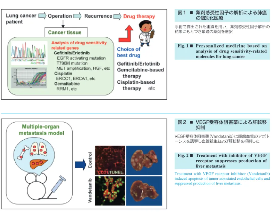
当研究所附属分子標的がん医療研究開発センターのコア 分野として,再発や転移性腫瘍を含む難治性がんへの取り 組み,制がんへの応用ならびに探索的がん医療を指向する 橋渡し研究に重点をおく。
Research Direction and Activities
The mission of the division centers on laboratory and clinical research to develop the novel strategies and modalities for diagnosis and treatment of cancer in the gastrointestinal and respiratory tracts. Research projects are based on molecular and cellular characteristics of individual tumor types that are relevant to metastatic potential, recurrence and outcome. Our current efforts are focused on:
①Molecular mechanism underlying oncogenic signaling networks
(1) Deregulated Wnt/β-catenin signaling
(2) Glycogen synthase kinase 3β(GSK3β)-mediated signaling
②Development of tailored chemotherapy by pharmacogenetics
③ Translational research of DNA methylation markers
④Establishment of tissue material resources of human gastrointestinal cancer
We are intending to translate as much the achievements created from these studies as possible to the fields responsible for diagnosis and treatment of cancer patients in clinical setting.
図1 ■ グリコーゲン合成酵素キナーゼ3β(GSK3β)は Wntシグナルに依存しない新しいがん標的である Glycogen synthase kinase 3β(GSK3β) supports and promotes tumor cells' survival and proliferation, and protects them from apoptosis in cancers developed in the major digestive organs, the results warrant proposing this kinase as a novel target in cancer treatment (PCT/JP 2006/300160).
図2 ■ RNAトランス因子CRD-BPはmRNAの安定性を修 飾してWnt,NF-κBとc-Myc経路をリンクする RNA trans-factor CRD-BP is a previously unrecognized transcription target of β-catenin/Tcf complex, and stabilizes mRNA of β-TrCP (β- transducin repeats-containing protein), NF-κB and c-Myc. CRD-BP is a novel cancer target that integrates multiple oncogenic signaling pathways
Division of Functional Genomics
機能ゲノミクス研究分野
がんの発症・悪性化の分子メカニズムを理解し,がんを 克服するためには,その原因となる遺伝子変異の同定が極 めて重要である。しかしヒトのがんでは,多くの変異の蓄 積とそのヘテロな形質ゆえに,現在でも原因遺伝子の同定 が容易ではない。これに対し,レトロウイルス感染マウス では,ウイルスが感染細胞のゲノムに挿入し,挿入部位の 遺伝子変異や周辺遺伝子の発現異常によって,がんを誘発 するため,ウイルス挿入部位を解析することで,原因遺伝 子を容易に同定することができる。本研究分野では,ウイ ルス感染マウスを用いて,がん関連遺伝子を網羅的に同定 し,その機能や相互作用の解析を通して,新しいがん分子 標的の探索や先進的ながんの遺伝子診断法の確立を目指し ている。また,重要な標的遺伝子については,逆遺伝学的 手法で新たな疾患モデルマウスを作製し,個体レベルでの がんの病態解析や治療法の開発に活用することも目標にし ている。現在の主な研究テーマは次のとおりである。
1)ゲノム不安定性を示す変異マウスを利用した新しいが ん抑制遺伝子の単離と機能解析
2)ヒストンのメチル化酵素および脱メチル化酵素と発が んとの関係
3)タンパク質のメチル化を制御する酵素群の新しい標的 基質の探索
4)ノックアウトマウスを用いた新しいがん関連遺伝子の 個体レベルでの機能解析
A detailed knowledge of the genes and signaling pathways mutated in cancer will be required to develop the novel target- based cancer therapeutics. However, the heterogeneity and complexity of genomic alterations in most human cancers hamper straightforward identification of cancer-causing mutations. We use the retrovirus-infected mice as model systems for identifying new cancer genes efficiently. Retroviruses induce tumors through activation of proto-oncogenes or inactivation of tumor suppressor genes as a consequence of retroviral integrations into host genome. Thus the viral integration sites provide powerful genetic tags for cancer gene identification. We are exploring the novel molecular targets for cancer treatment based on functional characterization of the cancer genes isolated by high-throughput screens using retroviral insertional mutagenesis. Once these genes are identified, we use gene knockout and transgenic mice to understand how these genes function in tumorigenesis, and to develop new animal models for human cancer. Our current projects are as follows.
1) Isolation of novel tumor suppressor genes using retroviral insertional mutagenesis in mice with genomic instability 2) "The histone code" and cancer
3) Identification of novel non-histone substrates for protein methyltransferases and demethylases
4) Functional analysis of the novel cancer genes using conditional knockout mice
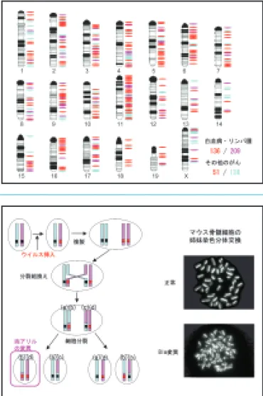
図1 ■ ヒトのがんに関わる遺伝子の多くは,ウイルス挿入変異の標 的となっている
英国サンガー研究所Cancer Gene Census に登録されているヒトの白血病・リンパ腫 に関係する遺伝子(ピンクで示す),その他のがんに関係する遺伝子(ブルーで示す)の マウスにおける相同遺伝子のうち,ウイルス挿入の標的となる遺伝子を赤で示す。
Fig. 1■ Most of human cancer genes are shown to be the targets of retroviral insertional mutagenesis in mice
We mapped the mouse orthologs of human genes involved in leukemia/lymphoma (shown in pink) and in other cancers (shown in blue) registered in Cancer Gene Census Database.
Among them, a number of genes (shown in red) have been identified as the targets of retroviral insertional mutagenesis in mice.
図2 ■ ブルーム症候群モデルマウスを利用したがん抑制遺伝子の効 率的な単離
ブルーム症候群はヒトの劣性遺伝病であり,その患者は,ゲノム不安定性により様々 ながんを発症する。原因遺伝子Blmの変異マウスは,姉妹染色分体交換,分裂組換え,
LOHの頻度の上昇が観察される。Blm変異マウスを利用してウイルス挿入変異を行う と,分裂組換えなどにより両アリルへの変異導入効率が上昇するため,がん抑制遺伝 子が標的になりやすくなる。
Fig. 2■ Efficient isolation of candidate tumor suppressor genes using retrovirus-infected Bloom syndrome model mice
Bloom syndrome is a recessive genetic disorder associated with genomic instability that causes affected people to be prone to cancer. The mutant mice for Bloom (Blm) gene showed increased rate of sister chromatid exchange, somatic recombination and loss of
Division of Tumor Dynamics and Regulation
腫瘍動態制御研究分野
細胞増殖因子は極微量ながら細胞の増殖・分化や細胞 死,さらに遊走や形態形成など多彩な細胞機能を調節する タンパク質である。HGF(hepatocyte growth factor: 肝 細胞増殖因子)は,当初,肝細胞の増殖促進を指標として 発見・単離・クローニングされた増殖因子であり,Metチ ロシンキナーゼを受容体として生理活性を発揮する。
HGFは発生過程においては上皮−間葉相互作用を介した 器官の形態形成を担う一方,成体においては肝臓をはじめ とする様々な組織・臓器の再生を担っている。また,がん 細胞のダイナミックな動態,すなわち浸潤や転移に関与し ている。本研究所における私達の研究室は2007年4月にス タートし,HGFとMet受容体を中心として組織再生(肝再 生など)の制御機構の研究,HGFによる疾患治療の研究,
がん−間質相互作用を介したがん悪性化機構とNK4による 制がん研究などを行っている。がんは「never healing wound(修復しない傷)」とたとえられる。多くのがんはダ イナミックな組織の修復・再生を担う生物学的な仕組みを 巧妙に使って勢力拡大−成長や浸潤・転移−に至る。私達 は生化学・分子生物学を基盤として,HGF-Met系を分子 標的とする制がん研究や再生制御の研究などオリジナルな 研究成果を発信したいと考えている。
Hepatocyte growth factor (HGF) was originally discovered as a mitogenic protein for mature hepatocytes. HGF exerts various biological activities cell proliferation, migration, anti-apoptosis, and morphogenesis in diverse biological processes. The receptor for HGF is Met tyrosine kinase. HGF plays critical roles in dynamic morphogenesis and regeneration of various tissues such as the liver. In cancer tissues, however, activation of the Met/HGF receptor is tightly associated with malignant behavior of cancer, i.e., invasion and metastasis, thus HGF-Met system is emerging target in the molecular target therapy of cancer. HGF is a stromal- derived mediator in tumor-stromal interaction. Our group started research from the April in 2007. We are studying on 1) regulation of tumor invasion-metastasis by the HGF-Met and therapeutic approach with NK4 (HGF-antagonist and angiogenesis inhibitor), 2) negative (suppressive) mechanisms for the Met receptor function and their biological significance in the termination of tissue regeneration and organ homeostasis, 3) clinical application of HGF for treatment of diseases, and 4) drug discovery based on structure of HGF-Met complex. HGF-Met system makes a way for dynamic reconstruction of tissues via epithelial-stromal interactions for regeneration of wounded tissues, whereas it is utilized for acquisition of malignancy of cancers. The simile that
"cancer is never-healing wound" seems pertinent from the aspect of HGF-Met.
図1 ■ HGF(肝細胞増殖因子)とNK4の生物機能
HGFは697個のアミノ酸からなるタンパク質でMetチロシンキナーゼを受容体と し,発生過程における器官形成や肝臓をはじめとする組織の再生を担う一方,多く のがん細胞の動態(浸潤・転移)を促す。私達はNK4をHGF-アンタゴニストとし て見いだすとともに,血管新生阻害作用をも有する2機能性分子であることを明ら かにした。
図2 ■ NK4の制がん作用の概略
NK4はHGF-Met系に対するアンタゴニスト(競合的阻害分子)としてがんの浸潤・
転移を阻害すると同時に,腫瘍血管新生阻害を介してがんの成長を抑制する。すな わち,がんの動態を止める"凍結作用"と腫瘍血管新生を阻害することによってがん の成長を抑制する"休眠作用"を発揮することによって,がんの生物学的な悪性形質 を抑制する。
Fig. 2■ Anti-cancer action of NK4
Fig. 1■ Biological functions of HGF (hepatocyte growth factor) and NK4 HGF is a heterodimer protein composed of 697 amino acids and the receptor for HGF is Met tyrosine kinase. HGF plays key roles in morphogenesis and tissue regeneration such as the liver. In cancer tissues, HGF plays a critical role in tumor invasion and metastasis as a mediator in tumor-stromal interaction. We discovered NK4 as the antagonist against HGF-Met. NK4 has dual functions as HGF-antagonist and angiogenesis inhibitor.