CANCER
RESEARCH INSTITUTE
金沢大学がん進展制御研究所概要 2013
金沢大学がん進展制御研究所概要目次
はじめに
Preface… ……… 1
沿 革 Historical Chart……… 2 〜 3 歴代所長
Successive Directors… ……… 4
機 構 Organization……… 5
職 員 数
Number of Staff……… 5
研 究 活 動
Research Activitiesがん幹細胞研究プログラム Cancer and Stem Cell Research Program
… ……… 6 〜 7遺伝子・染色体構築研究分野…
Division of Molecular Genetics… ……… 8腫 瘍 遺 伝 学 研 究 分 野 …
Division of Genetics……… 9腫 瘍 分 子 生 物 学 研 究 分 野 …
Division of Oncology and Molecular Biology……… 10がん幹細胞探索プロジェクト
Exploratory Project on Cancer Stem Cells……… 11がん微小環境研究プログラム
Cancer Microenvironment Research Program… ……… 12 〜 13細 胞 機 能 統 御 研 究 分 野 …
Division of Molecular Virology and Oncology… ……… 14分 子 生 体 応 答 研 究 分 野 …
Division of Molecular Bioregulation… ……… 15免 疫 炎 症 制 御 研 究 分 野 …
Division of Immunology and Molecular Biology… ……… 16腫 瘍 動 態 制 御 研 究 分 野 …
Division of Tumor Dynamics and Regulation……… 17がん分子標的探索プログラム Cancer Molecular Target Exploration Program
… ……… 18 〜 19分 子 病 態 研 究 分 野 …
Division of Cancer Cell Biology… ……… 20シ グ ナ ル 伝 達 研 究 分 野 …
Division of Molecular Cell Signaling… ……… 21腫 瘍 制 御 研 究 分 野 …
Division of Translational and Clinical Oncology……… 22機 能 ゲ ノ ミ ク ス 研 究 分 野 …
Division of Functional Genomics……… 23がん分子標的医療開発プログラム
Cancer Therapeutics Development Program……… 24腫 瘍 内 科 研 究 分 野 …
Division of Medical Oncology……… 25中央実験施設
Central Research Resource Branch… ……… 26 〜 29基 礎 統 計
Foundation Statistics決算額(運営費交付金)等……… 30
Settlement of accounts for Each Year
(
Subsidy from the National Government) 教 育 活 動
Educational Activities大学院生・研究生数
Graduate Students and Research Students… ……… 31交流協定校
Partner Universities and Faculties… ……… 31各種シンポジウム開催状況
Research Activities……… 32 〜 33所 在 地 Campus Locations
金沢大学がん進展制御研究所概要目次
Cancer Research Institute Contents
本研究所は,がんに関わる研究所としては文部科学省唯一 の機関として,昭和 42 年に臨床研究部門を含む8研究部門 制で設立されました。その後,部門増設等を経て平成9年に 3大部門制に拡大改組するとともに,新規抗がん治療法の開 発を目指す“分子標的薬剤開発センター”を設置しました。
この間,本研究所では,がん転移に関わるタンパク質分解酵 素 MT1-MMP の発見をはじめ,がんの病態成立に密接に関 与しているケモカインやアポトーシスの機能解明を始めとし た基礎研究に大きな成果をあげて参りました。
平成 18 年には,抗がん剤・放射線治療への抵抗性の克服 を目指す「がん幹細胞研究センター」と,先進的ながんの診断・
治療法の開発を目指す「分子標的がん医療研究開発センター」
の設置などの改組を行ない,白血病幹細胞の維持に関わる重 要な分子機序を明らかにしました。さらに平成 22 年には,
がんの悪性化にともなう転移・再発,および薬剤耐性機構の 研究を進めるために,「がん幹細胞研究プログラム」「がん微 小環境研究プログラム」「がん分子標的探索プログラム」「が ん分子標的医療開発プログラム」の4プログラム制へと改組 して現在に至っております。現在は,がん幹細胞制御機構,
肺がんの薬剤耐性獲得機構,慢性炎症による発がん機構など の研究を通して,がんの転移や再発の本態解明と,革新的分 子医薬の開発を目指した研究を推進しています。
このような研究所の使命を一層明確にするために,平成 23 年 4 月より本研究所は「金沢大学がん進展制御研究所」へと 改称し,同時に文部科学省より「がんの転移・薬剤耐性に関 わる先導的共同研究拠点」として認定され,共同利用・共同 研究拠点としての活動を開始いたしました。社会の高齢化に ともないがん罹患者数は増加し,十数年後には国民の2人に 1人ががんで亡くなると言われており,がんの克服は健康長 寿社会の実現のために喫緊の課題です。そのために本研究所 では,医学・薬学・獣医学および理工学系の幅広い分野の研 究者が集結し,がんの悪性化機構の本態解明とその制御によ る先制医療の実現を目指した研究を推進しています。共同利 用・共同研究拠点の認定を契機に,さらに幅広い分野の研究 者との連携のもと,がんの「悪性進展」過程の国際的研究拠 点の形成を目指し,がんの克服につながる研究の推進に向け て研究所員一同が全力で取り組んでいます。
平成 25 年度の金沢大学がん進展制御研究所概要を刊行す るにあたり,一層のご理解とご支援をお願い申し上げます。
In 1967m Kanazawa University Cancer Research Institute was founded as the only Cancer Research Institute of the Ministry of Education, Culture, Sports, Science and Technology (MEXT). In 1997, our organization was rearranged and at the same time Center for the Development of Molecular-targeted Drugs was established.
Since its establishment, our institute produced epoch-making achievements in basic cancer research field, such as the discovery of proteinase MT1-MMP, elucidation of function of chemokines and apoptosis.
In 2006, Cancer Research Institute was reorganized and 2 centers were newly established, “Cancer and Stem Cell Research Center” and “Molecular and Cellular Targeting Translational Oncology Center”, which aim discovery of the role of cancer stem cells in drug resistance and development of innovative diagnostic and therapeutic strategy, respectively. We then discovered the molecular mechanism for maintenance of leukemia stem cells. In 2010, our Research Institute has been further reorganized to establish 4 programs to identify mechanisms of metastasis, relapse, and drug resistance. They are "Cancer and Stem Cell Research Program",
"Cancer Microenvironment Research Program", "Cancer Molecular Target Exploration Program", and "Cancer Therapeutics Development Program". Currently, Cancer stem cell biology, molecular mechanisms of drug resistance, and chronic inflammation and cancer are research fields that we are leading in the cancer research field.
In July 2010, our institute was authorized by the MEXT as the Joint Usage/Research Center on Metastasis and Drug Resistance, and started the Joint Usage/Research Center Program. In Cancer Research Institute, researchers from a variety of fields including natural science, engineering, and clinical medicine have assembled to establish a cutting-edge research locus, to prevail over metastasis and drug resistance. With the authorization as the Joint Usage/
Research Center, all members in the Institute are endeavoring to widen collaboration with researchers in a wide variety of fields, to establish an international center of excellence on metastasis and drug resistance and to eventually promote research for conquering these conditions.
With the publication of the 2013 Kanazawa University Cancer Research Institute Outline, I would like to request your continuous support and understanding.
はじめに Preface
沿 革
Historical Chart
■結核研究所 TuberculosisResearchInstitute 1940.12.6
TuberculosisResearchFacilitywasestablishedinSchoolofMedicirefor
"thestudyofchemotherapyoftuberculosis".
金沢医科大学に「結核の化学療法に関する研究」のため結核研究 施設が設置された。
1942.3.20
TuberculosisResearchInstitutewasestablishedbyexpandingtheFacility.
Threedepartments,DepartmentofPharmaceutics,DepartmentofMicrobial Immunology and DepartmentofChemistry,opened for"thebasicand appliedresearchforthepreventionandtreatmentoftuberculosis".
金沢医科大学附属結核研究所となり「結核の予防及び治療に関 する学理並びにその応用研究」を目的とし,薬理製剤,細菌免疫 及び化学の3研究部門に増設された。
1947.7.3
DepartmentofMedicalExamination and Treatmentopened in Izumi- honmachi,Kanazawa.
金沢市泉本町に診療部門が増設された。
1949.5.31
TheTuberculosisResearchInstitutewasattachedKanazawaUniversity.
金沢大学附置の結核研究所となった。
1963.3.18
Twodepartmentswererenamed;DepartmentofPharmaceuticstoDepartment ofPharmacology,DepartmentofMedicalExaminationandTreatmentto DepartmentofClinic.
薬理製剤部門が薬理部門に,診療部門が臨床部門に研究部門名 が変更された。
1963.4.1
DepartmentofPathophysiologyopened.
病態生理部門が増設された。
1964.4.1
ClinicalfacilityoftheDepartmentofClinicrenamedasTuberculosisResearch InstituteHospital.
臨床部門の診療施設が結核研究所附属病院に改称された。
1967.3.
TheDepartmentofClinicandtheTuberculosisResearchInstituteHospital movedtoYoneizumi-machi,Kanazawa.
臨床部門及び附属病院が金沢市米泉町に新築移転された。
■医学部附属癌研究施設 CancerResearchFacility,SchoolofMedicine 1961.4.1
CancerResearchFacilitywasestablishedinSchoolofMedicinefor"the basicbiologicalstudyofcancer".DepartmentofBiochemistryopened.
医学部に「癌の基礎生物学的研究」のため附属癌研究施設が新設 され,研究部門は生化学部門が設置された。
1964.4.1
DepartmentofVirologyopened.
ウイルス部門が増設された。
1966.4.5
DepartmentofMolecularImmunologyopened.
分子免疫部門が増設された。
■がん研究所 CancerResearchInstitute 1967.6.1
CancerResearch Institute wasestablished combining the Tuberculosis ResearchInstituteandtheCancerResearchFacility.Theinstitutestartedwith eightdepartments;MolecularBiology,Virology,MolecularImmunology, Immunology,Pathophysiology,Pharmacology,ExperimentalTherapeutics andClinic.
TuberculosisResearchInstituteHospitalwasrenamedasCancerResearch InstituteHospital.
「がんに関する学理及びその応用の研究」を目的に,結核研究所 と医学部附属癌研究施設が統合され金沢大学がん研究所となり,
分子生物,ウイルス,分子免疫,免疫生物,病態生理,薬理,
化学療法及び臨床の8研究部門が設置された。
結核研究所附属病院は,がん研究所附属病院に改称された。
1968.6.1
DepartmentofBiophysicsopened.
生物物理部門が増設された。
1969.4.3
A new buildingforbasicresearchdepartmentsmovedtoTakara-machi, Kanazawa.
基礎研究系の研究棟が金沢市宝町に新築移転された。
1977.4.18
DepartmentofSurgery opened.DepartmentofClinicwasrenamed as DepartmentofInternalMedicine.
外科部門が増設され,臨床部門が内科部門に研究部門名が変更 された。
1983.3.30
AnofficebuildingwasbuiltfortheCancerResearchInstituteHospital.
附属病院に管理棟(軽量鉄骨)及び渡り廊下が増築された。
1997.4.1
Tendepartmentswerereorganizedtobeconsistedofthreedepartments(14 divisions)andonecenter.DepartmentofMolecularOncology,Departmentof MolecularandCellularBiology,DepartmentofBasicandClinicalOncology andCenterfortheDevelopmentofMolecularTargetDrugsopened.
10部門を3大部門(14研究分野)1センターに改組し,腫瘍分子 科学,細胞制御,腫瘍制御の3大部門及び分子標的薬剤開発セ ンターを置く。
2001.4.1
TheHospitalwasmergedwiththeUniversityHospital.
附属病院は医学部附属病院と統合された。
2006.4.1
Threedepartments(14divisions)andonecenterwerereorganizedtobe consistedoftwodepartmentsandtwocenter.DepartmentofMolecular CancerCellBiology,DepartmentofCancerBiomedicine,CenterforCancer andStem CellResearchandMolecularandCellularTargetingTranslational OncologyCenteropened.
3大部門(14研究分野)1センターを2大部門2センターに改組 し,がん分子細胞制御研究部門,がん病態制御研究部門の2大 部門及びがん幹細胞研究センター,分子標的がん医療研究開発 センターを置く。
2010.3.
A new buildingforbasicresearchdepartmentsmovedtoKakuma-machi, Kanazawa.
基礎研究系の研究棟が金沢市角間町に新築移転された。
2010.4.1
Twodepartmentsandtwocenterswerereorganizedtobeconsistedoffour programs.CancerandStem CellResearchProgram,CancerMicroenviron- mentResearchProgram,CancerMolecularTargetExplorationProgram and CancerTherapeuticsDevelopmentProgramopened.
2大部門2センターを4プログラムに改組し,がん幹細胞研究 プログラム,がん微小環境研究プログラム,がん分子標的探索 プログラム及びがん分子標的医療開発プログラムを置く。
2010.7.
CancerResearchInstitutewasauthorizedbytheMinistryofEducation, Culture,Sports,ScienceandTechnologyoftheJapaneseGovernmentasthe JointUsage/ResearchCenteronMetastasisandDrugResistance.
「がんの転移・薬剤耐性に関わる先導的共同研究拠点」として文 部科学省より認定された。
■がん進展制御研究所 CancerResearchInstitute 2011.4.1
ThenameofCancerResearchInstituteinJapanesewaschanged.
TheJointUsage/ResearchCenterProgramstarted.
がん研究所は,がん進展制御研究所に改称された。
共同利用・共同研究拠点として活動を開始した。
歴 代 所 長
Successive Directors
■歴代研究所長・研究施設長 SuccessiveDirectors
1942.4.8~1954.3.31 石 坂 伸 吉 結核研究所長 ISHIZAKA,Shinkichi DirectorofTuberculosisResearchInstitute 1954.4.1~1954.6.30 戸 田 正 三 結核研究所長事務取扱 TODA,Shozo ActingDirectorofTuberculosisResearchInstitute 1954.7.1~1958.6.30 岡 本 肇 結核研究所長 OKAMOTO,Hajime DirectorofTuberculosisResearchInstitute 1958.7.1~1961.6.30 柿 下 正 道 〃 KAKISHITA,Masamichi 〃
1961.7.1~1962.6.30 斎 藤 幸一郎 〃 SAITO,Koichiro 〃 1962.7.1~1966.6.30 石 崎 有 信 〃 ISHIZAKI,Arinobu 〃
1966.7.1~1967.5.31 伊 藤 亮 〃 ITOU,Ryo 〃
1961.4.1~1967.5.31 岡 本 肇 癌研究施設長 OKAMOTO,Hajime DirectorofCancerResearchInstitute 1967.6.1~1967.8.14 岡 本 肇 がん研究所長事務取扱 OKAMOTO,Hajime ActingDirectorofCancerResearchInstitute 1967.8.15~1968.3.31 岡 本 肇 がん研究所長 OKAMOTO,Hajime DirectorofCancerResearchInstitute 1968.4.1~1971.3.31 石 川 太 刀 雄 丸 〃 ISHIKAWA,Tachiomaru 〃
1971.4.1~1975.1.30 伊 藤 亮 がん研究所長事務取扱 ITOU,Ryo ActingDirectorofCancerResearchInstitute 1975.1.31~1978.4.1 伊 藤 亮 がん研究所長 ITOU,Ryo DirectorofCancerResearchInstitute 1978.4.2~1982.4.1 越 村 三 郎 〃 KOSHIMURA,Saburo 〃
1982.4.2~1984.4.1 倉 田 自 章 〃 KURATA,Yoriaki 〃 1984.4.2~1988.3.31 波田野 基 一 〃 HATANO,Motoichi 〃 1988.4.1~1990.3.31 右 田 俊 介 〃 MIGITA,Shunsuke 〃 1990.4.1~1993.3.31 亀 山 忠 典 〃 KAMEYAMA,Tadanori 〃 1993.4.1~1997.3.31 高 橋 守 信 〃 TAKAHASHI,Morinobu 〃 1997.4.1~2001.3.31 磨 伊 正 義 〃 MAI,Masayoshi 〃 2001.4.1~2005.3.31 山 本 健 一 〃 YAMAMOTO,Ken-ichi 〃 2005.4.1~2009.3.31 佐 藤 博 〃 SATO,Hiroshi 〃 2009.4.1~2011.3.31 向 田 直 史 〃 MUKAIDA,Naofumi 〃 2011.4.1~2013.3.31 向 田 直 史 がん進展制御研究所長 MUKAIDA,Naofumi 〃
2013.4.1~ 大 島 正 伸 〃 MASANOBU,Oshima 〃
■歴代附属病院長 SuccessiveDirectorsoftheInstituteHospital
1964.4.1~1965.7.31 水 上 哲 次 結核研究所附属病院長 MIZUKAMI,Tetsuji DirectorofTuberculosisResearchInstituteHospital 1965.8.1~1966.2.1 石 崎 有 信 〃 ISHIZAKI,Arinobu 〃
1966.2.1~1967.6.1 倉 金 丘 一 〃 KURAKANE,Kyuichi 〃
1967.6.1~1982.4.20 倉 金 丘 一 がん研究所附属病院長 KURAKANE,Kyuichi DirectorofCancerResearchInstituteHospital 1982.4.20~1983.1.31 磨 伊 正 義 がん研究所附属病院長事務取扱 MAI,Masayoshi ActingDirectorofCancerResearchInstituteHospital 1983.2.1~1991.1.31 磨 伊 正 義 がん研究所附属病院長 MAI,Masayoshi DirectorofCancerResearchInstituteHospital 1991.2.1~1993.1.31 澤 武 紀 雄 〃 SAWABU,Norio 〃
1993.2.1~1997.1.31 磨 伊 正 義 〃 MAI,Masayoshi 〃 1997.2.1~2001.3.31 澤 武 紀 雄 〃 SAWABU,Norio 〃 2001.4.1~2001.9.30 澤 武 紀 雄 がん研究所附属病院長を命ずる SAWABU,Norio 〃
■附属がん幹細胞研究センター長 CenterforCancerandStemCellResearch
2006.4.1~2009.3.31 向 田 直 史 MUKAIDA,Naofumi 2009.4.1~2010.3.31 平 尾 敦 HIRAO,Atsushi
■附属分子標的がん医療研究開発センター長 MolecularandCellularTargetingTranslationalOncologyCenter 2006.4.1~2010.3.31 源 利 成 MINAMOTO,Toshinari
■名誉教授 ProfessorEmeritus
倉 田 自 章 高 橋 守 信 KURATA,Yoriaki TAKAHASHI,Morinobu 村 上 清 史 澤 武 紀 雄 MURAKAMI,Seishi SAWABU,Norio 原 田 文 夫 山 本 健 一 HARADA,Fumio YAMAMOTO,Ken-ichi
事務部長
Directorプ ロ グ ラ ム
Programがん進展制御 研究所
Cancer Research Institute
(所 長)
(Director)
共 同 利 用 施 設
Central Facilitiesが ん 微 小 環 境 研 究 プ ロ グ ラ ム
Cancer Microenvironment Research Programが ん 幹 細 胞 研 究 プ ロ グ ラ ム
Cancer and Stem Cell Research Programが ん 分 子 標 的 探 索 プ ロ グ ラ ム
Cancer Molecular Target Exploration Programがん分子標的医療開発プログラム
Cancer Therapeutics Development Program課 長
Chief腫 瘍 遺 伝 学 研 究 分 野
Division of Genetics遺 伝 子・ 染 色 体 構 築 研 究 分 野
Division of Molecular Genetics腫 瘍 分 子 生 物 学 研 究 分 野
Division of Oncology and Molecular Biologyがん幹細胞探索プロジェクト
Exploratory Project on Cancer Stem Cells細 胞 機 能 統 御 研 究 分 野
Division of Molecular Virology and Oncology分 子 生 体 応 答 研 究 分 野
Division of Molecular Bioregulation免 疫 炎 症 制 御 研 究 分 野
Division of Immunology and Molecular Biology腫 瘍 動 態 制 御 研 究 分 野
Division of Tumor Dynamics and Regulation機 構
Organization
職 員 数
Number of Staff
平成 25 年7月1日現在
分 子 病 態 研 究 分 野
Division of Cancer Cell Biologyシ グ ナ ル 伝 達 研 究 分 野
Division of Molecular Cell Signaling腫 瘍 制 御 研 究 分 野
Division of Translational and Clinical Oncology機 能 ゲ ノ ミ ク ス 研 究 分 野
Division of Functional Genomics企 画 総 務 係
General Affairs研 究 協 力 係
Research Cooperative Affairs会 計 係
Accountingヒトがん組織バンク
Human Cancer Tissue Bankマウス発がん組織バンク
Mouse Carcinogenesis Model Tissue Bankヒトがん細胞株バンク
Human Cancer Cell Line Bankがん創薬・ケミカルバイオロジーユニット
Cancer Chemical Biology & Drug Discovery Unit前臨床実験施設
Pre-clinical Research Facility
臨床治験施設
Clinical Research Facility腫 瘍 内 科 研 究 分 野
Division of Medical Oncology中央実験施設
Central Research Resource Branch
専 門 職 員
Chief●●●●●●●●●●
xxxxxxxxxxxxxxxxxxxxxxxxxxxxxxxxxxxxxx
xxxxxxxxxx
●●●●●
■
●●●●●●●●●● xxxxxxxxxx
●●●●●
xxxxxxxxxx
●●●●●
●●●●●●●●●●
xxxxxxxxxxxxxxxxxxxxxxxxxxxxxxxxxxxxxx
がん幹細胞研究プログラム
Cancer and Stem Cell Research Program
■
遺伝子・染色体構築研究分野 Division of Molecular Genetics
教授… 平尾 敦
Professor HIRAO, Atsushi
助教… 田所 優子
Assistant Professor TADOKORO, Yuko
助教… 星居 孝之
Assistant Professor HOSHII, Takayuki
■
腫瘍遺伝学研究分野 Division of Genetics
教授… 大島 正伸
Professor OSHIMA, Masanobu
助教… 大島 浩子
Assistant Professor OSHIMA, Hiroko
助教… 石川 智夫
Assistant Professor ISHIKAWA, Tomo-o
助教… 大田 久美子
Assistant Professor OHTA, Kumiko
助教… 小林 昌彦
Assistant Professor KOBAYASHI, Masahiko
特任助教… 中山 瑞穂
Assistant Professor NAKAYAMA, Mizuho
●●●●●●●●●●
xxxxxxxxxxxxxxxxxxxxxxxxxxxxxxxxxxxxxx
■
●●●●●●●●●● xxxxxxxxxx
●●●●●
xxxxxxxxxx
●●●●●
■
●●●●●●●●●● xxxxxxxxxx
●●●●●
■
がん幹細胞探索プロジェクト Exploratory Project on Cancer Stem Cells
准教授… 仲 一仁
Associate Professor NAKA, Kazuhito
■
腫瘍分子生物学研究分野 Division of Oncology and Molecular Biology
教授… 髙橋 智聡
Professor TAKAHASHI, Chiaki
助教… SHAMMA…AWAD
Assistant Professor
助教… 林 直之
Assistant Professor HAYASHI, Naoyuki
特任助教… 北嶋 俊輔
Assistant Professor KITAJIMA, Shunsuke
Division of Molecular Genetics
遺伝子・染色体構築研究分野
幹細胞とは,各組織あるいは細胞の源となる細胞であ り,多系統の細胞に分化する“多分化能”と幹細胞を再 び作る“自己複製能”を持つ細胞と定義される細胞である。
幹細胞プールが個体の生涯に亘って維持され続けるため には,自己複製能を適切に制御する必要がある。我々は,
これまで FOXO や mTOR 経路など,寿命制御に関わる 分子が幹細胞の自己複製に重要な役割を果たしているこ とを明らかにしてきた。このことは,幹細胞制御におけ る細胞内代謝の重要性を示唆するものである。
近年,がん組織中に,幹細胞的役割を持つ“がん幹細胞”
の存在が示され,がん治療の真の標的細胞として注目さ れている。正常幹細胞とがん幹細胞の共通および相違点 を見極めることによって,がんの根治を目指した新たな 治療法の開発に寄与できると考えられる。
Stem cells are defined as cells that have the ability to perpetuate through self-renewal, and develop into mature cells of a particular tissue through differentiation. Appropriate controls of stem cell functions are critical for maintaining tissue homeostasis.
We have revealed that genes that are involved in longevity, including FOXO and mTOR pathways, contribute to the maintenance of stem cell self-renewal capacity. Thus, signaling pathways for control of intracellular metabolism may play a critical role in stem cell regulation.
Recent evidence has demonstrated that in tumors only a minority of cancer cells has the capacity to proliferate extensively and form new tumors. These tumor-initiating cells, which are called cancer stem cells, are thought as a novel target for cancer therapy. The investigation of distinct and parallel roles in normal stem cells and cancer stem cells will contribute to the design of cancer therapy without damaging normal tissues.
Fig.1…
■…
Nutrient sensor signals 図1…■…
栄養センサーシグナルFig.2…
■…
mTOR and FOXO pathways in quiescent hematopoietic stem cells図2…
■…
静止期造血幹細胞における mTOR および FOXO 経路Fig.3…
■…
FOXOactivation for drug-resistance of leukemia stem cells 図3…■…
治療耐性白血病幹細胞における FOXO 活性化Fig.4…
■…
mTOR complex in leukemia stem cells 図4…■…
白血病幹細胞における mTOR 複合体機能Division of Genetics
腫瘍遺伝学研究分野
Aim and Projects on going
Accumulating evidence has indicated that cooperation of oncogenic mutations and host reactions are responsible for tumorigenesis. To elucidate the genetic mechanisms of tumorigenesis, we constructed mouse models and examined molecular pathogenesis of gastric tumors.
1) Wnt signaling and PGE2 pathway are important for gastric tumorigenesis. We constructed mouse model, in which both Wnt and PGE2 pathways are activated in the gastric mucosa, and found that transgenic mice develop gastric cancer (Oshima H, et al, Gastroenterology, 2006).
2) Sox17 represses Wnt signaling and downregulated in gastric and colon cancer, suggesting that Sox17 is a tumor suppressor.
Importantly, we found that Sox17 expression is strongly induced at early stage of tumorigenesis. It is thus possible that Sox17 plays a role in tumor development (Du YC, et al, Gastroenterology, 2009).
3) Gan mice develop gastric tumors by activation of Wnt and PGE2 pathway. When Gan mice were raised in a germ free condition, inflammatory responses and tumor development were suppressed significantly. Accordingly, it is possible that innate immune responses for bacterial infection play a role in tumorigenesis (Oshima H, et al, Gastroenterology, 2011).
4) Expression profile of microRNAs in Gan mouse tumors was examined. We found that miR-7 was downregulated in tumor tissues by inflammation-dependent mechanism. miR-7 is downregulated also in the inflamed human gastric cancer, and plays a tumor suppressor role (Kong D, et al, Oncogene, 2012).
図1…
■…
Wnt シグナルと Sox17 の相互作用による発がん胃がん発生マウスモデルの初期病変では,β-catenin の発現誘導が認められる。同 じ腫瘍細胞で Sox17 の顕著な発現誘導が観察され,協調的に発がん促進に作用す る可能性を示している。
In tumor cells at early stage of mouse gastric tumors, Wnt signaling is activated, and thus β-catenin is accumulated in nuclei. Importantly, Sox17 is simultaneously induced in tumor cells, suggesting a role in gastric tumorigenesis together with Wnt signaling.
図2…
■…
無菌化によるマウス胃がん発生の抑制…胃がん発生モデルマウスを無菌環境で飼育すると,胃がん発生が顕著に抑制される。
細菌感染刺激による自然免疫活性化が発がんの微小環境形成に重要である可能性 を示している。
Germfree gastric tumor mouse models showed significant suppression of gastric tumorigenesis. It is therefore possible that bacterial infection through innate immune response is required for construction of microenvironment and gastric tumorigenesis.
目的と研究課題
消化器がんの発生過程には,上皮細胞での遺伝子変異と 微小環境による影響が複雑に関与している。これらの相互 作用を個体レベルで解明する事を目的として,遺伝子改変 マウスモデルを作製し,病理学的および分子生物学的なア プローチにより研究を行なっている。
1)胃がん発生過程では,上皮細胞での Wnt シグナル亢 進と,間質細胞での PGE
2産生が重要と考えられてい る。双方のシグナルを同時に活性化したマウスモデル を作製した結果,Wnt と PGE
2の相互作用が胃がん発 生に作用する事を明らかにした(Oshima H, et al,
Gastroenterology, 2006)。2)Sox17 には Wnt シグナルを抑制する作用があり,悪 性胃がん大腸がん細胞で発現抑制されていることか ら,癌抑制遺伝子と考えられた。しかし,消化管腫瘍 の初期発生過程では発現が強く誘導されており,腫瘍 発生に何らかの作用を及ぼす可能性が考えられた(Du YC, et al, Gastroenterology, 2009)。
3)Wnt と PGE
2の相互作用により胃がんを発生する Gan マウスを無菌化すると,炎症性微小環境の形成 が抑制されて,胃がん発生が顕著に抑制されること を明らかにした。したがって,細菌感染刺激による 自然免疫の活性化が炎症性微小環境の構築に重要と 考えられた(Oshima H, et al,
Gastroenterology,2011)。
4) Gan マウスを用いて,炎症反応依存的に腫瘍組織で 発現変化する microRNA を網羅的に解析した結果,
miR-7 の発現が有意に低下していることを明らかに
した。ヒト胃がんでも miR-7 は炎症依存的に発現低
下し,それにより腫瘍原性が維持されることが明ら
かとなった(Kong D, et al, Oncogene, 2012)。
Division of Oncology and Molecular Biology
腫瘍分子生物学研究分野
ヒトがんにおける臨床的エビデンスが豊富ながん遺伝 子・がん抑制遺伝子を変異させたマウス・細胞を中心に,
シンプルで分子生物学的・遺伝学的な解析がしやすい in vivo・in vitro がんモデル系を組み立て,発がん・転移・
薬剤耐性・がん幹細胞を克服する突破口になる新規パスウェ イを探索する。具体的な取り組みは以下。
1) 数多くの増殖シグナルのアダプター分子となる RB 蛋 白質 (pRB) の不活性化は,多くのヒトがんの悪性進 展過程において観察される。pRB は,従来知られた 細胞周期や細胞分化の制御だけでなく,細胞老化,
DNA 損傷応答,DNA メチル化,蛋白質イソプレニ ル化,脂質代謝,ミトコンドリア機能あるいはサイ トカイン分泌を制御することによっても腫瘍原性や 悪性度を規定することを見出してきた。
2) がん細胞は正常細胞と較べると代謝様式が劇的に異 なる。それは,好気的解糖と脂質合成の亢進であり,
p53 と pRB が協調してこれを制御すると考えている。
その他,Ras や Myc 等のがん遺伝子も代謝制御に関 わる。様々ながん化シグナルによって誘導されるメ タボリック・リプログラミングが,がん細胞の悪性 の挙動に与える影響とその機構を探索する。
3) 悪性進展機構の深い理解に基づき,がん幹細胞が示 すと想定される様々な挙動の一部を安定的に表現す るin vitro がん幹細胞モデル系を組み立て,がんの幹 細胞様表現型に関連する遺伝子の探索および新しい がん標的薬の開発に応用する。
We innovate in vivo and in vitro cancer model systems that can be readily analyzed by genetic and molecular biology techniques.
This aims to find pathways critical for carcinogenesis, metastasis, drug resistance, and stem cell-like behaviors in cancer cells.
Below are ongoing projects in our laboratory.
1 ) The RB tumor suppressor gene product has been implicated in control of cell cycle and terminal differentiation. However, we propose pRB plays many more roles during tumor progression beyond such functions.We focus onpRB functions in chromatin instability, DNA damage response, cellular senescence, mevalonate pathway, lipid metabolism, mitochondrial function, chromatin remodeling and stem cell- like behaviors in cancer cells.
2 ) Analysis of oncogenic signals that induce malignant behaviors in cancer cells through metabolic reprogramming.
3 ) Development of in vivo & in vitro cancer stem cell models in an aim to develop novel drugs or chemicals that specifically target hypothetical cancer stem cells.
図1…
RB 蛋白質に集まる様々なシグナルと RB 蛋白質から発せら れる様々なシグナル。RB 蛋白質の多様な働きを説明する。
E2F ファミリーが最も有名な標的であるが,その他にも,
多様な標的蛋白質(100 種類以上)があることが知られる。
Fig.1
Cellular signals merged on the modulation of pRB functions, and effectors of pRB. This at least partially explains multi- faceted functions of pRB.
図2
がん抑制遺伝子の複合的変異によって誘導される幹細胞様 のがん細胞集団の蛍光多重染色像。
Fig.2
Stem cell-like cells appeared in cancers induced by the combinational suppression of tumor suppressor genes including Rb.
Exploratory Project on Cancer Stem Cells
がん幹細胞探索プロジェクト
近年,一部のがんで,がん細胞を生み出すもととなる「が ん幹細胞」の存在が報告されており,抗がん剤治療後の根絶 を免れたがん幹細胞は再発を引き起こす原因になると考え られている。例えば,慢性骨髄性白血病(CML)患者の治療 にはメシル酸イマチニブなどのチロシンキナーゼ阻害薬
(TKI)が用いられているが,TKI 抵抗性の CML 幹細胞の残 存は CML の再発の原因となる。
私たちは CML のマウスモデルを用いて CML 幹細胞を純 化し,CML 幹細胞の TKI 抵抗性にフォークヘッド転写因子 FOXO が重要な役割を担っていることを発見した(図1)。
また,この FOXO はがん微小環境細胞が作り出す TGF-β によって活性化されており,CML 幹細胞を移植したマウス に TGF-β 阻害薬を投与すると TKI 抵抗性の CML 幹細胞 を抑制できることを見いだした。従って,TGF-β-FOXO シグナルは TKI 抵抗性の CML 幹細胞の治療薬を開発する ための重要なターゲットであると考えられる(図2)。
現在,TGF-β-FOXO シグナルによる CML 幹細胞の TKI 抵抗性メカニズムの解明と,このメカニズムをターゲット にする新しい CML 治療薬の開発を目指した研究を実施し ている。
Although the discovery of the tyrosine kinase inhibitors (TKI) have significantly improved the prognosis of chronic myeloid leukemia (CML) patients, a complete cure is not possible due to the existence of a rare population of CML stem cells known to be resistant to TKI therapy. We have recently reported that Forkhead transcription factor (FOXO) is essential for the TKI-resistance of CML stem cells (Fig. 1). Furthermore, TGF-β originate from the microenvironment regulates FOXO activity in CML stem cells.
Importantly, a combined administration of TGF-β inhibitor and TKI leads to reduction of CML stem cells in vivo. Our results demonstrate a critical role for the TGF-β-FOXO pathway in the maintenance of TKI-resistant CML stem cells (Fig. 2).
The purpose of our current research is to clarify the molecular mechanisms governing TKI-resistance of CML stem cells via TGF-β-FOXO signaling pathway. The long-term outcome of our investigation will hopefully be the development of novel agents that can specifically suppress the effects of these TGF-β-FOXO signaling pathway, and thereby provide a novel avenue for curative CML patient therapy.
図1…
■…
CML 幹細胞の TKI 抵抗性制御におけるフォークヘ ッド転写因子 FOXO の役割野生型(Foxo3a
+/+),並びに Foxo3a ノックアウト(Foxo3a
-/-)マウス由来の CML 幹細胞を移植したマウスにチロシンキナーゼ阻害薬 (TKI) の投与を行った。
その結果,CML 幹細胞における FOXO 遺伝子の欠損は TKI 投与後の CML の 再発を軽減することが明らかとなった。従って,FOXO は CML 幹細胞の TKI 抵抗性の制御に必須な役割を担う。
Fig.1…
■…
FOXO plays an essential role for the tyrosine kinase inhibitor (TKI) resistance of CML stem cellsMice transplanted with wild-type (Foxo3a+/+) or Foxo3a-deficient (Foxo3a-/-) CML stem cells received TKI. FOXO deficiency promoted the survival of CML-affected mice after administration of TKI, indicating that FOXO is responsible for the maintenance of TKI-resistant CML stem cells.
Fig.2…
■…
TGF-β-FOXO signaling pathway maintains TKI- resistant CML stem cellsWe have recently reported that FOXO is crucial for the TKI resistance of CML stem 図2…
■…
TGF-β-FOXO シグナルによる CML 幹細胞の TKI 抵抗性制御メカニズム
CML 幹細胞(CML… stem… cells)は分化した CML 細胞(CML… cells)の供給源と
なる。CML 幹細胞は TKI に対して抵抗性を示し,根絶を免れた CML 幹細胞
は CML の再発の原因となる。FOXO は CML 幹細胞の TKI 抵抗性の制御に関
わっている。また,CML 幹細胞の FOXO はがん微小環境細胞の作り出す
TGF-β によって活性化される。従って,TGF-β-FOXO シグナルは TKI 抵抗
性の CML 幹細胞を治療するための重要なターゲットとなる。
●●●●●●●●●●
xxxxxxxxxxxxxxxxxxxxxxxxxxxxxxxxxxxxxx
xxxxxxxxxx
●●●●●
■
●●●●●●●●●● xxxxxxxxxx
●●●●●
xxxxxxxxxx
●●●●●
●●●●●●●●●●
xxxxxxxxxxxxxxxxxxxxxxxxxxxxxxxxxxxxxx
がん微小環境研究プログラム
Cancer Microenvironment Research Program
■
細胞機能統御研究分野 Division of Molecular Virology and Oncology
教授… 佐藤 博
Professor SATO, Hiroshi
准教授… 滝野 隆久
Associate Professor TAKINO, Takahisa
■
分子生体応答研究分野 Division of Molecular Bioregulation
教授… 向田 直史
Professor MUKAIDA, Naofumi
助教… 馬場 智久
Assistant Professor BABA, Tomohisa
●●●●●●●●●●
xxxxxxxxxxxxxxxxxxxxxxxxxxxxxxxxxxxxxx
■
●●●●●●●●●● xxxxxxxxxx
●●●●●
xxxxxxxxxx
●●●●●
■
●●●●●●●●●● xxxxxxxxxx
●●●●●
■
腫瘍動態制御研究分野 Division of Tumor Dynamics and Regulation
教授… 松本 邦夫
Professor MATSUMOTO, Kunio
助教… 中村 隆弘
Assistant Professor NAKAMURA, Takahiro
助教… 酒井 克也
Assistant Professor SAKAI, Katsuya
■
免疫炎症制御研究分野 Division of Immunology and Molecular Biology
教授… 須田 貴司
Professor SUDA, Takashi
助教… 今村 龍
Assistant Professor IMAMURA, Ryu
助教… 木下 健
Assistant Professor KINOSHITA, Takeshi
Division of Molecular Virology and Oncology
細胞機能統御研究分野
目的と研究課題
正常細胞においてがん遺伝子,がん抑制遺伝子の変異 が蓄積した結果としてがんが発生し,悪性化する。悪性 化したがんは組織内へ浸潤し,遠隔臓器へ転移する。我々 はがん化,悪性化そして転移性獲得の過程を分子レベル で明らかにすると共にその成果を診断・治療法へと応用 することを目指している。
がんの組織内への浸潤には組織・基底膜の破壊を伴う。
我々は 1994 年にがん転移の鍵を握るタンパク分解酵素 を発見し MT1-MMP と命名した (Nature, 1994)。MT1- MMP は細胞浸潤のみならず増殖・運動などの調節にも重 要な役割を果たしているとのデーターが蓄積しつつある。
Aim and Projects on going
Accumulation of mutation in ocogenes and tumor suppressor genes in normal cells results in malignant tumors. Malignant tumors invade into tissues and finally metastasize to distant organs. The goal of our project is to elucidate the molecular mechanism of tumor metastasis and develop diagnostic and therapeutic application.
Tumor invasion into tissue requires degradation of tissue basement membrane. We discovered a protease which is the key enzyme for tumor metastasis, and named it as MT1-MMP (Nature, 1994). Accumulating evidences indicate that MT1-MMP plays important roles in not only tumor invasion but also regulation of tumor growth and migration.
図1…
■…
上皮細胞のがん化に伴う MT1-MMP の発現と浸潤正常上皮細胞株 MDCK はがん遺伝子 (erbB2) によりトランスフォームし,がん細 胞の形態を示すとともに MT1-MMP を発現する。コラーゲンゲル内での培養では 正常細胞は凝集して増殖するのに対して MT1-MMP を発現するがん化した細胞は 浸潤性の増殖をする。MMP 阻害剤 BB94 の添加によりコラーゲンゲル内での浸潤 は抑制される。また,正常 MDCK 細胞は HGF 添加によりコラーゲンゲル内で管空 を形成する。この管空形成も MT1-MMP を阻害することにより完全に抑制される。
Fig. 1
■
Induction of MT1-MMP and Invasive Growth by Ocnogenic Transformation of Normal Epithelial CellsNormal epithelial MDCK cells were transformed with oncogne (erbB2), and showed tumor phenotype including MT1-MMP expression. Normal cells grow to form cysts in collagen gel, but transformed cells which express MT1-MMP show invasive growth.
Tumor invasive growth is suppressed by the addition of MMP inhibitor BB94. Normal MDCK cells form branching tubules upon addition of HGF, which is also suppressed by BB94
図2…
■…
細胞運動と MT1-MMPMT1-MMP を発現する HT1080 細胞をコラーゲン上で培養するとパキシリンで 可視化された細胞接着斑とアクチンの走行により細胞運動の状態が見える。
BB94 の添加により MT1-MMP を阻害すると細胞接着班の局在が変化し,極性 を喪失して細胞は静止状態となる。MT1-MMP は細胞接着斑のターンオーバー を促進することにより運動シグナルを増強している。
Fig. 2
■
Cell Migration and MT1-MMPHT1080 cells were cultured on collagen, which express MT1-MMP, and were stained for paxillin to visualize focal adhesion and actin. Addition of MT1-MMP inhibitor BB94 altered the localization of focal adhesion, reduced cell polarity and suppressed cell migration. MT1-MMP enhances motility signal by stimulating turnover of focal adhesion.
Division of Molecular Bioregulation
分子生体応答研究分野
目的,研究課題,最近の主要な成果
組織障害に対して,生体は炎症反応を行い,組織障害 を軽減するように働く。しかし,過剰な炎症反応は,
Helicobacter pylorii の慢性感染で見られるように,組 織障害を進行させ,時にがんを発症させる。
固形がん組織中の線維芽細胞・血管内皮細胞などのい わゆるストローマ細胞と白血球は,がん細胞との相互作 用を通して,ケモカインを始めとする炎症性サイトカイ ンを始めとする生理活性物質を産生する。産生された因 子は,がんの進展・転移に重要な役割を果たしている。本 研究分野では,
1) ケモカイン関連遺伝子欠損マウスを用いた解析から,
ケモカインががんの発症・進展に,種々の面から関 与していることを明らかにしつつある。
2) セリン/スレオニン・キナーゼ活性を保有する,原 がん遺伝子 Pim-3 の発現が,肝臓・膵臓におけるが ん病変で亢進していて,好アポトーシス分子 Bad の 不活性化を通して,がん細胞のアポトーシスを抑制 し,がんの進展に寄与している可能性を明らかにし た。このことは,Pim-3 を分子標的とした新たな抗 がん療法の可能性を示唆している。
Aims, Ongoing Projects, and Recent Achievements
Inflammatory responses occur upon tissue injuries, to reduce tissue damage. If inflammatory responses are exaggerated and prolonged as observed in chronic infection with Helicobacter pylorii, tissue injuries continue, leading sometimes to carcinogenesis.
By interacting with tumor cells, stroma cells and leukocytes can produce various bioactive substances including chemokines.
The produced molecules can affect tumor progression and metastasis. We are elucidating the interaction between tumor cells and stroma cells and obtained the following results recently.
1) By using mice deficient in chemokine-related genes, we are showing that chemokines can contribute to tumor development and progression by exerting various activities.
2) We revealed that the expression of a serine/threonine kinase, Pim-3, was aberrantly enhanced in malignant lesions of liver and pancreas. Moreover, aberrantly expressed Pim-3 can inactivate a proapoptotic molecule, Bad by phosphorylating its serine residue, and eventually prevent apoptosis of tumor cells.
Thus, Pim-3 may be a good molecular target for cancer treatment.
図1…
■…
ケモカインのがん病態における役割ケモカインは,①免疫担当細胞のがん病巣から所属リンパ節への移動過 程の調節,②腫瘍血管新生の誘導,③がん細胞の運動性亢進による転移 能の亢進以外に,がん細胞・ストローマ細胞からの種々の生理活性物質 の産生を誘導し,がん病態の形成に関与している。
Fig. 1
■
Roles of chemokines in tumor progression and metastasis processesVarious chemokines contributes to progression and metastasis through the following functions.
a . Regulation of immune cell trafficking b . Induction of neovascularization c . Enhancement of tumor cell motility
d . Induction of production of bioactive substances by tumor and stromal cells
図2…
■…
がん病変で発現亢進するセリン/スレオニン・キ ナーゼ Pim-3がん病変で発現亢進する Pim-3 は,好アポトーシス分子,Bad をリン酸 化し,不活性化することによって,がん細胞のアポトーシスを抑制して いる。
Fig. 2
■
Aberrant expression of a serine / threonine kinase, Pim-3 in malignant lesionsPim-3, aberrantly expressed in various malignant lesions, inactivates a pro- apoptotic molecule, Bad by phosphorylating its serine residue, and eventually prevent apoptosis of tumor cells.
Division of Immunology and Molecular Biology
免疫炎症制御研究分野
私たちの体を構成している一つ一つの細胞は,必要に応 じて自殺する能力を備えている。アポトーシス (枯死) とは,
この様な機能的,能動的細胞死の典型である。放射線など で遺伝子に多くの傷がついた時も,細胞はがん細胞になる 前に自殺することで,がんの出現を防いでいる。
一方,我々は,アポトーシス誘導蛋白 Fas リガンドに対 する中和抗体が,肝炎などの炎症性疾患の動物モデルで治 療効果を示すことを明らかにした。さらに慢性肝炎から肝 癌を発症する動物モデルで,この抗体を用いて肝炎の治療 を行うと,肝癌の発症も抑制された。これらの結果から,
我々は Fas リガンドのシグナル伝達経路が炎症性疾患の治 療薬,慢性炎症に伴う発がんの予防薬の標的になりうると 考え,研究を進めている。
最近の研究から,Fas リガンドに限らず,アポトーシス と炎症の双方に関わる蛋白が多数発見されている。すなわ ち,アポトーシスと炎症(の一部)の機構は共通の祖先的 生体防御システムから,機能的にも密接に関連しながら進 化したものと考えられる。この様な視点から,我々はアポ トーシスと炎症の双方に関連する蛋白因子を同定し,その 生体防御における役割とがんとの関わりを解明することを 目指している。
Each cell composing our body has an ability to kill itself when necessary. Apoptosis is a common type of such functional and active cell death. To prevent oncogenesis, cells often die by apoptosis when their genes were severely damaged.
On the other hand, we have demonstrated that a neutralizing antibody against Fas ligand (FasL), an apoptosis-inducing protein, has therapeutic potential in animal models of inflammatory diseases including hepatitis. Furthermore, using this antibody, we successfully prevented hepatic cancer development in an animal model of chronic hepatitis. Currently, we are exploring the signal transduction pathway of FasL, which is a potential target of drugs therapeutic for inflammatory diseases and/or preventive for cancer associating with chronic inflammation.
Recent studies have revealed that besides FasL, many other proteins have roles in both apoptosis and inflammation. We are exploring the function of such proteins, which could be important players in biodefense and cancer.
図1…
■…
慢性肝炎モデルにおける抗 Fas リガンド 抗体の治療効果肝臓にB型肝炎ウイルス抗原を発現するマウスに同抗原で免疫し たマウスのリンパ球を移植すると,慢性肝炎を発症し,一年以上 後にほぼ 100%肝癌を発症する。このモデルで,抗 Fas リガンド 抗体をマウスに投与すると,肝炎ばかりでなく肝癌も抑制された。
Fig. 1
■
Therapeutic effect of an anti-FasL antibody in an animal model of chronic hepatitis Transplantation of HBs antigen-primed lymphocytes into transgenic mice expressing HBs antigen in the liver caused chronic hepatitis, and after one year or more, led to hepatic cancer. Administration of an anti-FasL antibody not only ameliorated hepatitis, but also prevented cancer development.図2…
■…
抗 Fas リガンド抗体を投与しなかった場合(左)と投 与した場合(右)の慢性肝炎モデルマウスの肝臓感作リンパ球移植後 15 ヶ月。抗体非投与マウスの肝臓(左)は萎縮し,大小 の腫瘤(矢頭および矢印)が出来ている。これらの腫瘤が肝癌であることは組 織学的に確認した。これに対し,抗体投与マウスの肝臓(右)は大きさも組織 学的にもほぼ正常である。
Fig. 2
■
Livers from mice treated (right) or untreated (left) with an anti-FasL antibodyFifteen months after the lymphocyte transplantation. Untreated livers shrunk and carried multiple tumors (arrow heads and arrows). Histological analyses revealed that these tumors were hepatic cancer. On the other hand, treated livers were almost normal in size and histology.
Division of Tumor Dynamics and Regulation
腫瘍動態制御研究分野
細胞増殖因子は極微量ながら細胞の増殖・分化や細胞 死,さらに遊走や 3-D 形態形成など多彩な細胞機能を調 節するタンパク質である。HGF(hepatocyte growth factor: 肝細胞増殖因子)は,当初,肝細胞の増殖促進を 指標として発見された増殖因子であり,Met チロシンキ ナーゼを受容体として生理活性を発揮する。HGFは上皮−
間葉相互作用を介した器官の形態形成,成体においては 肝臓をはじめとする組織・臓器の再生を担う一方,がん 細胞のダイナミックな動態,すなわち浸潤・転移に関与 している。私達の研究室では HGF と Met 受容体を中心 として組織再生(肝再生など)制御の研究,がん微小環境 を介したがん悪性進展(浸潤・転移や薬剤耐性の獲得)に おける HGF-Met 系の意義,HGF-Met 阻害分子の創成と 制がんの基礎研究などを行っている。がんは「never healing wound(修復しない傷)」とたとえられる。多くの がんはダイナミックな組織の修復・再生を担う生物学的 な仕組みを巧妙に使って勢力拡大−成長や浸潤・転移,薬 剤耐性−に至る。私達は生化学・分子生物学を基盤として,
HGF-Met 系を分子標的とする制がん研究や再生制御の研 究などオリジナルな研究成果を発信したいと考えている。
Hepatocyte growth factor (HGF) was originally discovered as a mitogenic protein for mature hepatocytes. HGF exerts various biological activities, including cell proliferation, 3-D morphogenesis, migration, and anti-apoptosis in diverse biological processes. The receptor for HGF is Met tyrosine kinase. HGF plays critical roles in dynamic morphogenesis and regeneration of various tissues such as the liver. In cancer tissues, however, aberrant activation of the Met/HGF receptor is tightly associated with malignant progression of cancer, i.e., 3-D invasion, metastasis, angiogenesis, and drug resistance. Thus HGF-Met system is emerging hot target in the molecular targeted therapy of cancer. Our research projects include 1) regulation of tumor invasion-metastasis via HGF-Met pathway, 2) aberrant Met activation and drug resistance in cancer cells, 3) discovery of HGF-Met inhibitory molecules (NK4 and small synthetic) and anti-cancer approach with HGF-Met inhibitors, and 4) significance of suppressive mechanisms for the HGF-dependent Met activation in 3-D epithelial morphogenesis and tissue regeneration. HGF-Met system makes a way for dynamic 3-D reconstruction of tissues via epithelial-mesenchymal interactions for regeneration of wounded tissues, whereas it is utilized for acquisition of malignancy of cancers. The simile that "cancer is never-healing wound" seems pertinent from the aspect of HGF- Met.
図1…
■…
形態形成やがん浸潤・転移における HGFHGF は Met チロシンキナーゼを受容体とし,正常組織の上皮 3-D 形態形成や組織再生を担う一方,がん組織においてはがん 細胞の 3-D 浸潤や転移を促す。HGF-Met 系促進は再生治療に つながる一方,HGF-Met 系阻害は転移・薬剤耐性を克服する がん治療法開発につながる。
図2…
■…
3-D 形態形成と HGF-Met 阻害分子による浸潤阻止HGF は乳腺や腎尿細管などを含む上皮細胞の 3-D 管腔形成を 誘導する(上)。HGF に対する細胞の応答は 3-D… position によ って異なる。3-D… position 依存的な Met 活性化の仕組みが形態 形成や浸潤の仕組みの解明につながる。一方,HGF-Met 系に対 する選択的阻害分子はがん細胞の 3-D 浸潤を阻害する(下)。
HGF-Met 阻害分子はがん転移・薬剤耐性阻止につながると考え られる。私達は HGF-Met 阻害分子創成の研究も進めている。
Fig. 2
■
3-D morphogenesis and inhibition of tumor invasion HGF induces 3-D epithelial morphogenesis/tubulogenesis (upper).Fig. 1
■
Biological functions of HGF in regeneration, 3-D morphogenesis, and tumor invasion- metastasisHGF exerts biological actions through the Met, and plays roles in tissue regeneration and 3-D morphogenesis. In cancer tissues, HGF plays a definitive role in invasion, metastasis, and drug resistance.
Met activation by HGF become therapeutics for treatment of diseases, while inhibition of HGF-Met become anti-cancer therapeutics, leading to inhibition of metastasis and drug resistance.
●●●●●●●●●●
xxxxxxxxxxxxxxxxxxxxxxxxxxxxxxxxxxxxxx
xxxxxxxxxx
●●●●●
■
●●●●●●●●●● xxxxxxxxxx
●●●●●
xxxxxxxxxx
●●●●●
●●●●●●●●●●
xxxxxxxxxxxxxxxxxxxxxxxxxxxxxxxxxxxxxx
がん分子標的探索プログラム
Cancer Molecular Target Exploration Program
■
分子病態研究分野 Division of Cancer Cell Biology
■
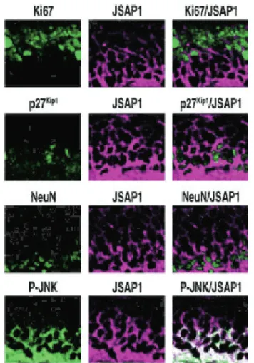
シグナル伝達研究分野 Division of Molecular Cell Signaling
教授… 善岡 克次
Professor YOSHIOKA, Katsuji
助教… 佐藤 時春
Assistant Professor SATO, Tokiharu
教授… 後藤 典子
Professor GOTOH, Noriko
●●●●●●●●●●
xxxxxxxxxxxxxxxxxxxxxxxxxxxxxxxxxxxxxx
■
●●●●●●●●●● xxxxxxxxxx
●●●●●
xxxxxxxxxx
●●●●●
■
●●●●●●●●●● xxxxxxxxxx
●●●●●
■
機能ゲノミクス研究分野 Division of Functional Genomics
教授… 鈴木 健之
Professor SUZUKI, Takeshi
助教… 石村 昭彦
Assistant Professor ISHIMURA, Akihiko
■
腫瘍制御研究分野 Division of Translational and Clinical Oncology
教授… 源 利成
Professor MINAMOTO, Toshinari
Division of Cancer Cell Biology
分子病態研究分野
癌と癌幹細胞に注目し,基礎研究から臨床へと連続す る研究の展開を目指している。最先端の分子生物学,細 胞生物学的手法,さらにはシステム生物学的理論を組み 合わせて,癌の早期発見や個々の患者に最適な治療法を 選択するための診断マーカーの抽出,そして新しい抗が ん剤開発のための新たな分子標的の発見を試み,トラン スレーショナルリサーチへと展開している。
1) 癌幹細胞−乳癌をモデル系として
マウス癌モデルや,ヒト乳癌の臨床検体を用いた癌 幹細胞の解析から,癌幹細胞内の新規分子標的や癌 の診断マーカーの探索を行っている。
2) 肺癌の診断マーカー及び分子標的の探索
世界的にも肺癌による死亡者数は,全癌死の一位を 占めている。増殖因子受容体シグナル伝達の解析に システム生物学的手法を取り入れ,肺癌の早期発見 や個々の患者に最適な治療法を選択するための診断 マーカーや新規分子標的の探索を行っている。
3) 正常の幹細胞と癌幹細胞をあやつる増殖因子受容体 シグナル伝達
癌という病気や,幹細胞の維持という生命現象を動 かしている主役の分子群として,増殖因子受容体型 チロシンキナーゼである,FGF 受容体や EGF 受容 体は重要である。これら代表的増殖因子受容体の細 胞内シグナル伝達の司令塔として,アダプター/ド ッキング分子 FRS2 ファミリー分子に注目している。
Our major research interest is to elucidate the molecular mechanisms regulating cancer cells, stem cells and cancer stem cells. Our team has two important research directions: One is to clarify the basic principles underlying biology and the other is to apply the knowledge extracted from the basic principles to translational medicine. In order to achieve the goal, we take challenging approaches of molecular biology and systems biology, in addition to conventional methods of molecular biology.
1) Molecular mechanisms of cancer initiation, progression and metastasis: breast cancer stem cells as key players
By analyzing the mouse cancer model or primary cancer cells derived from human specimens, we attempt to identify novel molecular targets and biomarkers for cancer.
2) Identification of new biomarkers and molecular targets of lung cancers by systems biology approach
Our hypothesis is that elucidation of the molecular mechanisms of addiction of lung epithelial cells to EGF RTK signaling leads us to identify new biomarkers and molecular targets of lung cancer. Our approach would certainly advance personalized medicine in the near future.
3) Signal transduction mechanisms through receptor tyrosine kinases (RTKs) for tumorigenesis and stem cell maintenance Fibroblast growth factor (FGF) and epidermal growth factor
(EGF) RTKs play major roles for a variety of physiological and pathological aspects of biology, including stem cell biology, and cancer biology. We focus on FRS2 family of adaptor/scaffolding docking proteins, as key intracellular signal regulators of these RTKs.
図1
癌幹細胞内ヘレギュリン -PI3 キナーゼパスウエイの活性化 は,様々なサイトカイン,増殖因子,細胞内因子を産生する。
Fig. 1
Activation of heregulin-phosphatidyl inositol (PI)-3 kinase pathway induces various cytokines, growth factors and cytoplasmic molecules that regulates cancer stem cells and their niche.
図2
EGF シグナル鍵分子 139 遺伝子を用いると,肺腺癌患者の 予後を精度高く予測できる
Fig. 2
The 139 key genes involved in EGF signaling accurately predict prognosis of lung cancer patients.