Original
*Author : Department of Otolaryngology, Saitama Medical University, 38 Morohongo, Moroyama, Iruma-gun, Saitama 350-0495, Japan Tel: + 81 492 76 1253, Fax: + 81 492 95 8061, E-mail: sugizaki@saitama-med.ac.jp
◯The authors declare that there are no conflicts of interest associated with the present study.
3D analysis of binocular eye movement during head tilt
Kazuki Sugizaki
1)*, Yasuo Koizumi
2), Mio Iwamura
2), Ryuichiro Araki
3),
Yasuhiro Kase
1), Tetsuo Ikezono
1), Toshiaki Yagi
4)1) Department of Otolaryngology, Saitama Medical University, 38 Morohongo, Moroyama, Iruma-gun, Saitama 350 - 0495, Japan
2) Department of Otolaryngology, Nippon Medical School, 1-1-5 Sendagi, Bunkyo-ku, Tokyo 113 - 8603, Japan 3) Community Health Science Center, Saitama Medical University, 38 Morohongo, Moroyama, Iruma-gun, Saitama
350 - 0495, Japan
4) University of Human Environments, 6 - 2 Kamisanhonmatsu, Motojyuku, Okazaki, Aichi 444-3505, Japan
【Objective】Ocular counter rolling (OCR) is one type of vestibulo-ocular reflex (VOR) originating in the otolith organ, and is one of the few effective tests of otolith function. The purpose of the present study was to assess the three-dimensional binocular eye movements that occur in response to the roll tilt. Furthermore, the study was intended to find out whether any asymmetry or disconjugacy of eye movements exists in 1 g environment.
【Methods】Ten healthy subjects (6 males and 4 females) participated in this experiment. The subjects were tilted to the left or to the right on the naso-occipital axis up to 35 degrees (deg.). The eye movements were recorded and an analysis of these eye movements was performed using a video - oculographic method.
【Results】In torsional eye movement, there was counter -rolling of the eyes in response to head tilt. In terms of horizontal eye movement, the left eye deviated towards the right during left ear down (LED) tilt, and the right eye deviated towards the left during right ear down (RED) tilt. There was no asymmetry in the performance of horizontal, vertical or torsional eye movements. There was no disconjugacy in the vertical or torsional eye movements, and in the horizontal eye movements disconjugacy was observed in which the lowermost eye to the tilt deviated towards the opposite side.
【Conclusion】In normal subjects in a 1 g environment, each of the three eye movements was symmetrical during head tilt. There was disconjugacy observed in the horizontal eye movements. The deviation of the lowermost eye towards the opposite relative to the tilt direction may be explained by the anatomical and/or physiological characteristics of the utricular nerve projection to the abducens nucleus.
J Saitama Medical University 2013; 39: 121 - 129
(Received August 17, 2012 / Accepted February 1, 2013)
Keywords: eye movements, ocular counter rolling, binocular, symmetry, disconjugacy
Introduction
Vestibulo-ocular reflex (VOR) occurs to stabilize images on the retina by causing ocular movement in the direction opposite to the head movement when
which the eyes roll in the direction opposite to the head roll when the head is tilted in either direction in the frontal plane. Hunter first reported this physiological behavior as long ago as 17861). The OCR is one type of
VOR originating in the otolith organ, primarily in the utricle, and is one of the few effective tests of otolith function2, 3). However, the difficulty of recording the
torsional eye movements has hampered the progress of OCR research, despite its early discovery. Although there are relatively few studies on the OCR4, 5), reports
on the binocular OCR are even fewer6). Therefore, it is
worthwhile to investigate not only the torsional, but also the horizontal and vertical binocular eye movements that occur during head tilt.
The purpose of the present study was to assess the three-dimensional eye movements in response to the roll tilt using a recently developed non-invasive recording system7, 8). Furthermore, the study was intended to find
out whether there is any asymmetry or disconjugacy of eye movements in 1 g environment, as reportedly there are under microgravity in human subjects9).
Materials and Methods
Ten healthy subjects without any history of inner ear disorders participated in this experiment. The subjects were six males and four females, ranging in age from 20 to 38 years. Before starting the experiment, we explained the purpose and method of the study and obtained the informed consent in a document according to the Declaration of Helsinki. The period of the experiment was from July 2007 to September 2008.
The subjects were seated on a chair in the center of a
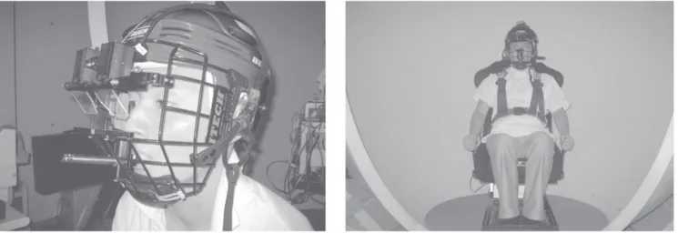
specially constructed dome-like structure. The subjects were asked to wear a whole head helmet equipped with a binocular Charge Coupled Device (CCD) camera, which was newly designed and constructed for this experiment. This helmet has a facemask, and subjects were instructed to bite a bar integrated into the facemask so as to keep the position constant between the camera and subject’s eye (Fig. 1). The subject’s body was further secured by a body harness and soft rubber for the purpose of a secure fit.
The whole head helmet that was used has two infrared CCD cameras with cold filters for see-through vision. The cold filter is made of 31 layers of mutually laminated TiO2 and SiO2. TiO2 has a high reflective
index, while SiO2 has a low reflective index. The cold
filter transmits visible light, but reflects infrared light of a wavelength ranging from 729 to 1134 nm. The images of both eyes, illuminated by infrared light and reflected by the cold filters, are transmitted to the two CCD cameras. In this process, the subjects can see the objects without any difficulty through the cold filters.
The subjects donned this whole head helmet described above. Before recording, the eye position was calibrated. The red light emitting diode, with a diameter of 5 mm, was mounted in the line of sight as the visual target. The subjects gazed at a pair of light emitting diodes, which were placed 5 degrees (deg.) apart horizontally and vertically from the central target. After turning off the target lights, the subjects were instructed to look forward in the dome. The inside of the dome was painted white and illuminated with no light, so that there were no object cues in the dome.
Fig. 1. Two infrared CCD cameras with cold filters for see -through vision were included in the whole head helmet with
facemask. The cold filter was made of 31 layers of mutually laminated TiO2 and SiO2. The subjects bit a bite bar
From the upright position, the subjects were tilted to the left or right on the naso-occipital axis up to 35 deg. and then were returned to the upright position. After a 3 - minute rest, the subjects were tilted in the opposite direction, again up to 35 deg. A constant tilting velocity of 1 deg. /second was applied, which is slower than the 3 deg. that is reported to have virtually no impact on the semicircular canal10).
The eye movements were recorded directly into the hard disk of the computer. During tilting, each eye was simultaneously and independently observed in the TV monitors. The 3D analysis was performed offline using the video image analysis system. The sampling interval of the eye movements was 30 Hz. The direction of the torsional eye movements, clockwise and counterclockwise, is described from the subject’s point of view. To eliminate artifacts, the eye position of the video images, such as the difference between the inner canthus and the edge of the monitor screen, was constantly monitored throughout the procedure.
Asymmetry score
We examined whether asymmetr y of the eye movements occurred during the head tilt. We compared the eye positions of the left eye and those of right eye at a given angle. The actual calculation was as follows: the eye position at the head tilt of A deg. (the left eye position at A deg. of LED tilt)-(the left eye position at A deg. of RED tilt) or (the right eye position at A deg. of LED tilt)-(the right eye position at A deg. of RED tilt). If this value (we defined as Asymmetr y score) increased correspondingly as the angle of the head tilt increased, it would indicate that there is asymmetry eye movement.
Disconjugacy score
We also examined whether disconjugacy of the eye movements occurred during the head tilt. We compared the eye positions during the LED tilt and those of RED tilt at a given angle. The actual calculation was as follows: the eye position at the head tilt of A deg. (the left eye position at A deg. of LED tilt) -(the right eye position at A deg. of LED tilt) or (the left eye position at A deg. of RED tilt) -(the right eye position at A deg. of RED tilt). If this value (we defined as the disconjugacy score) increased correspondingly as the angle of the head tilt increased, it would indicate that both eyes had disconjugate eye movements.
Statistical Analysis
The Jonckheere - Terpstra trend test was employed to assess monotonic trend of the eye position dependent on the angle of the head tilt using R version 2.15.2 for
Windows11). p<0.05 was considered statistically significant.
Results
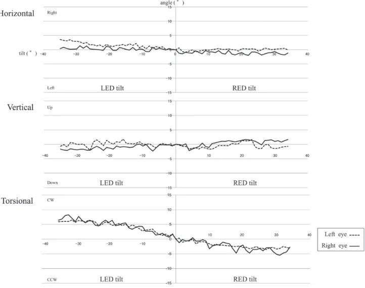
One example of three-dimensional analysis of eye movements during the LED tilt and the RED tilt per second is shown in Fig. 2. The horizontal eye movements show the counter-deviation to the tilting direction, especially in the lowermost eye. That is, the left eye tends to deviate to the right during the LED tilt and the right eye tends to deviate to the left during the RED tilt. The vertical eye movements did not exhibit any discernible ocular deviation in response to head tilt. In the torsional eye movements, clear counter rolling was demonstrated in both eyes in response to the LED and RED tilts.
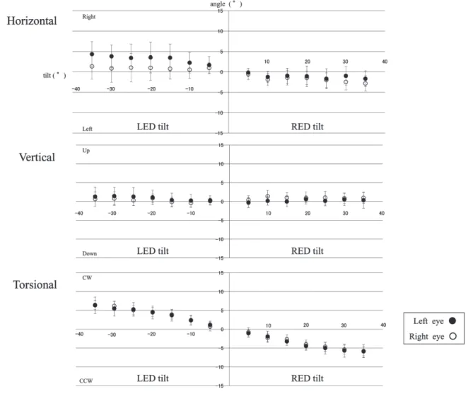
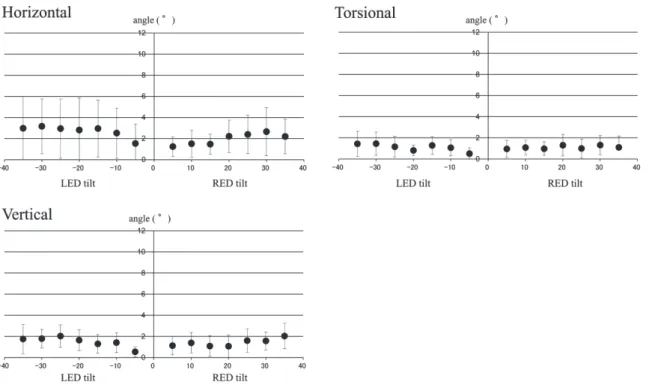
Fig. 3 shows the mean and standard deviations of three eye movements - horizontal, vertical, and torsional - in the 10 cases for every 5 deg. during the LED and RED tilts in both eyes. In the torsional eye movement, there was a statistically significant trend for the eye position on the angle of the head tilt (Jonckheere-Terpstra trend test, p < 0.001). In other words, as the head tilt increased, the eye torsional angle also increased correspondingly. On the other hand, in the case of horizontal eye movement, the left eye deviated towards the right during LED tilt, and the right eye deviated towards the left during RED tilt. A statistically significant trend for the horizontal eye position on the head tilt angle was identified (Jonckheere -Terpstra trend test, p = 0.008, p = 0.004). However, in the uppermost eye (the right eye during the LED and the left eye during the RED) there was no clear tendency of deviation (Jonckheere -Terpstra trend test, p = 0.61, p = 0.13). In the case of vertical eye movement, the left eye deviated up during LED tilt, a statistically significant trend for the vertical eye position on the head tilt angle was observed (Jonckheere-Terpstra trend test, p = 0.04). The other vertical eye movements (right eye during LED and RED tilts, the light eye during RED tilt) did not deviate to any direction.
the head tilt angle was identified (Jonckheere-Terpstra trend test, p = 0.02). This result indicated that there is asymmetry eye movement only in the vertical eye movements of left eye. In the other case, no asymmetry was observed.
Fig.5 shows the difference between the right and left eye position, which is mentioned above as the disconjugacy score, at ever y five deg. of tilt. We examined the disconjugacy score statistically, in the horizontal eye movements of the RED tilt and in the vertical eye movements of the LED tilt and in the torsional eye movements of the LED tilt, a significant trend for the disconjugacy score on the head tilt angle was identified (Jonckheere-Terpstra trend test, p = 0.01,
p = 0.001, p = 0.04) . The result indicated that there are disconjugate eye movements in those cases.
Discussion
There are numerous reports on the eye movements that occur during head tilt, especially on OCR, as an otolith-ocular reflex12 - 14). However, only a few studies
have been reported on the symmetry and conjugacy of eye movements during head tilt. Kanzaki et al.15)
studied the OCR symmetry for the purpose of assessing otolith function and found that the majority of normal subjects exhibited symmetrical eye movements during head tilt. In their report, torsion of the eyes below 2 deg either the right or the left tilt was considered abnormal,
Fig. 2. One example of the three -dimensional analysis of eyes during the left ear down (LED) tilt and the right ear down
and they also suggested that a difference of 5 deg. at 30 deg. of tilt and 4 deg. at a 45 deg. tilt between the RED and LED results suggested otolith dysfunction. Furthermore, Yashiro et al.16) reported that the OCR
asymmetry is quite exceptional in normal subjects, and the presence of this asymmetry may be related to a latent functional difference between the otolith organs on the two sides. In our study, none of the ten normal subjects showed a deviation of 4 deg. or more between the left and right eyes at a 30 deg. head tilt. The average difference was 1.9 deg. for the left eye and 1.4 deg. for the right eye, and this is consistent with the studies noted above, so there was no asymmetry in the OCR. In contrast to the situation with the numerous
studies on torsional eye movements, we did not find any studies on the symmetry of horizontal or vertical eye movements during head tilt. In the present study, we demonstrated that there was no asymmetr y in horizontal eye movements in normal subjects. On the other hand we found out the asymmetry eye movement only in the vertical eye movements of left eye. This result was demonstrated statistically, but those eye movements occurred with the eye on only one side. Therefore we consider there was no significant meaning in this result physiologically, and conclude that there is no asymmetry in the horizontal and vertical eye movements during head tilt. Thus, in normal subjects in a 1 g environment, each of the three types
Fig. 3. The mean (open and closed circles) and standard deviations (vertical bars) of horizontal, vertical, and torsional eye
of eye movement - horizontal, vertical, and torsional - is symmetrical during head tilt.
Pansell et al.14) observed no disconjugate horizontal
eye movements during head tilt. In our experiment, however, the left eye deviated to the right in the LED tilt, while the right eye deviated to the left in the RED tilt. There was a statistically significant trend for the horizontal eye position on the head tilt angle. We observed disconjugacy in that the lowermost eye to the tilt deviated towards the opposite side. However, a significant trend for the disconjugacy score on the head tilt angle was identified only in the horizontal eye movements of the RED tilt, not in those of LED tilt. We speculate that the large variance in the horizontal eye movements of LED tilt caused no significant trend for the disconjugacy score on the head tilt angle. If more subjects had participated in this study, the variance may
have been smaller than shown in this study’s results. A limitation of this study is that the number of subjects were relatively small. Uchino et al.17, 18) selectively
stimulated the primary afferents of the utricular nerve and demonstrated that they project directly into the ipsilateral abducens nucleus. In addition, Goto et al.19)
reported the horizontal eye movements evoked by the selective stimulation of the utricular nerve in cats. This study confirmed the anatomical connections from the utricular primary afferents to the ipsilateral abducens nucleus neurons. Thus, the deviation of the lowermost eye towards the opposite relative to the tilt direction which we observed may be explained by the above mentioned experimental findings.
The vertical skew was induced by dynamic head tilt with an upward deviation of the ipsilateral eye and downward deviation of the contralateral eye,
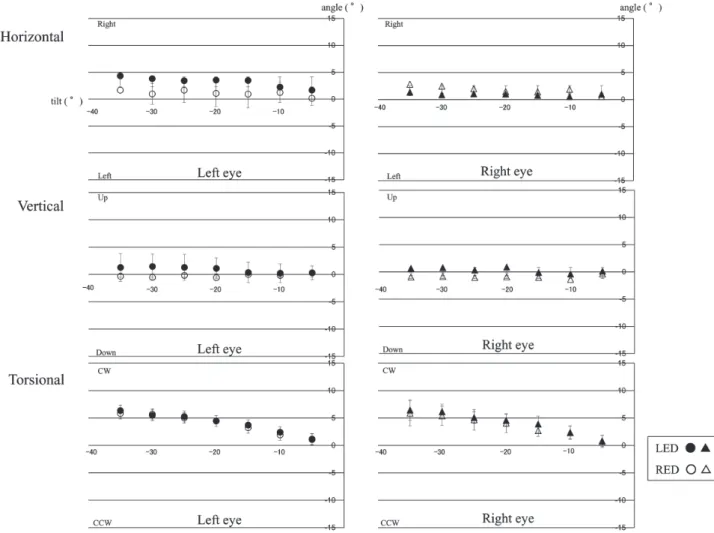
Fig. 4. The eye positions at every 5 deg. during the LED and RED tilts were superimposed as the values of eye deviation
as reported by Pansell et al.20). However, in a 2003
study14) by the same authors, disconjugate vertical eye
movement was not consistently observed among the test subjects. They observed cases of the right eye over the left eye and cases of the left eye over the right eye, even in the same tilt direction. As the authors mention in their discussion of these observations, the different observation in the more recent experiment was likely the result of an apparent disconjugacy between the left and right eyes caused by a slipping of the head mask along with the camera during the head tilt, which can occur even when a bite bar is used. To avoid this, we always monitored camera slippage relative to the eyes on TV monitor. In this study, in the vertical eye movement, the left eye deviated towards up during LED tilt. Markham et al.3) reported that the binocular
OCR during head tilt for 16 normal subjects in a 1 g environment was approximately symmetric in both tilt directions, LED and RED. They studied on the OCR responses in normal subjects under both dynamic and static conditions, and found that the mean OCR difference between the left and right eyes at 30 deg. of tilt were less than 1.5 deg. in the LED and RED tilt21).
In the present study, we examined up to 35 deg. of tilt and observed no disconjugacy (1.46 deg. in the LED,
1.32 deg. in the RED) in the torsional movements, consistent with these reports. In the vertical eye movements of the LED tilt and in the torsional eye movements of the LED tilt, a significant trend was identified between the disconjugacy score and the head tilt angle. However, because those eye movements occurred in only one side tilt, we consider there was no significant meaning in this result physiologically. We conclude that there is no disconjugacy in the vertical and torsional eye movements during head tilt.
Diamond et al.22) measured torsional eye movements
at 0 g and 1.8 g during parabolic flight, and observed a disconjugacy that was not observable 1 g environment. They found significantly higher scores of disconjugate eye torsion in the astronauts who had been sick in space. Karmali et al.9) repor ted ocular ver tical
disconjugacy occurred along with changes in gravity during parabolic flight. They speculated that the anatomical asymmetry that exists between the right and left otolith organs might be compensated for in the central nervous system when the subjects are in a 1 g environment, but this compensation is inappropriate in other gravity levels and results from ocular vertical disconjugacy. The results presented here show that the functions of the otolith organ may be different in 1 g
Fig. 5. A comparison of the eye positions during the LED and RED tilt at a given angle in three-dimensions. The difference
environment and microgravity. While normal subjects exhibit eye movements based on the physiological characteristics of the otolith organ in the normal physical (1 g), latent functions and/or anatomical differences may become evident in a non-physiological environment, as they did during the experimental changes in gravity.
In this study, we demonstrate the deviation of the lowermost eye towards the opposite relative to the tilt direction in horizontal eye movement, which has not mentioned until now. We speculate that this eye movement is one of VOR originating in the otolith organ. This phenomenon may occur to stabilize images on the retina, same as OCR. We consider these results in this study contribute the development of otolith organ research.
References
1) Hunter J. First description of ocular counterrolling in 1786. The use of the oblique muscles. Strabismus 2002;10:279 - 81.
2) Diamond SG, Markham CH. Ocular counterrolling as a test of otolith function. Acta Otolar yngol (Stockh) 1989;468(Suppl):267 - 70.
3) Diamond SG, Markham CH. Ocular counterrolling as an indicator of vestibular otolith function. Neurol 1983;33:1460 - 9.
4) Kuilman J. Nystagmography during counterrolling of the eyes in man. AMM Arch Otolar yngol 1958;67(4):424 - 6.
5) Miller EF. Evaluation of otolith organ function by means of ocular counterrolling measurements. In: Stahle J, editors. Vestibular Function on Earth and in Space. Oxford: Pergamon Press; 1970. p. 97 - 107. 6) Diamond SG, Markham CH, Simpson NE,
Cur thoys IS. Binocular counterrolling during dynamic rotation. Acta Otolaryngol 1979;87:490 - 8. 7) Yamanobe S, Taira S, Morizono T, Yagi T, Kamio T.
Eye movement analysis system using computerized image recognition. Arch Otolaryngol Head Neck Surg1990;116:338 - 41.
8) Yagi T. Nystagmus as a sign of labyrinthine d i s o r d e r s - T h r e e - d i m e n s i o n a l a n a l y s i s of N y s t a g m u s . C l i n E x p O t o r h i n o l a r y n g o l 2008;1:63 - 74.
9) Karmali F, Ramat S, Shelhamer M. Vertical skew due to change in gravitoinertial force: a possible consequence of otolith asymmetry. J Vestib Res 2006;16:117 - 25.
10) Miller EF. Counterrolling of the human eyes
produced by head tilt with respect to gravity. Acta Otolaryngol 1962;54:479 - 501.
11) R Development Core Team (2012), R: A language and environment for statistical computing. R Foundation for Statistical Computing, Vienna, Austria. ISBN 3 - 900051 - 07 - 0, URL http://www. R-project.org/.
12) Zingler VC, Kryvoshey D, Schneider E, Glasauer S, Brandt T, Str upp M. A clinical test of otolith function: static ocular counterroll with passive head tilt. Neuroreport 2006;17(6):611 - 5.
13) Schwor m HD, Pansell T, Lennerstrand G. Assessment of ocular counterroll during head tilt using binocular video oculography. Invest Ophthalmol Vis Sci 2002;43:662 - 7.
14) Pansell T, Yagg J, Schworm HD. Conjugacy of torsional eye movements in response to a head tilt paradigm. Invest Ophthalmol Vis Sci 2003;44:2557 - 64.
15) Kanzaki J, Ouchi T. Measurement of Ocular counter reflex with fundoscopic camera in normal subjects and in patients with inner ear lesions. Arch Otorhinolaryngol 1978;218:191 - 201.
16) Yashiro T, Ishii M, Igarashi M, Kobayashi T, Kaneta K, Kobayashi N, et al. Evaluation of ocular counterrolling-normal subjects and the cases with unilateral of bilateral vestibular dysfunction. Oto-Rhino-Laryngology, Tokyo 1996;39:56 - 63.
17) Uchino Y, Ikegami H, Sasaki M, Endo K, Imagawa M, Isu N. Monosynaptic and disynaptic connections in the utriculo-ocular reflex arc of the cat. J Neurophysiol 1994;71:950 - 8.
18) Uchino Y, Sasaki M, Sato M, Imagawa M, Suwa H, Isu N. Utriculoocular reflex arc of the cat. J Neurophysiol 1996;76:1895 - 903.
19) Goto F, Meng H, Bai R, Sato H, Imagawa M, Sasaki M, Uchino Y. Eye movements evoked by the selective stimulation of the utricular nerve in cats. Auris Nasus Larynx 2003;30,341 - 8.
20) Pansell T, Ygge J, Schworm HD. Dynamic changes of eye cyclo position during head tilt. Ann N Y Acad Sci 2002;956:564 - 7.
21) Markham CH, Diamond SG. Ocular counterrolling in r esponse to static and dynamic tilting: Implications for human otolith function. J Vestib Res 2002 - 2003;12:127 - 34.
頭部傾斜時の眼球運動の3次元解析 杉 崎 一 樹 1), 小 泉 康 雄 2), 岩 村 美 生 2), 荒 木 隆 一 郎 3), 加 瀬 康 弘 1), 池 園 哲 郎 1), 八木 聰明 4) 前庭眼反射は,頭部が動いた時に,その動きと反対方向に眼球を偏位させるもので,網膜上の像を安定 させる働きがある.眼球反対回旋運動は,頭部傾斜時に,眼球が傾斜と反対方向に回旋する現象であり, 耳石器,主として卵形嚢由来の前庭眼反射と考えられている.そしてこれは,数少ない耳石機能検査の1つである. 今回の研究の目的は,頭部傾斜時の眼球運動を非侵襲的記録装置で両眼同時に記録し,眼球運動を3次元的に 解析し,頭部傾斜との関連や,対称性,共同性を評価することである.対象は,内耳障害の既往のない健常人10人 (男性6名,女性4名)である.被検者は特別に設計されたドームの中心の椅子に座り,両眼CCDカメラ付きヘル メットを装着してもらった.被検者の体はシートベルトで椅子にしっかり固定され,正中位から,鼻後頭軸に対 して左右に35度まで傾斜され,傾斜時の眼球運動をカメラで記録した.その後,コンピュータに記録された画像 を,ビデオ画像解析システムを用いて3次元解析した.回旋眼球運動は,両眼とも反対回旋がみられ,眼球運動 と頭部傾斜角度との間に統計学的有意差を認めた(トレンド検定 p<0.001).水平眼球運動では,左傾斜時に左 眼が右へ,右傾斜時には右眼が左への偏位が認められ,傾斜方向の外側眼のみが内側へ偏位するという非共同 性がみられた.(トレンド検定,p=0.008, p=0.004)水平・垂直・回旋眼球運動とも,頭部傾斜に対して対称性 を認めた.頭部傾斜時の,傾斜方向の外側眼の内側への偏位は,過去に電気生理学的に報告されている卵形嚢刺 激の同側外転神経核への投射により説明できるのではないかと考えた.よって,この眼球運動は耳石器由来の前 庭眼反射と推測される.この結果は,過去に報告がなく,今後の耳石機能研究の発展の一助となると考える. 1) 埼玉医科大学 耳鼻咽喉科 〒 350-0495 埼玉県入間郡毛呂山町毛呂本郷 38 2) 日本医科大学 耳鼻咽喉科 〒 113-8603 東京都文京区千駄木 1-1-5 3) 埼玉医科大学 地域医学・医療センター 〒 350-0495 埼玉県入間郡毛呂山町毛呂本郷 38 4) 人間環境大学 〒 444-3505 愛知県岡崎市本宿町上三本松 6-2 〔平成 24 年 8 月 17 日受付 / 平成 25 年 2 月 1 日受理〕