九州大学学術情報リポジトリ
Kyushu University Institutional Repository
Fabrication of skeletal muscle tissue
constructs using coculture and microfabrication systems
ムド, アリフッザマン
http://hdl.handle.net/2324/2236206
出版情報:九州大学, 2018, 博士(工学), 課程博士 バージョン:
権利関係:
Fabrication of skeletal muscle tissue constructs using coculture and microfabrication systems
Submitted by MD ARIFUZZAMAN
TABLE OF CONTENTS
Chapter 1 ……….1
Introduction………1
1.1 Regenerative medicine ………..1
1.2 Tissue Engineering ……….. 3
Magnetic force based tissue engineering ………. 6
1.3 Heterotypic cellular communication………. 7
1.4 Synaptogenesis between muscle and nerves………. 8
1.5 Microfabrication of artificial tissues………. 9
Organ-on-Chip………. 9
1.6 Drug screening based on 3D tissues………... 10
1.7 References……… 12
Chapter 2 ………....18
Background ………. 18
2.1 Skeletal muscle tissue engineering (SMTE) ……… 18
2.1.1 Cell alignment by topography ……… 20
2.1.2 Cell alignment by surface patterning ……… 20
2.1.3 Cell alignment by mechanical stimulation ………. 21
2.1.4 Application of magnetic nanoparticle for skeletal muscle tissue engineering ……… 21
2.1.5 Stem cells for skeletal muscle tissue engineering ……… 22
2.2 Nerve muscle co-culture ……….. 23
2.3 Neuromuscular junction ……….. 25
2.3.1. AchR subtype ………. 26
2.3.2 Agrin ……….. 27
2.3.3 Role of neurotrophic factors and cytokines for the formation of NMJ ………. 27
2.4 Microfabrication of skeletal muscle tissues ……….. 28
2.5 Skeletal muscle tissue engineering for drug screening ……….. 31
i
2.6 Purpose of the study ……… 33
2.7 Thesis components ……… 33
2.8 References ……… .……..35
Chapter 3………. 45
Fabrication of muscle-neuron constructs by using C2C12/PC12 co-culture system for enhancement of contractile force generation ………45
3.1 Introduction ……….45
3.2 Materials and Methods ………...47
3.3 Result ………..52
3.4 Discussion ………64
3.5 Conclusion ………...68
3.6 References ………...69
Chapter 4 ……….72
Microfabrication of artificial skeletal muscle constructs………72
4.1 Introduction ……….72
4.2 Materials and Methods ………...74
4.3 Result ……….80
4.4 Discussion ………...84
4.5 Conclusion ………..90
4.6 References ……….92
Chapter 5 ……….96
Conclusion and Perspectives………96
5.1 Summary of the thesis ………96
5.2 Prospect………...98
5.3 References ……… 99
Acknowledgements ………100
ii
1
Chapter 1 Introduction
1.1 Regenerative medicine
Regenerative medicine is the potential tool to heal or replace tissues and organs damaged by different disease or trauma or due to the congenital defects. Now a day, regenerative medicine is the common term among physicians and scientists for treating different chronic diseases and acute insults e.g. different types of dermal wounds, cardiovascular diseases, traumas and also treatment for certain types of cancers [1-3]. Current therapy of transplantation of organs and tissues to treat tissue and organ failures sometimes difficult due to limited donor supply. Also, after transplantation there is a risk of immune rejection of transplanted tissue or organ grafts. But, these obstacles may potentially be solved through the use of regenerative medicine strategies.
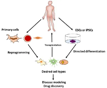
The process of regenerative medicine is given in Fig. 1-1. It requires two main strategies for generating patient-specific cells of a desired type. Pluripotent cells to be used for regenerative medicine can be either patient-derived (induced pluripotent stem cells (iPSCs)) or non-patient-derived (either embryonic stem cells (ESCs) or iPSCs). Pluripotent cells can be differentiated in vitro to a desired cell state (directed differentiation, right).
Alternatively, primary cells derived from a patient can be used to generate a desired cell type directly reprogramming (left). Cells of a desired type obtained by either of these methods can then be studied in vitro (bottom) or used for transplantation into patients (top).
The field of regenerative medicine covers a wide range of areas including the use of materials and de novo generated cells, as well as various combinations thereof, to take the place of missing tissue, effectively replacing it both structurally and functionally or to contribute tissue healing [4]. Normally, body’s innate immune system is involved to
2
promote regeneration, but adult humans possess limited regenerative capacity in comparison with lower vertebrates [5].
Fig. 1-1. Process of regenerative medicine
Since tissue engineering and regenerative medicine emerged as an industry about two decades ago, Food and drug administration (FDA) approved a number of therapies for commercial availability. Till today, the delivery of therapeutic cells that directly contribute to the structure and function of new tissues is a principle paradigm of regenerative medicine. Generally, autologous or allogenic and typically differentiated cells are used in these therapies those have proliferative capacity [6]. Besides cells, materials are often an important component of current regenerative medicine strategies because material can mimic the native extracellular matrix (ECM) of tissues and contribute to the structure and function of new tissues and locally present growth factors [7]. For example, 3D polymer scaffolds are used to promote expansion of chondrocytes and cartilage repair and provide scaffolds for fibroblasts in the treatment of venous ulcers [8]. Sometimes a material can alone provide cues for regeneration and graft or implant integration, as in the case of bioglass-based grafts that permit fusion with bone [9]. Incorporation of growth factors promote healing or regeneration into biomaterials can provide a local and sustained presentation of these factors, and this approach has been exploited to promote wound healing by delivery of platelet derived growth factors and bone formation via the delivery of bone morphogenetic protein (BMP-2) [10].
However, the study of regenerative medicine comprises a wide range of studies at both pre-clinical and clinical stages which includes: recapitulating organ and tissue
3
structure via scaffold fabrication, integrating grafts with the host via vascularization and innervation, and, altering the host environment to induce therapeutic responses through cell infusion and modulating the immune system [11]. Most regenerative medicine
strategies rely on an ample cell source, but identifying and obtaining sufficient numbers of therapeutic cells is often a challenge. Stem, progenitor and differentiated cells derived from both adult and embryonic tissues are widely being explored in regenerative medicine although adult tissue-tissue derived cells are the dominant cell types used clinically to date due to both their availability and safety [6].
There is a great chance in obtaining a large number of stem cells from adult tissues and in identifying stem cell populations are suitable for therapeutic use in tissues [12].
Stem cells play a vital role in the field of regenerative medicine. For example, coculture of hematopoietic stem cells (HSCs) with cells implicated in the HSC niche and in
microenvironments engineered to mimic bone marrow improve HSC stemness during expansion, enhancing stem cell numbers for transplantation such as direct contact of HSCs with mesenchymal stem cells (MSCs)grown in a 3D environment induces greater CD34+ expansion than with MSCs grown in 2D substrate [13]. Another example is that culture of skeletal muscle stem cells on substrate with mechanical properties similar to normal muscle leads to greater stem cell expansion, rescue impaired proliferative ability in stem cells from aged animals [14]. Also, Embryonic stem (ES) cells are characterized potential infinite sources of cells for regeneration and can be used for clinical application [15]. ES cells are derived from blastocyst stage embryos and have been shown to be pluripotent, giving rise to tissues from all three germ layers [16].
1.2 Tissue Engineering
Tissue engineering is an interdisciplinary field which comprises the principle of engineering and life sciences toward the development of biological substitutes that restore, maintain, and improve tissue function or whole organ [17]. Development of biological substitutes by using tissue engineering techniques based on three key elements: living cells, biomaterials that provide the base for cells to attach, and bioactive factors that stimulate cells to make proliferation and differentiation.
4
Tissue engineering approach includes isolation and purification of a cell source, seeding of the cells on scaffold, stimulation of the cell -seeded scaffold to develop a tissue equivalent and implantation of the construct in vivo.
Fig.1.2. Strategies of tissue engineering
Tissue engineering strategies require interaction and integration through incorporation of appropriate physical and cellular signals. Therefore, inclusion of modifying factors such as biologically active proteins and DNA are critical to success [18].
Two main approaches are utilized in this area to produce engineered tissue. First, scaffolding can be used as a cell support device upon which cells are seeded in vitro, cells are then encouraged to lay down matrix to produce the foundations of a tissue for transplantation. The second approach involves using the scaffold as a growth factor/drug delivery device. This strategy involves the scaffold being combined with growth factors, so upon implantation cells from the body are recruited to the scaffold site and from tissue upon and throughout the matrices. These two approaches are not mutually exclusive and can be easily combined [19]. The assembly of cells into tissues is highly orchestrated set of events that requires time scale ranging from seconds to weeks and dimensions ranging from 0.0001 to 10 cm. Until today different engineered tissues have been approved by the Food and Drug Administration (FDA). But, still there are many technical challenges to overcome before the creation of the shelf tissues that represent the translation of scientific discoveries into treatments for millions of patients. The successful large- scale production
5
of engineered tissues requires an adequate source of healthy expandable cells, the optimization of scaffolds, and the creation of bioreactors which mimic the environment of the body and that are amenable to scale up. Another type of challenge includes the preservation of the product so that it has a long shelf-life and successful use of various approaches to prevent tissue rejection.
The important prerequisite for effective tissue engineering is the cellular environment that allows the cells to function as they do in native tissue. Sometimes environment mimics several critical aspects of the in vivo niche through proper control of the chemical and mechanical setting as well as the chemical stimuli. Cell scaffolds generally serves several purposes including: a). cell attachment and perhaps migration; b). retention and
presentation of biochemical factors; c). porous environment for adequate diffusion of nutrients, expressed products and waste, and d). mechanical rigidity or flexibility [20].
The production of engineered tissues in vitro requires the use of cells to populate matrices and produce matrix resembling that of the native tissues. The source of the cells is also an important choice for scaffolds [21]. A range of cell types can now be combined with scaffolds to produce tissue engineered constructs. Major success in this field have been achieved from the use of patient specific primary cells to fabricate tissues for re- implantation. However, this strategy has some limitations because of the invasive nature of the cell collection and the potential for the cells to be in a diseased state [22]. Recently, different types of cell lines and stem cells including ES cells, bone-marrow derived mesenchymal stem cells (BM-MSCs) and umbilical- cord derived mesenchymal stem cells (UM-MSCs) are being extensively used for fabricating artificial tissues. Many researchers have chosen cell lines for tissue fabrication rather than primary cells as they are highly proliferative and easier to culture and transfect [23]. Cell lines applied in the field of tissue engineering includes: C2C12 cell lines for skeletal muscle tissue engineering, MC-3T3 cell lines for bone tissue engineering, human umbilical vein endothelial cord (HUVEC) cell lines for vascular tissue engineering, mouse fibroblast cell line (NIH-3T3) for skin tissue engineering etc. ES cells allow the production of type-matched tissues for each patient, either through stem cell banking or by the use of therapeutic cloning. ES cells have the ability to be maintained for long (theoretically indefinite) culture periods, therefore potentially providing large amounts of cells for tissues that could not be derived directly from a tissue source. Proof of the true pluripotent nature of ES cells is teratoma formation.
6
This property demonstrates the ability of stem cells to tissue-engineer multiple tissue types but also highlights the importance of using a terminally differentiated cell stock without latent stem cell-like properties. The use of stem cells will therefore require a method to ensure differentiation, either by demonstration of selection of only non-stem cells or by removal of all stem cells [24] and by in vivo demonstration of an absence of teratoma formation. On the other hand, mesenchymal stem cells (MSCs) specially bone marrow derived- MSCs (BM-MSCs) are very popular for engineering of bones and cartilages due to its highly osteogenic and chondrogenic potential [25].
Magnetic Force Based Tissue Engineering (Mag-TE)
In recent years, magnetic nanoparticles with a variety of nanometer and micrometer scales have applied in various biological and biomedical research fields, such as cell mechanical investigation, cell separation, cell targeting and cancer therapy [26-31]. Most of these studies have used two major characteristics of magnetic particles; large surface area for biochemical modification in accordance with targets, and magnetic attractiveness to high magnetic flux density. Thus, magnetic particle-labeled targets, for example cells, can be remotely controlled by an external magnetic field, which provide a useful physical manipulation tool compared with other techniques.
Previously, Ito et al. has proposed a novel tissue engineering methodology, named magnetic force-based tissue engineering (Mag-TE), based on the concept of constructing tissues using functionalized magnetic nanoparticles (Fig. 1-3) and magnetic force [32]. As functionalized magnetic nanoparticles, magnetic cationic liposomes (MCLs), which were prepared by encapsulating 10 nm magnetite nanoparticles into cationic liposomes were developed [33]. Because MCLs have positive surface charges, target cells can be labeled with MCLs via electrostatic interaction between MCLs and cell membrane, enabling the physical manipulation of MCL labeled cells by magnetic force. Sheet-like multilayered 3D constructs with a high cell density were successfully created by accumulating MCL-labeled cells in the presence of magnetic field without scaffold materials [34, 35]. Therefore, this thesis focused on the technological advances of magnetic tissue engineering and their application to the fabrication of 3D tissue constructs required for next-generation of regenerative medicine.
7
1.3 Heterotypic cellular communication
Proper functioning of biological tissues relies on the interaction of cells with other cell types and with their extracellular matrix [36]. These types of cellular communications can be unidirectional or bi-directional and may occur at both micro- and macro- scales. Cell-cell interactions control organ function in both tissue homeostasis and disease by regulating basic cellular functions such as survival/apoptosis, migration, proliferation, and differentiation [37]. Intercellular crosstalk is involved in both the innate and adaptive immune systems formation of new blood vessels, tumor growth and stem cell differentiation. During tissue regeneration, interactions between stem cells and other cell types within the “niche” environment are essential to preserve stem cell proliferative potential and multipotency [38-42].
Generally, co-culture models are used to anticipate the individual and collective effects of physical contact and soluble factors via paracrine signaling [43]. The simplest co-culture system requires physical contact between cells consists of a mixed monolayer of the cell types of interest [44]. Generally, monoculture systems provide us knowledge only about the cell growth environment, but not intercellular signaling factors. Cell-cell interactions are controlled by direct intercellular contact, as well as by signaling molecules secreted from cells [45]. Co-culture systems can be divided into direct and indirect systems depending on spatial arrangement in which the cells are cultured: direct co-culture system and indirect co-culture system. In direct co-culture systems, cells are mixed together in the culture environment and can make direct contact with each other. The force of cell-cell adhesion between different types of cells is strong and effective [46]. In direct co-culture system cells connect with each other in three different ways: gap junctions, tight junctions and desmosomes [47]. In indirect co-culture systems, two or more cell types are physically separated and cultured under the same conditions without direct cell-cell interaction.
Paracine signaling is only the way of this type of indirect cell-cell interaction. Secretion of different proteins such as growth factors and cytokines control cell behavior, proliferation, maturation and differentiation [48, 49]. In case of indirect co-culture system, signaling between different cell types occur through paracrine effects via soluble factors. These biochemical interactions can regulate cell fate and promote metabolism without the need for physical contact between distinct cell types. Also, this type of indirect co-culture system
8
can able to assess the influence of co-culture on growth behavior of one or both cell types without pre-labeling of the cell population.
Indirect co-culture system or cell-cell interactions between two different types of cells are important for engineering of complex tissues. This type of co-culture system is widely used in case of multilinage tissue engineering.
1.4 Synaptogenesis between muscle and nerve
Synaptogenesis between muscle and nerve is notably a coordinated process that involves a multitude of muscle as well as nerve factors. Most researchers supported to the
‘neurocentric model’ of synaptogenesis which hypothesized that the nerve was principally responsible for inducing synaptogenesis. This was supported by the observation that nerves can induce ectopic NMJs on denervated muscle. When the peroneal nerve of a rat was transected and transposed onto a denervated gastrocnemius muscle at a site outside its old endplate, the implanted nerve formed a functional NMJ with the muscle in two weeks [32-34]. Clearly the nerve supplied important signals that not only stimulated post- synaptic membrane development but also determined the location of the synapse on the sarcolemma.
However, this traditional hypothesis has been challenged in light of recent findings on the ability of muscle to regulate NMJ formation. It is well known that success rate of re- innervation of muscle is difficult. Chronically, re-innervation of denervated muscle is difficult [30]. Furthermore, innervated muscles become susceptible to further innervation by another nerve. In one study the peroneal nerves that were exchanged onto gastrocnemius muscles with the original nerve supply left intact failed to form ectopic NMJ while synaptogenesis occurred in gastrocnemius muscles with their original nerve supply divided [35]. All these examples highlighted the importance of the target muscle in the regulation of receptivity to incoming neurites. However, the most fascinating evidence for the importance of myogenic factor in NMJ formation was the observation that muscles are capable of accumulating the post-synaptic apparatus independent of neurons. Transgenic mice that lack nerves develop aneural AChR clusters on the muscle membrane suggesting that the muscles are pre-programed to form neuromuscular junctions and the aneural AChRs may provide critical cues to inducing synaptogenesis [20].
9
1.5 Microfabrication of artificial tissues
Microscale technologies are effective tools to face some challenges in tissue engineering [50]. Microelectromechanical systems (MEMS) which are an extension of the semiconductor and microelectronics industries, can be used to control features at length scales from <1 μm to > 1 cm [51]. These techniques are now facilitating fabrication of cell- material composites that can be used for tissue engineering.
Recent years, microfabrication has been widely used in biomedical and biological applications because of the emergence of techniques such as soft lithography to fabricate microscale devices without the use of expensive clean rooms and photolithographic equipment [52]. Microfabrication techniques and engineered biomaterials are being used for tissue engineering in a variety of applications: for example, by fabricating scaffolds with control over features such as shape and pore architecture, as templates for microtissue formation, or as improved bioreactors. Moreover, microscale control of cellular environments has been used to probe the influence of the spatial and temporal effects of specific cell-cell, cell-ECM and cell-soluble factor interactions on cell fate. The ability of this techniques to simultaneously test many environmental factors on cell behavior has been used to optimize culture conditions and material-cell interactions.
Microfabrication technologies are very important for fundamental studies of muscle biology and also can be incorporated into drug screening assays. These technologies have also been widely used for generating muscle actuators in the context of microrobotics and in miniaturized biological pumps. An important area of recent study involves coculture with cell types that either activate muscle or facilitate its function [53].
Organ-on-chip
Normally an organ-on-a-chip is a microfluidic cell culture device created with microchip manufacturing methods that contains continuously perfused chambers possessed by living cells to mimic tissue and organ-level physiology [54]. The goal of this technique is not to build a whole living organ but rather to synthesize fundamental functional units that recapitulate tissue and organ level function. By using this technology multicellular architecture can be recapitulated and tissue-tissue interfaces, physicochemical microenvironments and vascular perfusion of the body can be understood better which cannot be possible by using conventional 2D or 3D culture system [55].
10
The simplest example of organ-on-a chip system is a single, perfused microfluidic chamber composed of one kind of cultured cell (e.g. hepatocytes or kidney tubular epithelial cells). On the other hand, complex designs are developed by connecting two or more microchannels by porous membranes, lined in opposite sides by different cell types to recreate interfaces between different tissues. The word chip on an organ-on-a-chip system is related with original fabrication method which is a modified form of photolithographic etching used to manufacture computer microchips, which allows control of surface feature shapes and sizes on the same scale (nm to μm) that living cell sense and respond to their natural tissue milieu [56]. Later, this system was modified by pouring liquid polymer, such as poly-dimethylsiloxane (PDMS) and allowed it to polymerize into an optically clear rubber like material by creating a rubber stamp [57].
Now a day, with the development of organ-on-a chip technology, researchers can now easily investigate the basic mechanism of organ physiology and diseases more easily [58].
Researches have already been developed different organ-on-a chip system including liver, kidney, intestine, lung, blood vessel etc. But, all of these systems cannot be designated as a perfect model of organ because only one type of cell was cultured in one microchannel.
However, researchers are now trying to develop this system by incorporating two different or more types of cells in one microchannel. For example, formation of synapse at the neuromuscular junction was analyzed in chip containing mouse embryonic stem cell derived motor neurons and C2C12 myotubes. Moreover, myelination has also been studied on chip by co-culturing human embryonic stem cells derived schwann cells with human axons [59-66].
Till today, there are very few successes regarding organ-on-chip formation which can mimic specific organ level functions. Further improvement of this technology will open a new era of biological research specially to understand organ physiology, drug screening and toxicology research. Moreover, organs-on-chips developed from iPS cells isolated from different genetic subpopulations, disease subgroups or individual patients can facilitate drug discovery targeted to specific subpopulations or clinical trial design.
1.6 Drug screening based on 3D microtissues
As we have discussed previously that in vitro micro-technologies to engineer microscale versions of various organs and tissues are becoming popular among the researchers day by
11
day. These micro-tissues have been developed for transplantations of cornea, retina, bladder or heart to recapitulate organ level function for drug screening, drug discovery, personalized medicine and disease model [67-74]. Drug screening is a lengthy process including several stages from target identification which ultimately leads to discovery and optimization, preclinical validation and clinical trials. All of these stages are very important to get clinical approval for any drugs. For many years, animal testing by using different types of laboratory animals have been playing a crucial role in predicting pharmacokinetics as a preclinical test in drug discovery. But, number of issues are related regarding this type of experiments such as ethical consideration, species difference etc. To solve this problem in vitro drug screening based on 2D cell culture technology can be a good option. But, cultivated cells often don’t retain their original organ functions and morphologies.
However, with development of microfabrication technology, now a day, engineered micro tissues are very effective to investigate disease pathology and screen drugs against that disorder. An important step in this process is high-throughput screening (HTS) of small compound libraries for lead identification. At present, the majority of cell-based HTS is being carried out on cultured cells propagated in two dimensions (2D) on plastic surfaces optimized for tissue culture [75]. Thus, HTS based on 3D microtissues can be a powerful tool for future drug screening as it recapitulates most of the morphological features of the body.
In the past, cell based drug discovery highlighted chemical screens in well-characterized cell monolayers, and mostly in cancer drug discovery. But, in recent years, with the advancement of current microfabrication techniques, it is now possible to develop 3D tissues microenvironment which has made the drug screening process easier than before.
To achieve more success in drug discovery process, 3D cell culture model will need to take into account that the response to a broad spectrum of drugs. Also, complex microtissues should be developed by co-culturing different types of cells in a single chamber which can mimic the physiological microenvironment more clearly thus will help to screen the drugs more accurately.
12
References
[1] Jaklence, A., Stamp, A., Deweerd, E., Sherwin, A., and Langer, R. Progress in the tissue engineering and stem cell industry, are we there yet? Tissue Eng Part B Rev 18, 155, 2012.
[2] Bailey, A.M., Mendicino, M., and Au, P. An FDA perspective on preclinical development of cell- based regenerative medicine products. Nat Biotechnol 32, 721, 2014.
[3] Mendelson, A., and Frenette, P.S. Hematopoietic stem cell niche maintenance during homeostasis and regeneration. Nat Med 20, 833,2014.
[4] Bajaj, P., Schweller, R.M., Khademhosseini, A., West, J.L., and Bashir, R. 3D
biofabricationstrategies for tissue engineering and regenerative medicine. Annu Rev Biomed Eng 11, 247, 2014.
[5] Kami, D., and Gojo, S. Tuning Cell fate: From insights to vertebrate regeneration”.
Organogenesis 10, 231, 2014.
[6] Fisher, M.B., and Mauck, R.L. Tissue engineering and regenerative medicine: Recent innovations and the transition to translation. Tissue Eng Part B Rev 19,1, 2013.
[7] Huebsch, N., and Mooney, D.J. Inspiration and application in the evolution of biomaterials”. Nature 462, 426, 2009.
[8] Harding, K., Sumner, M., and Cardinal, M. A prospective multicenter, randomised controlled study of human fibroblast-derived dermal substitute (Dermagrafts) in patients with venous leg ulcers. Int Wound J 10, 132, 2013.
[9] Oryan, A., Alidadi, S., Moshiri, A., and Maffulli, N. Bone regenerative medicine: Classic options, novel strategies, and future directions. J Orthop Surg 9, 18, 2014.
[10] Walsh, G. Biopharmaceutical benchmarks. Nat Biotechnol 28, 917,2010.
[11] Garreta, E., Oria, R., Tarantino, C., Roca M.P., Prado, P., Avilés, F.F., Campistol, J.M., Samitier, J., and Montserrat, N. Tissue engineering by decellularization and 3D bioprinting.Mater Today 20,166, 2017.
[12] Lane, S.W., Williams, D.A., and Watt, F.M. Modulating the stem cell niche for tissue regeneration. Nat Biotechnol 32, 795, 2014.
[13] Zhang, C.C., and Lodish, H.F. Murine hematopoietic stem cells change their surface Phenotype during ex vivo expansion. Blood 105, 4314, 2005.
[14] Kim, B.S, and Mooney, D.J. Scaffolds for engineering smooth muscle under cyclic mechanical strain conditions. J Biomech Eng 122 ,210, 2000.
13
[15] Evans, M. Discovering pluripotency: 30 years of mouse embryonic stem cells. Nat Rev Mol Cell Biol 12, 680, 2011.
[16] Keller, G. Embryonic stem cell differentiation: Emergence of a new era in biology and Medicine. Genes Dev 19, 1129, 2005.
[17] Langer, R., and Vacanti, JP. Tissue Engineering. Science 260,920, 1993.
[18] Brittberg, M., Lindahl, A., Nilson, A., Ohlsson, C., Isaksson, O., and Peterson, L.
Treatment of deep cartilage defects in the knee with autologous chondrocyte transplantation. N Engl J Med 331, 889, 1994.
[19] Ripamonti, U. Soluble, insoluble and geometric signals sculpt the architecture of mineralized tissues. J Cell Mol Med 8, 169, 2004.
[20] Griffith, L.G., and Naughton, G. Tissue engineering--current challenges and expanding opportunities. Science 295, 1009, 2002.
[21] Howard, D., D Buttery, L., Shakesheff, K.M., and Roberts, S.J. Tissue engineering:strategies, stem cells and scaffolds. J Anat 213, 66, 2008.
[22] Francioli, S.E., Martin, I., Sie, C.P., Hagg, R., Tommasini, R., Candrian, C., Heberer, M., and Barbero, A. Growth factors for clinical-scale expansion of human articular chondrocytes: relevance for automated bioreactor systems. Tissue Eng 13, 1227, 2007.
[23] Mao, A.S. and Mooney, D.J. Regenerative medicine: current therapies and future directions. Proc Natl Acad Sci 112, 14452, 2015.
[24] Berthiaume ,F., Maguire, T.J., and Yarmush, M.L. Tissue engineering and regenerative medicine: history, progress, and challenges. Annu Rev Chem Biomol Eng 2, 403, 2011.
[25] Ashton ,B.A., Allen, T.D., Howlett ,C.R., Eaglesom, C.C., Hattori, A., and Owen, M.
Formation of bone and cartilage by marrow stromal cells in diffusion chambers in vivo.Clin Orthop Relat Res 151, 294,1980
[26] Mannix, R.J, Kumar, S., Cassiola, F., Montoya-zavala, M., Feinstein, E., Prentiss, M., and Ingber, D. Nanomagnetic actuation of receptor-mediated signal transduction. Nat Nanotechnol 3, 36, 2008.
[27] Hughes, S., El Haj, A.J., and Dobson, J. Magnetic micro- and nanoparticle mediated activation of mechanosensitive ion channels. Med Eng Phys 27, 754, 2005.
14
[28] Ito, A., Jitsunobu, H., Kawabe, Y., Ijima, H., and Kamihira, M. Magnetic separation of cells in coculture systems using magnetite cationic liposomes. Tissue Eng Part C 15, 413, 2009.
[29] Wilhelm, C., Bal, L., Smirnov, P., Galy-Fauroux, I., Clement, O., Gazeau, F., and Emmerich, J. Magnetic control of vascular network formation with magnetically labeled endothelial progenitor cells. Biomaterials 28, 3797, 2007.
[30] Ito, A., Shinkai, M., Honda, H., and Kobayashi, T. Heat-inducible TNF-alpha gene therapy combined with hyperthermia using magnetic nanoparticles as a novel tumor- targeted therapy. Cancer Gene Ther 8, 649, 2001.
[31] Kawai, N., Futakuchi, M., Yoshida, T., Ito, A., Sato, S., Naiki, T., Honda, H., Shirai, T., and Kohri, K. Effect of heat therapy using mangnetic nanoparticles conjugated with cationic liposomes on prostate tumor in bones. Prostate 68, 784, 2008.
[32] Ito, A., Shinkai, M., Honda, H., and Kobyashi, T. Medical application of functionalized magnetic nanoparticles”. J Biosci Bioeng 100, 1, 2005.
[33] Shinkai, M., Yanase, M., Honda, H., Wakabayashi, T., Yoshida, J., and Kobayashi, T.
Intracellular hyperthermia for cancer using magnetite cationic liposomes: in vitro study. Jpn J Cancer Res 89, 463, 1998.
[34] Ito, A., Hayashida, M., Honda, H., Hata, K., Kagami, H., Ueda, M., and Kobyashi, T.
Construction and harvest of multilayred keratinocyte sheets using magnetite nanoparticles and magnetic force. Tissue Eng 5, 873, 2004.
[35] Ito, A., Hibino, E., Kobyashi, C., Terasaki, H., Kagami, H., Ueda, M., Kobyashi, T., and Honda, H. Construction and delivery of tissue-engineered human retinal pigment epithelial cell sheets, using magnetite nanoparticles and magnetic force. Tissue Eng 3, 489, 2005.
[36] Danielle, R.B., and Helen, H.L. Studying cell-cell communication in co-culture.
Biotechnol J 8, 395, 2013.
[37] Kook, Y.M., Jeong, Y., Lee, K., and Koh, W.G. Design of biomimetic cellular scaffolds for co-culture system and their application. J Tissue Eng 8, 40, 2017.
[38] Lehuen, A., Diana, J., and Zaccone, P. Immune cell crosstalk in type 1 diabates . Nat Rev Immunol 10, 501, 2010.
[39] Walzer, T., Dalod, M., and Vivier, E., and Zitvogel, L. Natural killer cell-dendritic cell crosstalk in the initiation of immune response. Expert Opin Biol Ther 5, 49, 2005.
15
[40] Rafii, S., Butler, J.M., and Ding, B.S. Angiocrine functions of organ-specific endothelial cells. Nature 529, 316, 2016.
[41] Zhong, J., Guo, B., and Xie, J. Crosstalk between adipose derived stem cells and chondrocytes: when growth factors matter. Bone Res 4, 15036, 2016.
[42] Raymond, K., Deugnier, M.A., Faraldo, M.M., and Glukhova, M.A. Adhesion within the stem cell niches. Curr Opin Cell Biol 21, 623, 2009.
[43] Lu, H.H., and Wang, I.E. Multi-scale Co-culture Models for Orthopaedic Interface Tissue Engineering. Biomedical Nanostructures, NY: John Wiley & Sons Inc ,2007, pp.
357-373.
[44] Proffen, B.L., Haslauer, C.M., Harris, C.E., and Murray, M.M. Mesenchymal stem cells from the retropatellar fat pad and peripheral blood stimulate fibroblast migration, proliferation and collagen gene expression. Connect Tissue Res 54,14, 2013.
[45] Manz, B.N., and Groves, J.T. Spatial organization and signal transduction at intercellular junctions. Nat Rev Mol Cell Biol 11, 342, 2010.
[46] Harris, T.J.C., and Tepass, U. Adherens junctions: from molecules to morphogenesis.
Nat Rev Mol Cell Biol 11, 502, 2010.
[47] Shaw, R.M., Fay, A.J., and Puthenveedu, M.A. Microtubule plus-end-tracking proteins target gap junctions from the cell interior to adherens junctions. Cell 128, 547, 2007.
[48] Shirasaki, Y., Yamagishi, M., and Suzuki, N. Real-time single-cell imaging of protein secretion.Sci Rep 4, 4736, 2014.
[49] Lander, A.D. How cells know where they are. Science 339, 923, 2013.
[50] Andersson, H., and van den Berg, A. Microtechnologies and nanotechnologies for single cell analysis. Curr Opin Biotechnol 15, 44, 2004.
[51] Whitesides, G.M., Otsuni, E., Takayama, S., Jianx, X.Y., and Ingber, D.E. Soft lithography in biology and biochemistry. Annu Rev Biomed Eng 3, 335, 2010.
[52] Uzel, S.G., Pavesi, A., and Kamm, R.D. Microfabrication and microfluidics for muscle tissue models. Prog Biophys Mol Biol 115, 279, 2014.
[53] Chung ,B.G., Kang, L., and Khademhosseini, A. Micro- and nanoscale technologies for tissue engineering and drug discovery applications.Expert Opin Drug Discov 2, 1653, 2007.
[54] Bhatia, S.N., and Ingber, D.E. Microfluidic organs-on-chips. Nat Biotechnol 32, 760, 2014.
16
[55] Greek, R., and Menache, A. Systemic reviews of animal models: methodology versus epistemology. Int J Med Sci 10, 206, 2013.
[56] Duffy, D.C., McDonald, J.C., Schuelle, O.J., and Whitesides, G.M. Rapid prototyping of microfluidic systems in poly(dimethylsiloxane). Anal Chem 70, 4974, 1998
[57] Singhvi R. Engineering cell shape and function. Science 264, 696, 1994.
[58] Jang, J., and Dong, W.C. Recapitulation of microtissue models connected with rel-time readout systems via 3D printing technology. J Thorac Dis 9, 233, 2017.
[59] Lee, P.J., Hung, P.J., and Lee, L.P. An artificial liver sinusoid with a microfluidic endothelial-like barrier for primary hepatocyte culture. Biotechnol Bioeng 97, 340, 2007.
[60] Jang, K.J., and Suh, K.Y. A multi-layer microfluidic device for efficient culture and analysis of renal tubular cells. Lab Chip 10, 36, 2010.
[61] Esch, M.B. On chip porous polymer membranes for integration of gastrointestinal tract epithelium with microfluidic ‘body-on-a-chip’ devices. Biomed Microdevices 14, 895, 2012.
[62] Huh, D. Reconstituting organ-level lung functions on a chip. Science 328, 1662, 2010.
[63] Liu, M.C. Electrofluidic pressure sensor embedded microfluidic device: a study of endothelial cells under hydrostatic pressure and shear stress combinations. Lab Chip 13, 1743, 2013.
[64] Allen, J.W., and Bhatia, S.N. Formation of steady-state oxygen gradients in vitro:
application to liver zonation. Biotechnol Bioeng 82, 253, 2003.
[65] Park, H.S., Liu, S., McDonald, J., Thakor, N., and Yang, I.H. Neuromuscular junction in a microfluidic device. Conf IEEE Eng Med Biol Soc 2013, 2833, 2013.
[66] Ziegler, L., Grigoryan, S., Yang, I.H., Thakor, N.V., and Goldstein, R.S. Efficient
generation of schwann cells from human embryonic stem cell-derived neurospheres.
Stem Cell Rev 7, 394, 2011.
[67] Nishida, K., Yamato, M., Hayashida, Y., Watanabe, K., Yamamoto, K., Adachi, E., Nagai, S., Kikuchi, A., Maeda, N., and Watanabe, H. Corneal reconstruction with tissue- engineered cell sheets composed of autologous oral mucosal epithelium. N Engl J Med 351, 1187, 2004.
17
[68] Mandai, M., Watanabe, A., Kurimoto, Y., Hirami, Y., Morinaga, C., Daimon, T., Fujihara,M., Akimaru, H., Sakai, N., and Shibata, Y. Autologous induced stem-cell- derived retinal cells for macular degeneration. N Engl J Med 376, 1038, 2017.
[69] Atala, A., Bauer, S.B., Soker, S., Yoo, J.J., and Retik, A.B. Tissue-engineered autologous bladders for patients needing cystoplasty. Lancet 367, 1241, 2006.
[70] Memon, I.A., Sawa, Y., Fukushima, N., Matsumiya, G., Miyagawa, S., Taketani, S., Sakakida ,S.K., Kondoh, H., Aleshin, A.N., and Shimizu, T. Repair of impaired
myocardium by means of implantation of engineered autologous myoblast sheets. J Thorac Cardiovasc Surg 130, 1333, 2005.
[71] Miyagawa, S., Domae, K., Yoshikawa, Y., Fukushima, S., Nakamura, T., Saito, A., Sakata, Y., Hamada, S., Toda, K., and Pak, K. Phase I clinical trial of autologous stem cell–sheet transplantation therapy for treating cardiomyopathy. J Am Heart Assoc 6, e003918, 2017
[72] Inoue, H., and Yamanaka, S. The use of induced pluripotent stem cells in drug development. Clin Pharmacol Ther 89, 655, 2011.
[73] Esch, E.W., Bahinski, A., and Huh, D. Organs-on-chips at the frontiers of drug discovery. Nat Rev Drug Discov 14, 248, 2015.
[74] Rouwkema, J., and Khademhosseini, A. Vascularization and angiogenesis in tissue engineering: beyond creating static networks. Trends Biotechnol 34,733, 2016.
[75] Langhans , S.A. Three-dimensional in vitro cell culture models in drug discovery and drug repositioning. Front pharmacol 9, 6, 2018.
18
Chapter 2
Background
2.1 Skeletal muscle tissue engineering
Skeletal muscle is one of the most abundant and dynamic and tissues of the human body. In humans, skeletal muscle comprises approximately 40% of total body weight, and contains 50–75% of all body proteins. Muscle is mainly composed of water (75%), protein (20%), and other substances including inorganic salts, minerals, fat, and carbohydrates (5%). In general, muscle mass depends on the balance between protein synthesis nutritional status, hormonal balance, physical activity/exercise, and injury or disease.
The various protein compartments (structural, contractile, and regulatory) have received significant scientific attention because of their huge contribution to mobility, exercise capacity, functioning, and health. The architecture of skeletal muscle is characterized by a very particular and well-described arrangement of muscle fibers which is also referred to as myofibers or muscle cells and associated connective tissue. At the whole muscle level, the size of a muscle is determined mostly by the number and size of individual muscle fibers although pathological infiltration by fat and connective tissue may alter this relationship [1, 2].
Satellite cells are the adult stem cells of skeletal muscle. These cells are located between the sarcolemma and the basal lamina and contribute to muscle growth, repair, and regeneration. Satellite cells are activated by myogenic factors and differentiate and proliferate into muscle fibers [3-5]. An individual muscle is surrounded by a layer of connective tissue known as the epimysium. Group of fibers within that muscle are arranged in bundles and surrounded by another layer of connective tissue known as the perimysium.
A single muscle fiber (with approximate dimensions of 100 lm in diameter and 1 cm in length) is surrounded by a cell membrane or sarcolemma. Associated with the sarcolemma there is a complex of several proteins that is physically connected to the internal myofilament structure; particularly to the actin protein present in the thin filament.
However, the self-repair capacity of adult skeletal muscle is very week in its ability to
19
restore significant tissue loss caused by traumatic injury, congenital defects, prolonged denervation, or functional damage due to various types of myopathies. Conventional surgical treatments which includes both local or distant autologous muscle transposition yield a limited degree of success. However, transplantation of satellite and myoblast cells has been proposed to increase the regenerative capacity of skeletal muscle. Although, different intramuscular injection of allogenic myoblasts sometimes causes
immunerejection [6-9].
With the development of tissue engineering technology, now it is possible to get successful therapeutic remedy in case of severe muscle loss. Now a day it is an appealing treatment option where engineered tissue substitutes could be used for the functional and aesthetic reconstruction of damaged skeletal muscle [8]. However, in vitro fabrication of artificial skeletal muscle constructs ensure following therapeutic benefits compared with traditional surgical transplantation or cellular replacement therapy: a) the ability to
preengineer custom tissue architecture for precise structural repair at the site of injury , b) the ability to precondition tissue implants for specific mechanically or metabolically
demanding host environments such as sarcopenic muscle in the elderly or site of traumatic injury and c) localized delivery of concentrated angiogenic and anti-apoptotic paracrine factors upon implantation [10]. However, to engineer dense skeletal muscle tissues with adequate dimensions, uniform cell alignment and reproducible architecture is still challenging. A biomimetic functional engineered skeletal muscle tissue should manifest native like structural properties and, specifically, contain densely packed and uniformly aligned myofibers throughout a relatively large tissue volume. High muscle cell density and alignment was previously achieved by constraining the cell growth within thin cylindrically shaped collagen gels [11] and self-organization of cells in scaffold- free myooids under passive tension [12]. Recently, one approach is very popular for engineering of biomimetic skeletal muscle tissue is the use of biocompatible hydrogels. But, the main problem for muscle tissue fabrication with this method is the difficulty in fabricating tissue constructs with arbitrary 3D shapes.
However, there are several methods for skeletal muscle tissue engineering, which includes cell alignment by topography, surface patterning, mechanical stimulation, magnetic or electrical field etc. The brief description is given bellow:
20
2.1.1 Cell alignment by topography
Cellular behaviors are significantly influenced by topographical features such as size and geometry on the cellular response. Among these topographical features, parallel grooves are among the most studied patterns to elongate muscle cells in one direction. Primary studies of this technique determined how the cells sense their environment and which caused the cells to undergo development. Thus, grooved patterns with different widths and depths were tested. Evans et al. generated micropatterened grooves with depths ranging from 40nm to 6mm and widths ranging from 5 to 100 mm on silicon substrates by etching with conventional photolithographic methods and studied myoblast alignment and direction. On the other hand, Clark et al. developed nanosized grooves with a width of 130nm and a depth of 210 nm also induced myoblast alignment [13-15].
2.1.2 Cell alignment by surface patterning
Surface patterning is a general term used to describe the modification of a biomaterial’s surface by patterning techniques. This technique is based on the use of an elastomeric master that is easy to mold or emboss and can be used directly as substrate for biological applications or as mold. Among the elastomers used, PDMS is the most popular elastomer for biological applications, and the construction of a PDMS master is related to another mold prepared by conventional photolithography approaches [16]. Cell patterning has been mostly used to study cell behavior, such as cell migration, proliferation, cell–cell interactions, and drug screening, in a 2D environment. However, this approach is also appealing for the creation of 3D tissue-like constructs via cell-sheet-based tissue engineering. Indeed, various methods exist for the harvesting of prepatterned cell sheets.
For example, Nagamine et al. used a fibrin gel to embed aligned myotubes into a 3D hydrogel system [17]. Similarly, Huang et al. transferred aligned myotubes from a parallel micropattern of poly (2-hydroxyethyl methacrylate) (pHEMA) to a type I collagen gel overlaid on the micropattern. After 3 days of culture, the collagen sheet was rolled around a biodegradable polymericmandrel to fabricate a tubular muscle-like construct with aligned myotubes.
By patterning hydrophilic polymer on thermo responsive surface and by using a
plunger coated with gelatin to harvest the different cell sheets of human skeletal myoblasts, Takahashi et al. showed that an anisotropic cell sheet placed on the top of four random cell sheets stacked together induced the myoblasts and the ECM alignment in the whole
21
construct [18]. Recently, Guillaume-Gentil et al. described a method to fabricate and harvest micropatterned heterotypic cell sheets by local electrochemical dissolution of a polyelectrolyte coating [19]. Such methods introduce the possible creation of cocultured harvestable cell sheets and the growth of more complex tissue constructs via cell-sheet- based tissue engineering.
2.1.3 Cell alignment by mechanical stimulation
Lack of stimulation and mechanical force causes muscle degeneration which is seen in disabled individual or during skeletal muscle atrophy. Although the role of mechanical stimulation has been widely studied in gene regulation, endogenous protein regulation, accumulation, and metabolic products [20, 21]. it has been less studied as a tool in SMTE.
However, it has been reported that under continuous uniaxial strain, avian myoblasts and L6 rat skeletal muscle cells cultured on an elastic substratum differentiated into myotubes oriented parallel to the direction of strain, whereas under stretching/ relaxation cycles, the myotubes were aligned perpendicular to the stretch direction [22-24]. Other studies of myoblasts encapsulated in a collagen hydrogel and treated by continuous uniaxial strain also showed the formation of myotubes parallel to the direction of the strain [25- 27].
2.1.4 Application of magnetic nanoparticles for skeletal muscle tissue engineering As, we have discussed before that Mag-TE technique is very fruitful for fabrication of functional tissue constructs, it has huge amounts of applications for the fabrication of artificial skeletal muscle tissues. Previously, it was investigated that static magnetic field alone can induce the differentiation of myoblasts. Yamamoto et al. reported that C2C12 cells were elongated along the axis of a magnetic field after endocytosis of magnetic nanoparticles. Moreover, by using this method of Mag-TE, which promotes tissue
organization under magnetic field, Akiyama et al. fabricated 3D tissue architecture whereas, Yamamoto et al. fabricated 200μm thick skeletal muscle tissues [28-31].
To fabricate a 1.9mm thick skeletal muscle tissue, Yamamoto et al. combined the application of a magnetic field to C2C12 cells loaded with magnetic nanoparticles to induce tissue organization with the use of cell culture in a perfused hollow fiber reactor that allowed the maintenance of high cell density by supplying oxygen and nutrients. Later, by applying Mag-TE technique, Ito et al. fabricated functional skeletal muscle tissue constructs by using electrically stimulated C2C12 cells. Also, with the help of Mag-TE technique, Ikeda
22
et al. developed an in vitro drug testing system based on the contractile force produced by 3D skeletal muscle tissue constructs [32-34].
2.1.5 Stem cells for skeletal muscle tissue engineering (SMTE)
Satellite cells are the native stem cell precursor to skeletal muscle and the most common starting source for engineering of skeletal muscle tissue. But, use of satellite cells require some difficulties including: laborious isolation procedure, time consuming and variable between research laboratories. Acquiring enough satellite cells from a small skeletal muscle biopsy to produce a sufficiently sized tissue is challenging, yet a large skeletal muscle biopsy would not be clinically feasible because of the potential irreversible injury at the biopsy site. Senescence of satellite cells as they are expanded in vivo causes them to lose proliferative capacity, which reduces their ability to contribute to myogenesis over time [35, 36].
Several studies were performed on satellite cell-based SMTE in VML animal models [37, 38]. Machingal et al. created and tested the first tissue-engineered muscle repair (TEMR) construct [39]. The researchers created the TEMR by performing an enzymatic dissociation isolation of rat muscle to obtain muscle progenitor cells (MPCs), which is a more general category of Pax7+ cells that includes satellite cells. They cultured the MPCs to obtain rat myoblasts, and seeded the myoblasts onto a porcine bladder acellular matrix (BAM) scaffold to produce the TEMR.
Adipose derived stem cells (ADSCs) are an abundant, adult, multipotent, mesenchymal stem cell (MSC) variety with myogenic potential. MSCs are a general category of nonhematopoietic stem cells with specific cluster of differentiation (CD) protein surface markers that reside in many tissue types, including adipose tissue. MSCs have the capacity to differentiate into tissues of mesodermal lineage, including cartilage, bone, adipose tissue, and skeletal muscle. First ADSCs derived engineered skeletal muscle construct was developed to treat VML injury in a murine model and compared it to MPC derived artificial skeletal muscle construct [40-43].
Another important candidate for skeletal muscle tissue engineering is induced pluripotent stem cell (iPSC). iPSCs are produced in the laboratory by culturing adult cells and forcing them to upregulate transcription factors that induce pluripotency. They are pluripotent and capable of unlimited self-renewal in culture, which is a major advantage
23
over satellite cells. Previously, Takahashi and Yamanaka determined four essential transcription factors which must be transduced into starting cells using a viral vector to reprogram the cells into iPSCs: KLF4, c-MYC, OCT4, and SOX2. However, application of iPSCs for skeletal muscle tissue engineering is quite new. Recently, functional human skeletal muscle tissue construct has already been engineered by using human pluripotent stem cells (hPSCs).Although, three dimensional (3D) human induced pluripotent stem cells (hiPSCs) derived artificial skeletal muscle tissues have also been engineered by incorporating hiPSCs derived vascular cells and motor neurons [44- 46].
So, it is seen that satellite cells, ADSCs and iPSCs are particularly promising candidates for SMTE. Each offers potential advantages while having high proliferative capacities and significant myogenic differentiation potentials. However, ensuring proper myogenic differentiation of alternative stem cells either in vitro or in vivo will be particularly crucial because unlike satellite cells, they are capable of differentiating into multiple tissue types.
2.2 Nerve-muscle coculture
Effective models of mammalian tissues must represent correct interactions between co- cultured cell types in order to produce culture microenvironments as like as in vivo system.
In case of skeletal muscle, the formation of such a culture model, integrating multiple relevant cell types within a biomimetic scaffold, would be of significant benefit for investigations into the development, functional performance and pathophysiology of skeletal muscle tissue.
The strategy of nerve muscle coculture was very old. The first nerve-muscle co-culture system was developed over a century ago [47]. Conventional cell culture techniques have shown that motor neurons can elicite end-plate potentials in co-cultured myotubes.
Although, co-culture between mouse embryonic stem cell (ESC) derived motor neurons and C2C12 myotubes shown that electrical activation of neurons will trigger post-synaptic potentials in associated muscle fibers in vitro [48]. Moreover, functional analysis of primary rat skeletal muscle myotubes on microscale cantilevers has demonstrated that activation of co-cultured rat embryonic ventral horn motor neurons through application of glutamate induced contractile activity in underlying myotubes and the contractile activity was blocked through addition of acetylcholine receptor inhibitor D-tubocurarine [49].
Southam et al. developed a nerve-muscle co-culture system in order to better model the
24
compartmentalized nature of the in vivo peripheral nervous system [50]. Also, microfluidic nerve-muscle coculture model have been developed where fluids are manipulated at the submillimeter scale. By using this approach, multicompartment culture chambers can be produced which not only enable neuronal processes and muscle cells to be fluidically isolated from cell bodies, but also allows the cells to be monitored by live cell imaging [51].
Nerve-muscle coculture system provides us valuable informations regarding the generation of in vitro functional synapses between motor neurons and muscle cell types and also to investigate acetylcholinesterase production and cycling at the NMJ [52].
Besides, 2D nerve-muscle coculture system, 3D muscle constructs by applying nerve- muscle coculture have also been developed by several research groups. 3D co-culture of fibrin based skeletal muscle tissue constructs with organotypic neuronal slices was previously been shown to promote up-regulation of important NMJ markers [53]. On the other hand, Larkin et al. developed 3D skeletal muscle tissue constructs by incorporating neuronal cells and found improved myosin heavy chain (MHC) expression patterns appeared to progress from neonatal isoforms towards a more developmentally mature phenotype [54]. Dhawan et al. also developed a fibrin based constructs seeded with muscle-derived cells (MDCs) and implanted in close proximity to the transected femoral nerve and found enhanced contractile function of cocultured constructs when compared with aneural constructs [55]. Recently, a new co-culture system was developed by supporting synaptic contact between aligned myotubes and motor neuron neurospheres derived from mouse neural stem cells which was able to improve contractile functions compared with muscle-only controls and spontaneous twitch activity was interrupted by the treatment with AchR blocker curare [56]. This type of 3D co-cultured skeletal muscle tissue constructs has considerable benefits for studying neuromuscular physiology and pathology.
Recently, work has been published demonstrating the production of a co-culture system supporting synaptic contact between aligned myotubes and motor neuron neurospheres derived from mouse neural stem cell [57]. These cultures were also able to promote improved contractile properties compared with muscle-only controls and spontaneous twitch activity that was interrupted by treatment with the AChR blocker curare. Innervated in vitro 3D skeletal muscle models are also very amenable to mechanical strain [58] and electrical stimulation [59] which are key signals likely governing the
25
development of the neuromuscular system and as such could be used to understand how these signals alter neuromuscular maturation. 3D skeletal muscle constructs with a neuronal input also accurately replicate the in vivo niche will be used to manipulate and further understand the basis of muscle plasticity.
2.3 Neuromuscular junction
The neuromuscular junction (NMJ)is a synapse formed between motoneurons and skeletal muscle fibers that is covered by schwann cells (SCs). It is essential for controlling skeletal muscle tissue contraction. After arrival of action potentials (AP), the motor neuron terminals release acetylcholine (Ach) which activates acetylcholine receptors of muscle fibers to depolarize the muscle cell and trigger calcium release from the sarcoplasmic reticulum to initiate muscle contraction. The formation of NMJ involves the differentiation of presynaptic nerve terminals, postsynaptic muscle membranes, and terminal Schwann cells (tSCs). After innervation of nerve terminals into the muscle fibers, they induce new clusters and disperse those in non- synaptic areas.
Generally, NMJs are formed in the middle region of muscle fibers. Initially, AchR clusters appear like oval plaques that are often innervated by more than one axon. In matured stage, the plaques become perforated and eventually appear as pretzel-shaped arrays or branches of AchR. Branches are filled with junctional folds that are usually perpendicular to the long axis of the arrays, and AChRs are concentrated at the shoulder areas of junctional fold crests.
NMJ development requires extensive communication among the three components of the tripartite synapse: presynaptic motoneurons, postsynaptic muscle fibers, and SCs. In fact, the synaptic cleft is filled with synaptic basal lamina that is enriched with many proteins from these cells for NMJ development and maintenance. NMJs occupy less than 0.01–0.1% of the entire muscle surface. How this small area of muscle membrane becomes differentiated to possess a high concentration of AChRs, a hallmark of the NMJ, has riveted neuroscientists over many generations. Nerve terminals secrete positive factors such as agrin to concentrate AChRs at the NMJ by promoting the transcription of genes of AChR subunits and other proteins for NMJ structure and function in synaptic nuclei, AChR transport to the postjunctional membrane, AChR clustering, or anchoring and AChR stability. They also release ACh, a negative signal that suppresses these machineries, to
26
eliminate supernumerary AChR clusters from extrasynaptic regions [60-63].
The development of NMJ can be categorized into two phases: a) the primary specialization phase (before birth) and b) the secondary specialization phase (after birth).
In the primary specialization phase, varicose protrusions are developed in the nerve terminals which lined up with depressions in the muscle membrane to form primary synaptic clefts. ACh clusters are formed on the post-synaptic membrane and the pre- synaptic schwann cells which are capped off the synaptic cleft. On the other hand, during secondary synaptic specialization, rudimentary synapses further differentiate. The pre synaptic membrane on the nerve terminal and active zones develop on the membrane [64].
In the post synaptic membrane, secondary synaptic folds form and the AchR become concentrated at the crests. The pre and post synaptic apparatus become spatially aligned and the synaptic ECM further matures. Initially each post-synaptic membrane and its AchR clusters are innervated by multiple nerves from different motor units and each muscle fiber may contain multiple NMJ.
Another interesting point that, aneural AchR clusters play an important role for the regulation of NMJ formation. Aneural AchR clusters governs the competing axon towards itself to form new synapses. Moreover, dennervated muscle grafts with intact end plates are more amenable to forming NMJ after implantation compared to grafts with endplate zone excised, suggests that aneural AchR clusters on muscles potentially prime muscle for innervation by interacting with incoming neurites [65]. However, the final location of NMJ likely results from a dynamic interplay between the incoming neurite and muscle membrane and it can be said that the gross location of the synapse is likely set by the aneural AChR.
2.3.1 AChR subtype
AChRs are receptor gated sodium channels that are important in producing endplate potentials needed to stimulate an action potential in response to ACh. nicotinic AChR on muscle can be broadly classified into the embryonic isotype containing the γ subunit and the mature isotype containing the ɛ subunit [66]. At birth the γ AChR subunit is present at very high levels while ɛ AChR subunit is relatively low [67]. However, over the course of two to three weeks, the ˠ subunit decreases to undetectable levels while the ɛ subunit rises to its peak [68]. It was found that approximately 30% of agrin induced AChR clusters
27
contained a combination of ɛ and ˠ subunits while 70% contained only ˠ subunits [69]. One study showed that both three dimensional myotube cultures and neural-muscle co-cultures expressed ɛ AChR subunit mRNA while two dimensional myotube cultures failed to show any ɛ AChR mRNA [70]. The absence of ɛ subunits in two dimensional cultures suggests that ɛ mRNA expression is also sensitive to the scaffold that the muscles are immobilized in.
2.3.2 Agrin
Agrin is an important biomolecule which have the capacity of AChR clustering and is believed to be the main mediator of the post synaptic organization and differentiation [71]
[72]. It is a heparin sulfate proteoglycan that is widely present in schwann cells, muscle and motoneurons. There are many subtypes of agrin, among them neural agrin is secreted by motoneurons into the synaptic cleft where it is deposited with the extracellular matrix at the synaptic site [73]. It was found that deficiency of neural agrin resulted aberrant NMJ formation with dispersed AChRs and poor juxtaposition between nerve endings and AChRs [74]. Also it was found that, gene transfection of denervated muscle with neural agrin induced extensive AchR clusters at extra synaptic sites and assembly of key molecules of the post synaptic apparatus at the receptor cluster site [75].
Agrin clusters AChR through cytoskeletal remodeling. Agrin stabilizes AChR clusters by capturing cytoskeletal filaments at sub-synaptic sites and linking the filaments to the receptors. In addition to affecting post-synaptic development, agrin has also been found to regulate the electrophysiological behaviors of muscle cells and the maturation of the excitation-contraction (E-C) coupling mechanism. Agrin aggregates sodium channels at AChR clusters and upregulates the expression of mature isoforms of voltage gated sodium channels [76-78]. Agrin treated three dimensional muscle constructs display superior contractility and tetanic force generation in response to direct stimulation [79].
2.3.3 Role of neurotrophic factors and cytokines for the formation of NMJ
Large number of neurotrophic factors and cytokines e.g. bFGF, ciliary neurotrophic factor (CNTF), brain-derived neurotrophic factor (BDNF), nerve growth factor (NGF) etc. are involved for the retention of NMJ leading to polyinnervated muscle fibers. Some growth factors must be delivered during the synapse elimination phase in order to retain synapses such as CNTF [80]. On the post-synaptic membrane, BDNF and NT4 inhibit agrin induced