Development of multifunction fluorescence materials based on
aminonaphthalimide
2020. 03
Lei Wang
The Graduate School of
Natural Science and Technology (Doctor Course)
OKAYAMA University
Development of multifunction fluorescence materials based on aminonaphthalimide March 2020 Lei Wang
Development of multifunction fluorescence materials based on
aminonaphthalimide
2020. 03
Lei Wang
The Graduate School of
Natural Science and Technology (Doctor Course)
OKAYAMA University
Contents
Summary ...1
Chapter 1 ...4
1.1 Principle of fluorescence ...5
1.2 Photophysical mechanisms for modifying fluorescence ...6
1.2.1 Intramolecular charge transfer (ICT) ...6
1.2.2 Excited-state intramolecular proton transfer (ESIPT) ...9
1.3 References ...13
Chapter 2 ...16
2.1 Introduction ...17
2.2 Synthesis ...20
2.3 Absorption spectra ...22
2.4 Fluorescence properties ...26
2.4.1 Fluorescence behavior in solution ...26
2.4.2 Photophysical parameters ...32
2.4.3 Fluorescence behavior in the solid state ...34
2.5 Conclusion...36
2.6 Experimental section ...37
2.7 References ...43
Chapter 3 ...46
3.1 Introduction ...47
3.2 Synthesis ...51
3.3 Electronic spectra ...52
3.3.1 Absorption and fluorescence spectra in solution ...52
3.3.2 Solid-state fluorescence ...57
3.4 Theoretical calculations...58
3.5 Fluorescence responses to metal ions ...60
3.6 Conclusion...64
3.7 Experimental section ...65
3.8 References ...67
Chapter 4 ...70
4.1 Introduction ...71
4.2 Synthesis ...73
4.3 Electronic Spectra ...74
4.4 Effects of deprotonation on the electronic spectra ...81
4.5 Response to metal ions ...82
4.6 Conclusion...87
4.7 Experimental section ...88
4.8 References ...93
List of publications ...96
Acknowledgements ...97
Summary
Although numerous fluorophores were reported in the previous studies, development of multifunctional fluorophores and understanding the fundamental photophysical mechanisms are still desired to construct future analytical tools and functional luminophores. In this thesis, the author concentrates on constructing multifunctional fluorophores based on aminonaphthalimide which serve as fluorescent probes for microenvironments and a specific analyte. The followings are the summaries of the present studies.
In Chapter 2, synthesis of a series of amino-substituted 1,8-naphthalimide derivatives at the 2-, 3-, and 4-positions of the naphthalene ring (2APNI, 3APNI, and 4APNI, respectively, see Figure 1 for chemical structures) and their fluorescence behavior in various solvents were described.
Figure 1. Chemical structures of 2APNI, 3APNI and 4APNI.
Upon increasing solvent polarity, the fluorescence band of 2APNI only slightly red-shifted from 420 nm in hexane to 445 nm in DMSO and the fluorescence color almost unchanged in various solvent. Thus, 2APNI was insensitive to the solvent polarity due to the presence of intramolecular hydrogen bonding and effective conjugation between the amino moiety and the imide part. In contrast, the fluorescence band significantly red-shifted from 429 nm (blue) to 564 nm (orange-yellow) for 3APNI and from 460 nm (blue) to 538 nm (yellow) for 4APNI with increasing solvent polarity from hexane to MeOH. It was first reveled that 2APNI displayed no response to microenvironment of the solvents. The solid fluorescence behavior of the three isomers were also studied.
In Chapter 3, synthesis and fluorescence properties of 2,3-naphthalimide derivatives incorporating trifluoroacetamido and methansulfonamido functionalities at the 1- position of the naphthalene ring (1ANI-TfAc and 1ANI-Ms, respectively, see Figure 2 for chemical structures) are described.
Figure 2. Chemical structures of 1ANI-TfAc and 1ANI-Ms.
In toluene and MeCN, 1ANI-TfAc displayed a single fluorescence emission band at ca. 415 nm which was assigned to the normal fluorescence. In contrast, 1ANI-Ms exhibited dual emission bands at around 423 and 538 nm which were respectively assigned to normal fluorescence and intramolecular excited state proton transfer (ESIPT) fluorescence. In DMSO, the emission band of 1ANI-TfAc appeared at ca. 506 nm which is obviously different from those observed in toluene and MeCN. The 506- nm emission band was identical with that of amidate form, 1ANI-TfAc−, which was produced by adding a base to the solution of 1ANI-TfAc in MeCN. Therefore, the emission band was assigned to the amidate form 1ANI-TfAc− which was produced by spontaneous deprotonation in DMSO.In the case of 1ANI-Ms, dual emission bands were observed at 411 and 511 nm which were respectively assigned to amide form and the amidate form 1ANI-Ms−. Additionally, their fluorescence responses to metal ions were studied in the presence of a base in MeCN. After the addition of various metal ions, 1ANI-TfAc− exhibited no significant change in absorption and fluorescence spectra. In contrast, 1ANI-Ms− displayed an appreciably enhanced emission band at 480 nm only in the presence of Ca2+, and the fluorescence color changed from yellow to cyan. Therefore, 1ANI-Ms− showed unique selectivity for Ca2+ among the metal ions used. Thus, 1ANI-Ms would serve as fluorescent probe for Ca2+ ions.
In Chapter 4, synthesis of a series of 2,3-naphthalimide derivatives incorporating methansulfonamido and trifluorosulfonamido functionalities (ANI-Ms’s and ANI-Tf’s,
respectively, see Figure 3 for chemical structures) at the 5-, and 6-positions of the naphthalene ring and their fluorescence properties are described.
Figure 3. Chemical structures of ANI-Ms’s and ANI-Tf’s.
Upon increasing solvent polarity from toluene to MeCN, the emission band of ANI-Ms’s red-shifted from 418 to 445 nm for 5ANI-Ms and 405 nm to 422 nm for 6ANI-Ms due to their ICT features. In DMSO, ANI-Ms’s showed dual emission bands at 457, 636 nm for 5ANI-Ms and 439, 598 nm for 6ANI-Ms. The longer wavelength band and the shorter wavelength band were respectively assigned to their protonated and deprotonated forms arising from the partial deprotonation in the solvent. In the case of ANI-Tf’s, a single emission band appeared at ca. 400 nm in toluene which was assigned to the normal fluorescence. Similarly, dual emission bands were observed at 412, 552 nm for 5ANI-Tf and 402, 525 nm for 6ANI-Tf in MeCN. The shorter and longer wavelength emission bands were respectively assigned to their protonated and deprotonated forms. In DMSO, a single emission band was observed for 5ANI-Tf (552 nm) and 6ANI-Tf (523 nm). The emission band was assigned to their deprotonated forms. Moreover, the fluorescence responses of ANI-Ms’s and ANI-Tf’s to metal ions in the presence of base was also investigated in MeCN. After adding various metal ions, 6ANI-Ms−and 6ANI-Tf− displayed no appreciable change in their fluorescence spectra.
In contrast, in the absorption spectra of 5ANI-Ms− and 5ANI-Tf−, a new broad band was appeared at 521 nm for 5ANI-Ms−and 552 nm for 5ANI-Tf− after addition of Cu2+. Accordingly, the fluorescence of 5ANI-Ms− and 5ANI-Tf− was efficiently quenched by Cu2+ and their solution color changed from yellow to pink for 5ANI-Ms and colorless to pink for 5ANI-Tf. The color changes were clearly detected by naked eyes.
Therefore, 5ANI-Ms and 5ANI-Tf showed selective response to Cu2+ among the metal ions used and they would serve as fluorometric and colorimetric probe for Cu2+.
Chapter 1
Background
1.1 Principle of fluorescence
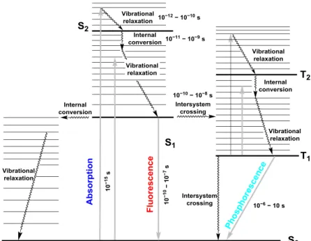
Conventionally, photophysical processes, such as light absorption and emission, are described by the classical Jablonski energy diagram[1], which was first proposed in 1933 by the Polish physicist Alexander Jablonski[2]. Later, the Jablonski diagram was further improved by the French physicists Jean Perrin and Francis Perrin[3]. Therefore, the modified descriptions are referred as Perrin–Jablonski diagram (Figure 1-1).
Figure 1-1. Perrin–Jablonski diagram for absorption, fluorescence and phosphorescence involving vibrational relaxation, internal conversion and intersystem crossing processes.[4]
As shown in Figure 1-1, after absorption of energy (light), a molecule is promoted from the singlet ground state S0 to excited singlet states Sn (n ≥ l). The electronic transition process is very fast in ca. 10–15 s time domain. Within the time of the electronic transition, the excited molecule keeps original nuclear positions and geometry because the molecule does not have enough time for molecular vibrations (10–10–10–12 s). The state is referred as the Franck–Condon state. Excited molecule in higher excited state, Sn (n > 1), will go back to the first excited singlet state S1 through internal conversion in 10–12–10–10 s time domain. Subsequently, the excited molecule in S1 state returns to the S0 state by giving off fluorescence in 10–10–10–7 s time domain.
Generally, fluorescence emission is produced from the S1 state.
There are another competitive photophysical processes with fluorescent decay. One possible non-radiative pathway is internal conversion for which the possibility is generally not high due to large energy gap between S1 and S0 states. Another possible non-radiative pathway is intersystem crossing from S1 state to triplet state Tn. Phosphorescence is photoemission given off through a transition from the first excited triplet state T1 to S0 state.
1.2 Photophysical mechanisms for modifying fluorescence
Many fundamental photophysical mechanisms have been employed to construct multifunctional fluorophores, such as intramolecular charge transfer (ICT)[5], excited- state intramolecular proton transfer (ESIPT)[6], photoinduced electron transfer (PET)[7], Förster resonance energy transfer (FRET)[8] and aggregation-induced emission (AIE)[9]. In this section, the author focuses on ICT and ESIPT fluorophores and their applications as the background of the present studies.
1.2.1 Intramolecular charge transfer (ICT)
Chromophores incorporating an electron-donating group (D) and an electron- accepting functionality (A) connected with a conjugated π-system, possess intramolecular charge transfer (ICT) character. The electron distribution of an ICT chromophore is different between in the ground state and in the excited state, thus, dipole moment of the molecule in the excited state (μg) is much greater than that of the ground state (μg) (Figure 1-2).[10]
Figure 1-2. Photo-induced charge separation of ICT fluorophores.
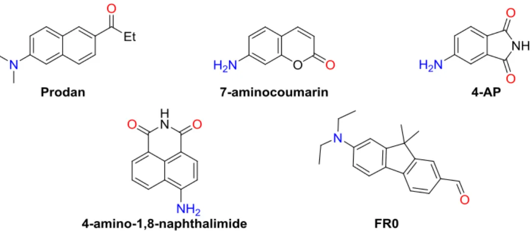
In a condensed medium, natures of excited molecules are affected by the local environment, i.e., solvation. Consequently, the ICT molecules in the excited state are more stabilized in more polar solvents. It is, thus, generally expected that fluorescence spectra of ICT fluorophores red-shift upon increasing solvent polarity, displaying significant positive solvatofluorochromism and multicolor fluorescence depending on microenvironment of the solvents. Therefore, ICT fluorophores have been widely used as solvent-sensitive fluorescent probes.[5,11] Representative ICT fluorophores (see Figure 1-3 for chemical structures) are 4,6-propionyl-2-(N,N- dimethyl)aminonaphthalene (PRODAN)[12], 7-aminocoumarin[13], 4-aminophthalimide (4AP)[14], 4-amino-1,8-naphthalimide[15] and 7-diethylamino-9,9-dimethyl-9H- fluorene-2-carbaldehyde (FR0)[16] and their derivatives.
Figure 1-3. Chemical structures of representative ICT fluorophores.
4-Aminophthalimide (4AP) and its derivatives are typical ICT fluorophores and they have been widely employed as environment-sensitive fluorescence probes (see Figure 1-4 for chemical structures). Their fluorescence properties are known to be sensitive toward the local environments, such as solvent polarity and hydrogen bonding.[14]
Figure 1-4. Chemical structures of 4AP based environment-sensitive probes.[14]
Furthermore, a fluorometric and colorimetric probe 4- trifluoroacetylaminophthalimide (4TAP) for iodide ions was reported.[17] After addition of halide ions, the probe displayed selective response to iodide ions accompanied by fluorescence color changes from blue to green upon 254 nm irradiation in MeCN. The response was induced by formation of the amidate anion 4TAP− (Figure 1-5).
Figure 1-5. Response of 4TAP for iodide ions upon 254 nm irradiation in MeCN.[17]
4-Amino-1,8-naphthalimide is one of the most common ICT fluorophores, and its derivatives have attracted considerable attention due to their extensive applications as DNA targeting, coloring reagents, fluorescent probes and bio-imaging, as well as antitumor drugs or agents (Figure 1-6).[18,19] 3-Amino-1,8-naphthalimide is an isomer of 4-amino-1,8-naphthalimide with ICT character and its derivatives have been broadly employed as chemo-sensors and anticancer agents (Figure 1-6).[19,20]
Figure 1-6. Chemical structures of amino-substituted 1,8-naphthalimide based ICT fluorophores and their applications.
Recently, fluorescence behavior of a series of amino-substituted 2,3-naphthalimide derivatives (ANIs, see Figure 1-7 for chemical structures) were systematically
investigated in various solvents.[21] Upon increasing solvent polarity, the fluorescence spectra of 1ANI showed no appreciable fluorescence response. Thus, 1ANI is insensitive to solvent polarity due to the presence of intramolecular hydrogen bonding and effective conjunction interaction between the amino fuctionality and the imide moiety.
Figure 1-7. Chemical structures of amino-substituted 2,3-naphthalimides (ANIs).[21]
In contrast, 5ANI and 6ANI showed significant solvent-dependent fluorescence behavior with marked positive solvatofluorochromism arising from their ICT features in the excited state. Multicolour fluorescence covering entire visible region was observed for 5ANI and 6ANI by changing solvent polarity. These results revealed that the substitution positions of the amino functionality significantly affected the fluorescence properties of ANIs.
1.2.2 Excited-state intramolecular proton transfer (ESIPT)
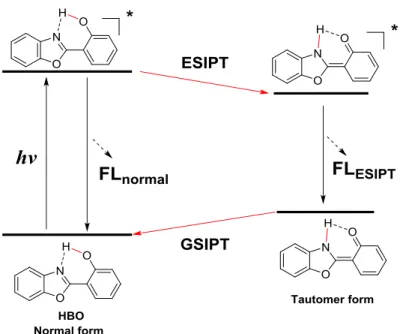
Excited-state intramolecular proton transfer (ESIPT) is an important photochemical process.[6,22] Generally, the structure of an ESIPT fluorophore incorporates proton donor and acceptor moieties. In the excited state, the proton migrates from the donor part to the acceptor moiety. This phototautomerization process is extremely fast (k >1012 s−1)[6]. Dual emission bands are often observed in ESIPT fluorophore (Figure 1-8). ESIPT fluorescence band generally displays very large Stokes shift (6,000–10,000 cm−1) because S0-S1 energy gap is quite different between the normal and tautomer forms.[23] Additionally, intensity ratio of the normal and ESIPT emission bands can be tuned by chemical modification of the fluorophores. Thus, the fluorescence color tuning of ESIPT fluorophores is possible. Based on the effective fluorescence color tuning, ESIPT fluorophores are of potential materials for light
emitting devices.[24-26]
Figure 1-8. Schematic description of an ESIPT cycle.
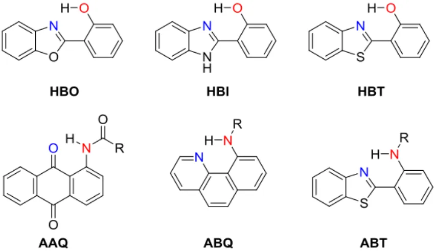
According to the types of proton donor (-OH or -NH), ESIPT fluorophores are categorized into OH-type and NH-type (see Figure 1-9 for chemical structures).
Representative OH-type ESIPT fluorophores are 2-(2’-hydroxyphenyl)benzoxazole (HBO)[27], 2-(2’-hydroxyphenyl)benzimidazole (HBI)[28], 2-(2’- hydroxyphenyl)benzothiazole (HBT)[29]. On the other hand, conventional NH-type ESIPT fluorophores are 1-(acylamino)anthraquinones (AAQ),[30] 10- aminobenzo[h]quinoline (ABQ)[31] and 2-(2’-aminophenyl)benzothiazole (ABT)[32].
Figure 1-9. Chemical structures of representative ESIPT fluorophores.
Conventionally, ESIPT fluorescence was mainly focused on OH-type ESIPT fluorophores and numerous papers have been published. N-H type ESIPT fluorescence phenomenon was first observed for 1-(acylamino)anthraquinones (AAQ) in 1991.[30]
Since then, N-H type ESIPT fluorophores have been attracted more attention because their fluorescence properties can be easily controlled and tuned by modification of the substituent on the amino functionality.
Due to the presence of the acidic proton and intramolecular hydrogen bonding, ESIPT fluorophores are sensitive to external stimuli, such as pH, solvent polarity and ionic species.[6,33] Therefore, ESIPT fluorophores have been widely used as fluorescence probes. Fluorescence response of 3-trifluoroacetylaminophthalimide (3TAP) to halide ions and alkaline metal ions was investigated (Figure 1-10). Under 254 nm irradiation, 3TAP showed selective response to Li+–I– ion pair displaying fluorescence color changes from orange-yellow to sky-blue in MeCN. Thus, 3TAP would be served as a fluorescent probe for Li+–I– ion pair.
Figure 1-10. ESIPT and Li+–I– ion-pair response of 3TAP.
Various ESIPT-based fluorescent probes for ionic species, small neutral molecules and biomacromolecules have been developed.[6] For example, fluorescein-derived HBI and HBT (FH-1[34] and FH-2[35] in Figure 1-11) were employed as fluorescent pH probes with large Stokes shifts. The HBT-based derivative HN (Figure 1-11) was used as a small and neutral molecular fluorescent probe for the detection of glutathione (GSH) and the imaging in HeLa cells.[36]
Figure 1-11. Examples of chemosensors using ESIPT fluorophores.
1.3 References
[1] M. Sauer, J. Hofkens and J. Enderlein, Handbook of Fluorescence Spectroscopy and Imaging: from Ensemble to Single Molecules, Wiley-VCH, Weinheim, Germany, 2010.
[2] A. Jablonski, Efficiency of anti-Stokes fluorescence in dyes, Nature, 1933, 131, 839-840.
[3] B. Valeur and J.-C. Brochon, New Trends in Fluorescence Spectroscopy: Applications to Chemical and Life Sciences, Springer, Berlin, Germany, 2001.
[4] J. R. Lakowicz, Principles of Fluorescence Spectroscopy, Springer, Boston, US, 2006.
[5] Z. R. Grabowski, K. Rotkiewicz and W. Rettig, Structural changes accompanying intramolecular electron transfer: focus on twisted intramolecular charge-transfer states and structures, Chem. Rev., 2003, 103, 3899-4032.
[6] C. Sedgwick, L. Wu, H.-H. Han, S. D. Bull, X.-P. He, T. D. James, J. L. Sessler, B. Z. Tang, H. Tian and J. Yoon, Excited-state intramolecular proton-transfer (ESIPT) based fluorescence sensors and imaging agents, Chem. Soc. Rev., 2018, 47, 8842-8880.
[7] M. D. Ward, Photo-induced electron and energy transfer in non-covalently bonded supramolecular assemblies, Chem. Soc. Rev., 1997, 26, 365-375.
[8] S. Jang, M. D. Newton and R. J. Silbey, Multichromophoric Főrster resonance energy transfer, Phys.
Rev. Lett., 2004, 92, 218301.
[9] Y. Hong, J. W. Y. Lam and B. Z. Tang, Aggregation-induced emission, Chem. Soc. Rev., 2011, 40, 5361-5388.
[10] B. Valeur and M. N. Berberan-Santos, Molecular Fluorescence: Principles and Applications, Wiley- VCH, Weinheim, Germany, 2012.
[11] P. De Silva, H. Q. N. Gunaratne, T. Gunnlaugsson, A. J. Huxley, C. P. McCoy, J. T. Rademacher and T. E. Rice, Signaling recognition events with fluorescent sensors and switches, Chem. Rev., 1997, 97, 1515-1566.
[12] T. Parasassi, E. K. Krasnowska, L. Bagatolli and E. Gratton, Laurdan and Prodan as polarity- sensitive fluorescent membrane probes, J. Fluoresc., 1998, 8, 365-373.
[13] G. Jones, W. R. Jackson, S. Kanoktanaporn and A. M. Halpern, Solvent effects on photophysical parameters for coumarin laser dyes, Opt. Commun., 1980, 33, 315-320.
[14] G. Saroja, T. Soujanya, B. Ramachandram and A. Samanta, 4-Aminophthalimide derivatives as environment-sensitive probes, J. Fluoresc., 1998, 8, 405-410.
[15] R. M. Duke, E. B. Veale, F. M. Pfeffer, P. E. Kruger and T. Gunnlaugsson, Colorimetric and fluorescent anion sensors: an overview of recent developments in the use of 1,8-naphthalimide- based chemosensors, Chem. Soc. Rev., 2010, 39, 3936-3953.
[16] A. Kucherak, P. Didier, Y. Mély and A. S. Klymchenko, Fluorene analogues of prodan with superior
fluorescence brightness and solvatochromism, J. Phys. Chem. Lett., 2010, 1, 616-620.
[17] H. Okamoto, H. Konishi, M. Kohno and K. Satake, Fluorescence response of a 4- trifluoroacetylaminophthalimide to iodide ions upon 254 nm irradiation in MeCN, Org. Lett., 2008, 10, 3125-3128.
[18] S. Saha and A. Samanta, Influence of the structure of the amino group and polarity of the medium on the photophysical behavior of 4-amino-1, 8-naphthalimide derivatives, J. Phys. Chem. A, 2002, 106, 4763-4771.
[19] S. Banerjee, E. B. Veale, C. M. Phelan, S. A. Murphy, G. M. Tocci, L. J. Gillespie, D. O.
Frimannsson, J. M. Kelly and T. Gunnlaugsson, Recent advances in the development of 1,8- naphthalimide based DNA targeting binders, anticancer and fluorescent cellular imaging agents, Chem. Soc. Rev., 2013, 42, 1601-1618.
[20] R. M. Duke and T. Gunnlaugsson, 3-Urea-1,8-naphthalimides are good chemosensors: a highly selective dual colorimetric and fluorescent ICT based anion sensor for fluoride, Tetrahedron Lett., 2011, 52, 1503-1505.
[21] M. Fujii, M. Namba, M. Yamaji and H. Okamoto, Solvent-induced multicolour fluorescence of amino-substituted 2,3-naphthalimides studied by fluorescence and transient absorption measurements, Photochem. Photobiol. Sci, 2016, 15, 842-850.
[22] V. S. Padalkar and S. Seki, Excited-state intramolecular proton-transfer (ESIPT)-inspired solid state emitters, Chem. Soc. Rev., 2016, 45, 169-202.
[23] C. Chen, Y. Chen, A. P. Demchenko and P. Chou, Amino proton donors in excited-state intramolecular proton-transfer reactions, Nat. Rev. Chem., 2018, 2, 131-143.
[24] J. Massue, D. Jacquemin and G. Ulrich, Molecular engineering of excited-state intramolecular proton transfer (ESIPT) dual and triple emitters, Chem. Lett., 2018, 47, 1083-1089.
[25] G. M. Farinola and R. Ragni, Electroluminescent materials for white organic light emitting diodes, Chem. Soc. Rev., 2011, 40, 3467-3482.
[26] J. E. Kwon and S. Y. Park, Advanced organic optoelectronic materials: Harnessing excited-state intramolecular proton transfer (ESIPT) process, Adv. Mater., 2011, 23, 3615-3642.
[27] Mordzinski and A. Grabowska, Intramolecular proton transfer in excited benzoxazoles, Chem. Phys.
Lett., 1982, 90, 122-127.
[28] H. K. Sinha and S. K. Dogra, Ground and excited state prototropic reactions in 2-(o-hydroxyphenyl) benzimidazole, Chem. Phys., 1986, 102, 337-347.
[29] T. Elsaesser and B. Schmetzer, Excited-state proton transfer in 2-(2'-hydroxyphenyl) benzothiazole:
formation of the anion in polar solvents, Chem. Phys. Lett., 1987, 140, 293-299.
[30] T. P. Smith, K. A. Zaklika, K. Thakur and P. F. Barbara, Excited state intramolecular proton transfer in 1-(acylamino)anthraquinones, J. Am. Chem. Soc., 1991, 113, 4035-4036.
[31] H. Tseng, T. Lin, C. Chen, T. Lin, Y. Chen, J. Liu, C. Hung, C. Chao, K. Liu and P. Chou, A new class of N–H proton transfer molecules: wide tautomer emission tuning from 590 nm to 770 nm via a facile, single site amino derivatization in 10-aminobenzo[h]quinoline, Chem. Commun., 2015, 51, 16099-16102.
[32] C.-L. Chen, H.-W. Tseng, Y.-A. Chen, J.-Q. Liu, C.-M. Chao, K.-M. Liu, T.-C. Lin, C.-H. Hung, Y.- L. Chou, T.-C. Lin and P.-T. Chou, Insight into the amino-type excited-state intramolecular proton transfer cycle using N-tosyl derivatives of 2-(2′-aminophenyl)benzothiazole, J. Phys. Chem. A, 2016, 120, 1020-1028.
[33] Z. Liu, W. He and Z. Guo, Metal coordination in photoluminescent sensing, Chem. Soc. Rev., 2013, 42, 1568-1600.
[34] V. S. Patil, V. S. Padalkar, K. R. Phatangare, V. D. Gupta, P. G. Umape and N. Sekar, Synthesis of new ESIPT-fluorescein: Photophysics of pH sensitivity and fluorescence, J. Phys. Chem. A, 2012, 116, 536-545.
[35] V. S. Patil, V. S. Padalkar, A. B. Tathe and N. Sekar, ESIPT-inspired benzothiazole fluorescein:
Photophysics of microenvironment pH and viscosity, Dyes Pigm., 2013, 98, 507-517.
[36] X. Ren, F. Wang, J. Lv, T. Wei, W. Zhang, Y. Wang and X. Chen, An ESIPT-based fluorescent probe for highly selective detection of glutathione in aqueous solution and living cells, Dyes Pigm., 2016, 129, 156-162.
Chapter 2
Synthesis and fluorescence behavior of a series of amino-
substituted 1,8-naphthalimide derivatives
2.1 Introduction
Recently, development of multifunctional fluorophores has attracted much attention due to their potential applications in the fields of fluorescent probes[1-3], chemo- sensors[4-6], bio-imaging[7-9] and optoelectronic devices[10-12]. Particularly, push-pull fluorophores having electron-accepting (A) and electron-donating (D) functionalities in a single molecule are highly attractive because their photophysical behavior is significantly affected by local environment, e.g. solvent polarity. In solvents of different polarities, such fluorophores are expected to display appreciable solvatofluorochromism and multicolour fluorescence[13] due to the remarkable difference in the electron distribution between in the ground state and the excited state through the intramolecular charge transfer (ICT) characters in the emitting state. The ICT is a classic and crucial fluorescence mechanism to design and construct environment-sensitive fluorescent probes.
Figure 2-1 shows the mechanism for fluorescence modification of ICT fluorophores by micro-environments of solvents[14].
Figure 2-1. Mechanism of solvatofluorochromism of an ICT fluorophore.
Upon photoexcitation, ICT fluorophores produce the charge transfer excited state with larger dipole moment compared to the ground state. As a result, the interaction between the fluorophores and their surrounding environment, i.e. solvation, is significantly different from that of in the ground state. In more polar solvents, the excited state fluorophores are more stabilized and their emission bands red-shift to longer wavelength region. Thus, ICT fluorophores show significant solvent-dependent fluorescence behavior. Conventionally, 4-amino-1,8-naphthalimide[4], 4- aminophthalimide (4AP)[15], 4,6-propionyl-2-(N,N-dimethyl)aminonaphthalene (Prodan)[16] and 7-diethylamino-9,9-dimethyl-9H-fluorene-2-carbaldehyde (FR0)[17]
are known as representative ICT fluorophores (chemical structures are shown in Figure 2-2).
Figure 2-2. Chemical structures of representative ICT fluorophores.
It has been well established that amino-substituted 1,8-naphthalimide derivatives are important class of ICT fluorophores due to their unique photophysical properties.
Among the three possible amino-1,8-naphthalimide derivatives, most studied ones so far possess the amino functionality at the 3- and 4-positions of the naphthalene core
[4,7,18]. Pardo et al.[19] studied the effects of solvents on the photophysical properties of 3-amino-N-substituted-1,8-naphthalimide derivatives, indicating that their absorption and fluorescence spectra significantly red-shifted upon increasing the solvent polarity.
Alexiou et al.[20] studied the photophysical behavior of amino-substituted 1,8- naphthalimide and naphthalic anhydride derivatives demonstrating that the alkylamino substituted 1,8-naphthalimides and naphthalic anhydrides exhibited strong fluorescence with high fluorescence quantum yields. Samanta et al.[21] reported that the photophysical features of 4-amino-1,8-naphthalimide derivatives were strongly influenced by the nature of the substituted-amino groups and solvent polarity.
Recently, Okamoto et al. reported synthesis and fluorescence behavior of a series of amino-substituted 2,3-naphthalimide derivatives in various solvents (ANIs, the chemical structures are shown in Figure 2-3)[22]. The results indicated that 1ANI showed no appreciable fluorescence response to solvent polarity due to efficient intramolecular hydrogen bonding and effective conjunction between the amino group and the π-system. In contrast, 5ANI and 6ANI displayed marked positive solvatofluorochromism displaying multicolour fluorescence covering entire visible region upon changing the solvent polarityowing to their ICT character in the excited state. These observations clearly indicate that the positions of the amino functionality affect the fluorescence behavior of ANIs.
Figure 2-3. Chemical structures of amino-substituted 2,3-naphthalimides (ANIs)[22].
Based on the fluorescence characteristics of ANIs, it is expected that fluorescence behavior of amino-substituted 1,8-naphthalimides, the isomers of ANIs, could be also modified by the amino-substitution positions. Conventionally, 4- and 3-amino-1,8- naphthalimide derivatives have been utilized as the emitter of probes and sensors[4,7]. However, little systematic information is available on the photophysical properties of amino-substituted 1,8-naphthalimides in the literature. Furthermore, detailed photophysical properties of 2-amino-1,8-naphthalimide have not been investigated so far. Therefore, it would be of interest to reveal fluorescence behavior of the series of amino-substituted 1,8-naphthalimides to understand the effects of the amino- substitution positions on their photophysical properties.
In this chapter, preparation of the series of amino-substituted N-propyl-1,8- naphthalimide (APNIs, chemical structures are shown in Figure 2-4) and systematic investigation of their fluorescence behavior to reveal the effects of the substitution positions on their fluorescence behaviors are described.
Figure 2-4. Chemical structures of amino-substituted 1,8-naphthalimide derivatives (APNIs) investigated in the present study.
2.2 Synthesis
Detailed synthetic routes to 2APNI, 3APNI and 4APNI were depicted in Scheme 2-1.
All new compounds were characterized by 1H NMR, 13C NMR and IR spectroscopy, as well as elemental analysis.
Scheme 2-1. Synthetic routes to 2APNI, 3APNI and 4APNI.
2APNI was first synthesized by the multi-step reaction sequence. N,N'- Diphenyloxalimidoyl dichloride 1 was prepared according to the previously reported procedures[23-26], i.e., a reaction of aniline with oxalyl chloride in the presence of phosphorus pentachloride (PCl5) in dry toluene. Compound 1 was used directly in the
next reaction without further purification. Then, a Friedel-Crafts reaction between the intermediate 1 and 2-methoxynaphthalene 2 in the presence of AlCl3 in dry benzene afforded diketone 3 in 48% yield.[27,28] Next, oxidation reaction of diketone 3 with hydrogen peroxide (H2O2) under an alkaline condition[29] produced 2-methoxy-1,8- naphthalic anhydride 4 in excellent yield (94%). Subsequently, reaction of anhydride 4 with propylamine (PrNH2) in acetic acid gave 2-methoxy-N-propyl-1,8-naphthalimide 5 in quantitative yield. The methoxy group of compound 5 was converted into the phenolic hydroxy group to produce compound 6 through cleavage of the ether moiety with boron tribromide (BBr3) in dry CH2Cl2 (93%). Then, compound 6 was reacted with trifluoromethanesulfonic anhydride (Tf2O) in the presence of Et3N to obtain 2- tirifluoromethanesulfonate 7 in 88% yield. Compound 8 was synthesized by Buchwald–Hartwig reaction[30] of compound 7 with t-butyl carbamate using Pd2dba3 as catalyst,9,9-dimethyl-4,5-bis(diphenylphosphino)xanthene (XantPhos) as a ligand and caesium carbonate (CsCO3) as a base in dry dioxane. Finally, the Boc-protecting group of compound 8 was removed with trifluoroacetic acid (CF3COOH) in CH2Cl2 to give the desired 2APNI in 61% yield.
3APNI was prepared by a two-step reaction starting from 3-nitro-1,8-naphthalic anhydride 9. Compound 9 was reacted with PrNH2 in acetic acid to give 3-nitro-N- propyl-1,8-naphthalimide 10 in 93% yield, followed by a reduction of the nitro functionality in the presence of H2 and Pd/C to produce 3APNI in 64% yield.
4APNI was prepared starting from 4-amino-1,8-naphthalimide 11. The imide proton of compound 11 was removed by sodium methoxide in DMF to produce sodium salt of 4-amino-1,8-naphthalimide, then the sodium salt was reacted with 1- bromopropane to afford the target compound 4APNI in 61% yield.
2.3 Absorption spectra
Spectral behavior of APNIs was investigated by UV-vis absorption spectroscopy, steady-state and time-resolved fluorescence measurements in solvents of various polarities. Absorption spectra of APNIs were shown in Figure 2-5 and the corresponding photophysical parameters, such as maximum absorption wavelengths (𝜆maxAbs) and molar absorption coefficients (ε), are collected in Table 2-1.
Figure 2-5. Normalized absorption spectra of 2APNI (a), 3APNI (b) and 4APNI (c) in various solvents. The vertical black bars present calculated transition wavelengths and oscillator strengths at the TD-DFT PBE0/6-311+G(d,p) level in toluene.
Table 2-1. Photophysical parameters of APNIs in various solvents
APNIs Solvent 𝜆Absmax(nm)a (log ε)
𝜆FLmax
(nm)a
Δṽb
(cm−1) ΦFLc τFLd
(ns) kRe
(107 s−1) kNRf
(107 s−1)
2APNI
Hexane 405 (−g) 420 880 − g − g − g − g
Toluene 414 (4.05) 433 1060 0.27 1.71 15.8 42.7
DCM 413 (4.05) 427 790 0.25 −h −h −h
AcOEt 414 (4.05) 432 1000 0.20 −h −h −h
MeCN 413 (4.05) 435 1220 0.26 1.74 14.9 42.5
DMSO 420 (4.11) 445 1340 0.30 −h −h −h
MeOH 418 (4.10) 442 1290 0.23 1.86 12.4 41.4
solid state − g 511 − g 0.075 − g − g − g
3APNI
Hexane 394 (−g) 429 2070 −g −g −g −g
Toluene 407 (3.69) 464 3020 0.72 11.7 6.2 2.4
DCM 408 (3.71) 475 3460 0.73 −h −h −h
AcOEt 418 (3.43) 495 3720 0.53 −h −h −h
MeCN 417 (3.68) 510 4370 0.49 17.7 2.8 2.9
DMSO 438 (3.70) 543 4410 0.68 − h − h − h
MeOH 425 (3.48) 564 5800 0.23 12.4 1.9 6.2
solid state − g 571 − g 0.011 − g − g − g
4APNI
Hexane 389 (−g) 460 3970 −g −g −g −h
Toluene 400 (4.09) 482 4250 0.98 8.7 11.2 0.2
DCM 405 (4.09) 492 4370 0.94 −h −h −h
AcOEt 414 (4.04) 500 4150 0.70 −h − h −h
MeCN 416 (4.05) 517 4700 0.58 10.1 5.7 4.2
DMSO 437 (4.07) 532 4090 0.72 −h −h −h
MeOH 433 (4.11) 538 4510 0.39 7.3 5.4 8.4
solid state − g 562 − g 0.018 − g − g − g
a The spectral measurements were performed under aerated conditions and the concentrations of APNIs were 5×10⁻⁶ M.
b Δṽ represents Stokes shift determined by Δṽ = (𝜆Absmax−1 − 𝜆maxFL −1) ×107 cm−1.
c The fluorescence quantum yields (ΦFL) were obtained by using coumarin 153 in MeCN as the reference (ΦFL = 0.56)[31].
dτFL represents fluorescence lifetime.
e The radiative rate constants (kR) were calculated by equation kR = ΦFL× τFL−1.
f The non-radiative rate constants (kNR) were calculated by equation kNR = ΦNR× τFL−1, ΦNR= 1−ΦFL.
g Not determined due to low solubility in hexane.
h Not determined.
2APNI showed a structured absorption band in the wavelength region of 405–420 nm in various solvents (Figure 2-5a) accompanied by large ε values (log
ε = 4.05–4.10, Table 2-1). The band is thus assigned to π–π* transition. The absorption band only slightly red-shifted from 405 nm in hexane to 420 nm in DMSO. The solvatochromic shift (∆𝜆maxAbs) of 15 nm indicated that 2APNI was almost insensitive to solvent polarity. Similar solvent independent behavior was also observed for 1ANI due to intramolecular hydrogen bonding and effective conjunction between the amino moiety and the π system[22].
In contrast, 3APNI displayed broad absorption bands in the 394–438 nm wavelength region (log ε = 3.43–3.71, Table 2-1) in different solvents (Figure 2- 5b) showing significant red-shift from 394 nm in hexane to 438 nm in DMSO,
∆𝜆maxAbs = 44 nm. Similarly, broad absorption band was observed for 4APNI in the wavelength region of 389–537 nm (log ε = 4.04–4.11, Table 2-1) in various solvents (Figure 2-5c). The absorption band of 4APNI appreciably red-shifted from 389 nm in hexane to 537 nm in DMSO (∆𝜆maxAbs = 48 nm).
Upon increasing the solvent polarity, the absorption bands of 3APNI and 4APNI considerably red-shifted (∆𝜆maxAbs = 44 nm for 3APNI and 48 nm for 4APNI). In contrast, 2APNI showed only slight red-shift of the absorption band (∆𝜆maxAbs = 15 nm) in various solvents indicating that 2APNI was insensitive to solvents polarity. The differences in the solvent sensitivities of APNIs were attributed to different polar characteristics among the three APNI isomers in the ground state.
To get an insight into the detailed absorption characteristics of APNIs, their electronic features in the ground and excited states were analysed by theoretical calculations by DFT method by using the PBE0/6-311+G(d,p) level. The calculated ground-state dipole moment (μg) of 4APNI (6.28 D) and 3APNI (4.89 D) were larger than that of 2APNI (4.92 D). The vertical transition wavelengths (T) and oscillator strengths (f) were also calculated by using time-dependent density functional theory (TD-DFT) method at the TD-PBE0/6-311+G(d,p) level by considering solvent effects of toluene with polarizable continuum model. The calculated results are summarized in Table 2-2. The calculated electronic transitions of APNIs were compared with their experimental absorption spectra
(Figure 2-5). The frontier molecular orbitals of APNIs, highest occupied molecular orbital (HOMO) and lowest unoccupied molecular orbital (LUMO), are illustrated in Figure 2-6.
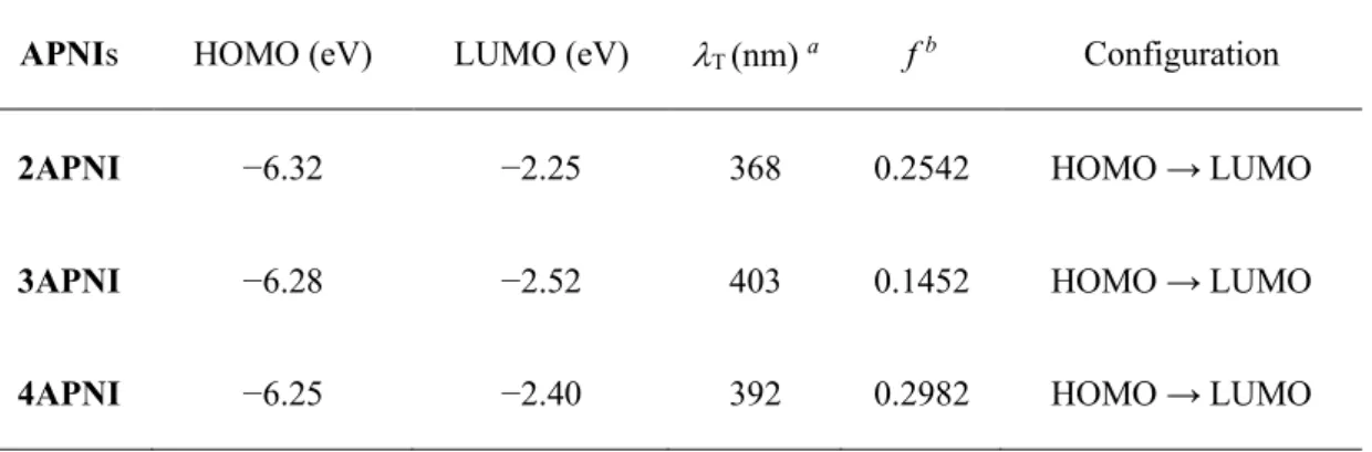
Table 2-2 Electronic transition of APNIs calculated at the TD-DFT PBE0/6-311 +G(d,p) level in toluene.
APNIs HOMO (eV) LUMO (eV) T (nm) a f b Configuration
2APNI −6.32 −2.25 368 0.2542 HOMO → LUMO
3APNI −6.28 −2.52 403 0.1452 HOMO → LUMO
4APNI −6.25 −2.40 392 0.2982 HOMO → LUMO
a Calculated wavelength for S0 to S1 transition.
b Calculated oscillator strength.
The calculated transition wavelengths are in good agreement with the experimental absorption bands (Figure 2-5). The first absorption band (S0 → S1
transition) of APNIs is assigned to the electronic transition of HOMO → LUMO with large f values. In the case of 2APNI, the calculated transition wavelength (368 nm) is shorter than that for the experimental absorption band (414 nm in toluene). The HOMO and LUMO of 2APNI distribute over the entire π- conjugated system of the naphthalimide and the amino moiety (Figure 2-6).
Additionally, the calculated results provide an evidence that the amino group of 2APNI is effectively conjugated with the naphthalimide part.
For 3APNI, the calculated transition wavelength (403 nm) is close to the experimentally observed absorption band at 407 nm in toluene. The HOMO localized on the aminonaphthalene moiety whereas the LUMO extend over the entire naphthalimide system (Figure 2-6). Therefore, S1 state of 3APNI has an ICT character. In the case of 4APNI, the calculated transition wavelength (392 nm) is consistent with the experimental absorption band at 400 nm in toluene with a largest f value (0.2982). Both HOMO and LUMO of 4APNI located over
entire aminonaphthalimide moiety (Figure 2-6), indicating the effective conjugation between the amino group and the π-conjugation system of the naphthalimide moiety. Thus, the S0 → S1 transition of 4APNI is assigned to the π → π* transition from polar S0 state to polar S1 state.
Figure 2-6. Frontier molecular orbital surfaces of APNIs (PBE0/6-311 +G(d,p)).
2.4 Fluorescence properties
2.4.1 Fluorescence behavior in solution
Fluorescence spectra of APNIs in various solvents are displayed in Figure 2-7 and the photophysical data, such as fluorescence maximum wavelength (∆𝜆maxFL ) and fluorescence quantum yield (ΦFL), are summarized in Table 2-1. The ΦFL values were determined by using coumarin 153 in MeCN (ΦFL = 0.56)[31] as the reference.
2APNI showed a structured fluorescence emission band in the wavelength region of 420–445 nm and slightly red-shifted from 420 nm in hexane to 445 nm in DMSO upon increasing solvent polarity (∆𝜆FLmax = 25 nm, Figure 2-7a, Table 1). The ΦFL
values of 2APNI were determined to be in the range of 0.20–0.30 independent to the solvents used.
3APNI and 4APNI displayed broad fluorescence bands in solvents of different polarities. The emission bands of 3APNI appreciably red-shifted from
429 nm in hexane to 564 nm in MeOH (∆𝜆maxFL = 135 nm, Figure 2-7b, Table 2- 1). Upon changing solvent polarity, 3APNI, thus, exhibited a marked positive solvatofluorochromism accompanied by the fluorescence color change from blue in hexane to orange-red in MeOH. The ΦF values of 3APNI decreased with increasing solvent polarity in the range of 0.49-0.73 in aprotic solvents. In MeOH, fluorescence of 3APNI was more effectively quenched to show ΦF = 0.23. It is considered that intermolecular hydrogen bonding interaction between 3APNI molecules and protic solvent molecules promoted non-radiative process resulting in reduced ΦF value.
Figure 2-7. Normalized fluorescence spectra of 2APNI (a), 3APNI (b) and 4APNI (c) in various solvents.
4APNI also showed solvent-dependent emission band from 460 nm in hexane to 538 nm in MeOH showing appreciable positive solvatofluorochromism (∆𝜆FLmax = 78 nm, Figure 2-7c, Table 2-1). Consequently, the fluorescence color altered from blue in hexane to yellow in MeOH. The ΦF
values of 4APNI was determined to be in the region of 0.58-0.98 in aprotic solvents and 0.39 in MeOH. The fluorescence behavior of 3APNI and 4APNI was sensitive to solvent polarity due to their ICT character.
Excitation spectra of APNIs were obtained to confirm the emitting species (Figure 2-8). Because the excitation spectra were consistent with the absorption spectra, the emission bands are unambiguously assigned to APNIs.
Figure 2-8. Absorption (red lines) and fluorescence excitation (black lines) spectra of 2APNI (a), 3APNI (b) and 4APNI (c) in various solvents.
The existence of intramolecular hydrogen bonding in 2APNI was confirmed by FT-IR spectroscopy (Figure 2-9). IR spectra of 3APNI and 4APNI are shown in comparation to that of 2APNI.
Figure 2-9. FT-IR spectra of 2APNI (black), 3APNI (red) and 4APNI (blue).
2APNI showed two absorption bands at 3378 and 3283 cm−1 arising from asymmetrical and symmetrical N-H stretching vibrations, respectively. In contrast, the vibration bands were respectively observed at 3474 and 3368 cm−1 for 3APNI, and 3435 and 3352 cm−1 for 4APNI. Obviously, two N-H stretching vibration bands (ν(N-H)) of 2APNI were significantly shifted to lower wavenumber region compared to those of 3APNI and 4APNI. The differences in the ν(N-H) bands were due to the intramolecular hydrogen bonding in 2APNI.
These IR spectral data of APNIs provide an experimental evidence that the existence of intramolecular hydrogen bonding in 2APNI.[32] Therefore, 2APNI exhibited solvent-independent fluorescence behavior even in protic solvent.
The solvent-dependent electronic spectral behavior of APNIs were further analyzed by using the Lippert–Mataga equation (eqn 1)[33,34],
∆ṽ = ṽ
A− ṽ
F= [2(μ
e– μ
g)
2/ hc(a
0)
3]∆f + k (1)
∆f = [(ε
r−1) / (2ε
r+1)] – [(n
2−1) / (2n
2+1)] (2)
where, ∆ṽ is the Stokes shift determined by difference between maximum absorption and emission wavenumbers, ṽA and ṽF respectively denote maximum absorption and emission wavenumber, μg and μe respectively denote dipole moment in the ground state and excited state, h is the Planck’s constant, c is the light velocity in vacuum, k is a constant, a0 is the radius of the Onsagar cavity[35]. Δf denotes orientation polarizability of solvent determined by equation (2), where εr and n are respectively the static dielectric constant and the refractive index of solvents.
The a0 of APNIs were obtained from their optimized geometries by the theoretical calculations at the DFT PBE0/6-311+G(d,p) level in vacuum, thus, the values were determined as half of the average distance between the nitrogen atom of the amino group and the two carbonyl oxygen atoms of the imide part[36]: 3.159 Å for 2APNI, 3.838 Å for 3APNI, and 3.620 Å for 4APNI.
As shown in Figure 2-10, linear correlations of Lippert-Mataga plots were observed for APNIs. Particularly, the experimental data of 3APNI in MeOH was not used for the linear fitting because fluorescence of 3APNI was significantly affected by intermolecular hydrogen bonding interaction with MeOH. From the slopes in the Lippert–Mataga plots of APNIs, the (μe − μg) values were evaluated to be 1.7 D for 2APNI, 5.9 D for 3APNI, and 2.4 D for 4APNI. The value obtained for 4APNI is consistent with that reported one for N-butyl-4-amino-1,8- naphthalimide (2.4 D) in the previous study[37]. By considering the calculated μg
values, the μe values of ANPIs were estimated to be 6.62 D for 2APNI, 10.79 D for 3APNI and 8.68 D for 4APNI. From the results of Lippert–Mataga plots and the calculated results. The fluorescence behavior of three isomers APNIs can be summarized into the following three points: (i) Upon increasing solvent polarity, emission bands of 2APNI only slightly red-shifted (∆𝜆maxFL = 25 nm) and moderate Stokes shifts (790−1340 cm−1) were observed. Correspondingly,
fluorescence color almost unchanged due to the small difference in dipole moment (μe − μg = 1.7 D) in the ground state and the excited state. (ii) 3APNI displayed appreciable red-shift in emission bands as well as very large Stokes shifts (2070−5800 cm−1) showing positive solvatofluorochromism. The solvent induced multicolour fluorescence was thus derived from the ICT character in the excited state. (iii) The emission band of 4APNI exhibited significantly red- shifted and very large Stokes shifts (3970−4700 cm−1) displaying positive solvatofluorochromism (∆𝜆maxFL = 78 nm). The (μe − μg) value (2.4 D) in 4APNI was much smaller than that for 3APNI (5.9 D). It is considered that as the absorption and fluorescence bands of 4APNI red-shifted in a parallel manner depending on solvent polarity, the difference in the Stokes shift was smaller than that for 3APNI. It is concluded that the solvent dependent shift in absorption and fluorescence spectra was caused by polar character both in the ground and the fluorescent states in 4APNI.
Figure 2-10. Lippert-Mataga plots for 2APNI (■), 3APNI (●), and 4APNI (▲).
2.4.2 Photophysical parameters
The fluorescence decay profiles of APNIs in toluene, MeCN and MeOH are displayed in Figure 2-11.
Figure 2-11. Fluorescence decay profiles of 2APNI (a-c), 3APNI (d-f) and 4APNI (g-i) in toluene, MeCN and MeOH.
The photophysical parameters, radiative rate (kR) and non-radiative rate (kNR), were evaluated by equations 3 and 4, and collected in Table 2-1.
k
R= Φ
FL× τ
F−1(3) k
NR= (1 − Φ
FL) × τ
F−1(4)
To further investigate the effect of solvent polarity on photophysical parameters of
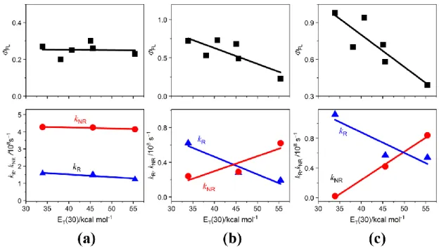
APNIs, the values of ΦFL, kR, and kNR obtained were plotted as a function of empirical solvent polarity parameters [ET(30)] in Figure 2-12. It was found that the values of ΦF, kR and kNR of 2APNI (Figure 2-11a, Table 2-1) are almost unchanged irrespective of changes of solvents, indicating that 2APNI was insensitive to solvent polarity. This may be due to the formation of intramolecular hydrogen bonding and effective conjugation interaction between the amino group and the naphthalimide moiety. Similar fluorescence properties were also observed for 1ANI which showed solvent- independent fluorescence behavior in various solvents.[22] In contrast, the photophysical parameters of 3APNI and 4APNI are significantly affected by the solvent polarity. The ΦFL values of 3APNI and 4APNI decreased upon increasing solvent polarity (Figures 2-12b and 12c). Subsequently, kR decreased with increasing solvent polarity, conversely, kNR increased as increasing solvent polarity (Figures 2-12b and 12c).
Figure 2-12. Photophysical parameters of APNIs plotted as a function of ET(30): fluorescence quantum yield ΦFL (■), radiative rate kR (▲), and non-radiative rate kNR (●). (a) 2APNI, (b) 3APNI, (c) 4APNI.
3APNI and 4APNI showed positive solvatofluorochromism in various solvents due to their ICT character in the excited state, it was expected that probability of non- radiative transition(s) from the excited statesto the ground state, such as the intersystem crossing (ISC) and/or internal conversion (IC), was enhanced in the ICT state. Non-
radiative process(es) accelerated by a charge transfer state was discussed for decay processes of charge-transfer complex between triplet benzophenone and anisol[38].
Additionally, kNR values of 3APNI and 4APNI in MeOH were larger than those of in aprotic solvents (Table 2-1). The non-radiative transition process of 3APNI and 4APNI significantly increased in the protic environment presumably by formation of intermolecular hydrogen bonding between the amino group or the imide moiety of APNIs and MeOH. IC process accelerated by intermolecular hydrogen bonding was also observed for aminophthalimide system[39].
Photostability of APNIs was also investigated in solution to clarify whether photodegradation is involved in the non-radiative decay. The solutions of APNIs in three solvents, toluene, MeCN, and MeOH, were irradiated at their absorption maximum wavelength (𝜆maxAbs) under the fluorescence measurement conditions and the absorption spectra were measured every 10 min (Figure 2-13). Under the fluorescence measurement conditions, the absorption intensity of APNIs were unchanged during 0- 60 min which indicated that APNIs are photochemically stable in the solvents.
Photodegradation can be ruled out for enhanced kNR observed for 3APNI and 4APNI in MeOH.
Figure 2-13. Changes in absorbance intensity of 2APNI (a), 3APNI (b) and 4APNI (c) during photoirradiation (0-60 min) in toluene, MeCN and MeOH.
2.4.3 Fluorescence behavior in the solid state
Solid-state fluorescence properties of three isomers APNIs were also investigated as displayed in Figure 2-14. Additionally, photophysical parameters in the solid state are collected in Table 2-1.
Figure 2-14. Solid-state fluorescence spectra of APNIs.
Solid-state fluorescence emission bands of APNIs appeared at 𝜆maxFL 511 nm for 2APNI, 571 nm for 3APNI and 562 nm for 4APNI. Compared to the cases of APNIs in solution, the fluorescence maxima 𝜆maxFL was observed in the longer wavelength region in the solid state. Thus, the differences in the fluorescence maximum wavelengths (Δ𝜆maxFL ) between in the solid state and in hexane solution are determined to be 91 nm for 2APNI, 142 nm for 3APNI and 102 nm for 4APNI. The solid-state fluorescence behavior of APNIs were considered to be due to the presence of intermolecular interactions with adjacent molecules, such as π–π stacking. Furthermore, the solid-state fluorescence quantum yield of APNIs was determined to be 0.075 for 2APNI, 0.011 for 3APNI and 0.018 for 4APNI. Clearly, the fluorescence quantum yields in the solid state are much smaller than those obtained in solution (Table 2-1).
Therefore, aggregation-induced fluorescence quenching is operative in solid state of APNIs. Unfortunately, as a single crystal of APNIs suitable for X-ray crystallographic analysis was not obtained, the detailed mechanism for the solid-state fluorescence quenching of APNIs was not clearly analyzed.

![Figure 2-1 shows the mechanism for fluorescence modification of ICT fluorophores by micro-environments of solvents [14]](https://thumb-ap.123doks.com/thumbv2/123deta/5820634.1034519/24.892.217.677.707.1102/figure-shows-mechanism-fluorescence-modification-fluorophores-environments-solvents.webp)