Fukushima Medical University
福島県立医科大学 学術機関リポジトリ
This document is downloaded at: 2021-11-08T00:15:41Z
Title Pre-eclampsia--still a disease of theories
Author(s) Schlembach, Dietmar
Citation Fukushima Journal of Medical Science. 49(2): 69-115
Issue Date 2003-12
URL http://ir.fmu.ac.jp/dspace/handle/123456789/142
Rights © 2003 The Fukushima Society of Medical Science
DOI
Text Version publisher
Vol. 49, No.2, 2003
[Review Article]
PRE-ECLAMPSIA-STILL A DISEASE OF THEORIES DIETMAR SCHLEMBACH
University of Texas Medical Branch, Dept. of Obstetrics and Gynecology, Reproductive Sciences, Galveston, Texas, USA and University of Erlangen-Nuremberg,
Dept. of Obstetrics and Gynecology, Erlangen and Nuremberg Germany (Received July 10, 2003, accepted September 19, 2003)
Abstract: Pre-eclampsia is still one of the leading causes of maternal and fetal morbidity and mortality. Despite active research for many decades, the etiology of this disorder exclusive to human pregnancy is an enigma. Recent evidence suggests there may be several underlying causes or predispositions leading to endothelial dysfunction and causing the signs of hypertension, proteinuria, and edema-findings that allow us to make the diagnosis of the "syndrome" of pre-eclampsia.
Itis obvious that a single mechanism responsible for the syndrome pre-eclampsia does not exist. Instead, several mechanisms can act together and even multiply each other. The search for the underlying cause of this disorder and for a clinical . marker to predict which women will develop pre-eclampsia is ongoing, with its
prevention being the ultimate goal.
Key words: Pre-eclampsia, vascular factors, oxidative stress, genetics, an- giogenesis
INTRODUCTION
Hypertensive disorders in pregnancy constitute a major risk factor for maternal mortality as well as fetal wastage and morbidity in the United States and in countries worldwide. They are the second leading cause of maternal mortality in the United States, representing almost 15% of pregnancy-related deaths and occur- ring in 3%-10% of pregnancies
i,2).This is especially true in underdeveloped nations. About 20% of perinatal mortality and morbidity are related to hyperten- sive disorders in pregnancy3).
Classifications of hypertensive disorders in pregnancy have varied in the past and led to some confusion in both the clinical management and research efforts
Correspondence to: Dietmar Schlembach, Division of Reproductive Sciences, Department of Obstetrics and Gynecology, University of Texas Medical Branch 301 University Blvd., Rte J- 62 Galveston, Texas 77555-1062 USA.
E-mail: [email protected]
69
70 D. SCHLEMBACH
toward the etiology of these disorders4l. Currently, a classification established by the National Institutes of Health Working Group on High Blood Pressure in Pregnancy2l is used in the United States and recommended by the International Society for the Study of Hypertension in Pregnancy:
1)
Chronic hypertension:
Hypertension present before pregnancy or first diagnosed before 20 weeks' gestation
2)
Preeclampsia-eclampsia:
Hypertension unique to pregnancy (blood pressure> 140 mmHg systolic or 90 mmHg diastolic)
Diagnosed after 20 weeks' gestation
Associated by new onset proteinuria
(~0.3g/24 h) Eclampsia, if seizures occur
3) Pre-eclampsia superimposed upon chronic or preexisting hypertension:
New onset or acutely worse proteinuria, a sudden increase in blood pressure, thrombocytopenia, or elevated liver enzymes after 20 weeks' gestation in a woman with preexisting hypertension
4) Gestational hypertension:
Hypertension first diagnosed after 20 weeks' gestation, not accompanied by proteinuria
a)
Transient hypertension
The hypertension resolves by 12 weeks' postpartum
b)Chronic hypertension
The hypertension does not resolve by 12 weeks' postpartum.
HISTORY
Hippocrates first described the condition when he wrote in one of his aphorisms
"convulsions take place from either repletion or depletion". Hippocrates had obser- ved the sudden and unexpected appearance of maternal grand-mal seizures, which occur when preeclampsia progresses to eclampsia, the word being derived from the Greek word for lightning.
Presumably eclampsia was first confused with epilepsy and not described as a separate entity until 1739. The use of the term has been attributed to Gutsch in 1776, but this has not been well documented. Nonetheless, it was many years more before it was universally accepted as separate from epilepsy or hysteria.
Even so, it was not until about 150 years ago, when protein could be measured in the urine and by the introduction of blood pressure measurements at the turn of the 20
thcentury, that the forerunner to eclampsia became apparent.
The triad of hypertension, proteinuria, and edema was termed preeclampsia.
Because of the toxins that were believed to be in the pregnant woman's body, this
disorder was also commonly called "toxemia of pregnancy," a term coined at least
150 years ago, but not currently used in today's nomenclature. Pre-eclampsia is now unanimously viewed as a multisystem disorder, as increases in blood pressure are rarely responsible for multi-organ dysfunctionS).
There is a vast diversity of additional symptoms and complications associated with pre-eclampsia. These can include cerebral edema6), neurological manifesta- tions (including headache, confusion, paralysis, coma, visual loss, and seizures)1>, liver capsule distensionS), renal failure9.10), pulmonary edema 10.11}, throm- bocytopeniaI2), coagulopathyI3), hemolysis, elevated liver enzymes, and low platelet count (HELLP) syndromeI4), and nausea IS).
EPIDEMIOLOGY
Pre-eclampsia is a pregnancy-specific syndrome that is the principal cause of maternal and fetal morbidity and mortality2.16), accounting for almost 15% of pregnancy-associated maternal deaths2).
In the United States, from the years 1979 to 1986, pre-eclampsia was the second leading cause of maternal deathl) and ranks between the second and third leading cause of maternal death in more recent yearsI6).
Risk and predisposing factors :
Numerous maternal factors can predispose women to the disorder pre-eclamp- sia; these may be genetic, behavioral, or environmental. The list of predisposing factors includes, besides the history of a previous pre-eclampsia, hypertension, diabetes, increased insulin resistance, increased testosterone, black race, and in- creased blood homocysteine concentration (Table
1).In industrialized countries, pre-eclampsia complicates 3%-5% of pregnanciesl7) and is more likely to occur at both extremes of reproductive age, but is greatest in women younger than 20 years of agel).
Primigravid women are at higher risk for pre-eclampsia IS). The incidence of pre-eclampsia in multiparous women is lower than in primiparous women, but higher if the multiparous woman has a different partnerI9.20). This finding supports the hypothesis that risk is reduced with repeated exposures to specific antigens from the same partner. However, the protective effect against pre-eclampsia of a previous pregnancy with the same partner was likely confounded by the time interval between births. Skjaerven et al.
21 )showed that the risk of pre-eclampsia in subsequent pregnancies was related to the time that had elapsed since the index pregnancy, not to a change of partners. When the birth interval was greater than 10 years, the risk of the multiparous woman was identical to that of a primiparous woman.
Also, recent information suggests that a short interval of sexual cohabitation
before conception is associated with an increased risk of pre-eclampsia22). Women
with longer durations of sexual cohabitation before conception may be exposed to
72 D. SCHLEMBACH
Table 1. Risk factors for pre-eclampsia Preconceptional and/or chronic risk factors
Nulliparity Primipaternity Teenage pregnancy
Previous pre-eclampsia Family history
Increasing maternal age
Chronic hypertension Renal disease Obesity
Insulin resistance Gestational diabetes Diabetes mellitus Type-1
Partner- related risk factors:
Limited sperm exposure Donor insemination
Partner who fathered a pre-eclamptic pregnancy in another woman
Maternal-specific risk factors : Interval between pregnancies Patient requiring oocyte donation
Presence of specific underlying disorders : Low maternal birth weight APC resistance (factor V Leiden) Protein S deficiency
Antiphospholipid antibodies Hyperhomocysteinaemia
Exogenous factors :
Smoking (risk decrease) Structural congenital anomalies
Stress Hydrops fetalis (hydropic placenta)
Urinary tract infection Hydatiform moles
Pregnancy-associated risk factors Chromosomal anomalies (trisomy 13, triploidy) Multiple pregnancy
paternal antigens and presumably become more tolerant.
Pre-eclampsia is more likely to occur in women with underlying hypertension or other chronic illnesses such as autoimmune disease, renal disease, and diabetes.
The risk of superimposed pre-eclampsia upon already existent hypertension is approximately 25%23). Women with a strong family history of hypertension are also more susceptible to this syndrome24).
Additionally, women with thrombophilias, both inherited and acquired, may be more likely to develop pre-eclampsia25- 28). An association with pre-eclampsia has been suggested for women who have antiphospholipid syndrome25,29,30), factor V Leiden mutation25- 27,32) (whereby this mutation especially seems to be associated with hemolysis, elevated liver enzymes, low platelets [HELLP] syndrome33- 35
»)activated protein C resistance25,26,36), and hyperhomocysteinemia26
,37).Women who are carriers of certain other inherited metabolic disorders, aside
from those that predispose to thrombophilia, also appear to be more likely to
develop pre-eclampsia. Specifically, women who are heterozygous carriers for
beta-oxidation disorders appear to be at a higher risk for pre-eclampsia as well as
other complications of pregnancy38). Mothers with long chain 3-hydroxyacyl-coen-
zyme A dehydrogenase (LCHAD) deficiency not only have a higher risk of pre-
eclampsia, and acute fatty liver of pregnancy, but also HELLP syndrome, as well as intrahepatic cholestasis and hyperemesis gravidarum38.39).
Other lipid abnormalities may be associated with the development of pre- eclampsia. Wetzka et al.
40 )found higher levels of triglycerides and lipoproteins in women with severe pre-eclampsia with or without HELLP syndrome.
Interestingly, these are also risk factors for other endothelial diseases, particu- larly atherosclerosis, and the late complications of diabetes mellitus41 ). Pre-eclamp- sia, atherosclerosis, and diabetes also share a common dyslipidemia. Increased triglycerides, decreased high density lipoproteins (HDL), and an increased concentra- tion of small dense low density lipoproteins (LDL) are characteristic of these disorders. In addition, although Chesley et
at.42) showed that pre-eclampsia does not cause cardiovascular disease, the work of Fisher et
at.43) indicates that pre- eclamptic women have a higher risk of cardiovascular disease in later life. This finding supports common risk factors for pre-eclampsia and atherosclerosis, with normal pregnancy being a screening test indicating the absence of these factors.
Pre-eclampsia occurs only when placental tissue is present, and more often with an excess of placental tissue, even without the presence of a fetus. Women with multiple gestations (multiple placentas), i.e., twins and triplets, are more likely to develop pre-eclampsia44), than women with a partial or complete molar pregnancy45).
Interestingly, women with hydropic or extremely edematous fetuses (and hydropic, edematous placentas) may also show signs and symptoms of pre-eclampsia46) that have actually been noted to resolve before delivery if the fetal hydrops resolves47).
ETIOLOGY / PATHOPHYSIOLOGY
Pre-eclampsia, a life-threatening disease unique to pregnancy has been called a disease of theories. Even today, the etiology of pre-eclampsia is unknown, widely speculated about, and studied. A completely satisfactory, unifying hypothesis has not emerged.
Itis likely that there may be several etiologies or underlying predis- positions with effects that result in the common group of signs and symptoms we can find with the syndrome pre-eclampsia.
Vasospasm or increased vascular reactivity and endothelial cell dysfunction may be the final common pathway of several different pathophysiologic mecha- nisms48). Nonetheless, the inciting organ in the syndrome of pre-eclampsia is the placenta49). The major underlying mechanism of pre-eclampsia is inadequate placentation with resultant placental ischemia50). According to this thesis, abnor- mal cytotrophoblast invasion of the spiral arteries of the uterus leads to failure of remodeling of these vascular channels into more spacious, lower resistance vessels.
As a result, uteroplacental blood flow is compromised5
1).The characteristic pathologic finding in the pre-eclamptic placenta results from
shallow or "inadequate" interstitial invasion by the cytotrophoblastic cells and
limited endovascular invasion52). Thus, the trophoblast fails to develop into vascu-
74 D. SCI-ILEMBACH
lar cells, as they do in normal pregnancy. In consequence, the spiral arteries of the uterus remain small and narrow, with high resistance to flow, resulting in a failure of the uterine blood supply to adequately nourish the placenta52,53). These vessels, in pre-eclamptic patients, are estimated to be only about
40%
of the diameter of those in normal pregnancy. This leads to placental hypoxia resulting in the villi of pre-eclamptic patients demonstrating abnormalities associated with growth in an environment of low oxygen tension.In the past, several pathophysiologic mechanisms have been proposed suggesting that pre-eclampsia has multifactorial origins (Fig. 1) :
1) An imbalance of vasodilative and vasoconstrictive substances54-56) resulting in peripheral vasoconstriction, which causes the reduced organ periusion3,57) 2) Oxidative stress caused by an increased production of free radicals or by a
deficiency of protective antioxidative substances58-60)
3) Immunologic defects such as anti phospholipid syndrome29,30) or angiotensin- 1 receptor antibody61,62)
4) An excessive maternal inflammatory reaction to pregnancy59,63-65) 5) Coagulopathies and thrombophilias25-28,31,32,36)
6) Genetic mutations may play an important role in the pathogenesis of pre- eclampsia66,67)
7) Alterations in angiogenesis due to hypoxia and/or alterations in levels of angiogenic factors leading to inadequate placentation and immature vessel forma tion49,68-80)
imbalance of vasoactive substances
oxidative stress
Fig.1. Etiologic and pathophysiologic mechanisms in the development of pre- eclampsia.
VASCULAR FACTORS
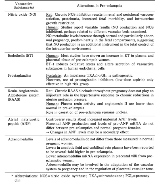
During pregnancy, extensive haemostatic changes occur in the utero-placental circulation and, in pregnancies complicated by pre-eclampsia, a restricted physiolog- ical adaptation of the utero-placental blood vessels leads to increased vascular resistance and reduced blood flow. Nearly every vasoactive substance (which additionally may interact with each other) has been investigated with respect to its possible involvement in the pathogenesis of pre-eclampsia (Fig. 2). This categoriza- tion includes the prostaglandins, the renin-angiotensin-aldosterone axis, nitric oxide (NO), atrial natriuretic peptide (ANP), endothelin, adrenomedullin, and vasopressin (Table 2).
Nitric O xide :
In the last years, extensive study of the nitric oxide (NO) system has been performed. Both, animal experiments and studies in pregnant women, have strong- ly suggested an important role for the nitric oxide synthase (NOS) system in pre- eclampsia. NO is a small, molecular weight mediator with diverse functions that include vasodilation, inhibition of platelet aggregation, and vascular remodeling.
t Testosterone t Arginase activity
~
.j. L-arginine
~
.j. L-arginine/ADMA
~
tADMA
~
Hormonal Shear
t Thromboxane .j. Prostacyclin
Platelet activation Vasoconstriction Proteinuria .j. Glomerular filtration
rate
.j. Prostacyclin Synthase activity
i Prostaglandin H2 i F1-lsoprostanes
Platelet activation Endothelial damage PARS activation Caspase-3 activation Cytochrome C
Oxidase Inhibition .j. MnSOD Activity
Lipid peroxidation .j. DDAH activity ....
~
o(E---- - - -Oxidized LDLtTNF-a
~
SYNDROME of PRE~ECLAMPSIA Fig. 2. Interactions involved in the pathophysiology and leading to the clinical
syndrome of pre-eclampsia. Abbreviations: ADM A = asymmetrical dimethylar- ginine; BI-I4 = tetrahydrobiopterin; DDAH =dimethylarginine dilllethylalllino- hydrolase; H,O, = hydrogen peroxide; LDL = low-density lipoproteins; (Mn) SOD=(lllanganese) superoxide dismutase; NO=nitric oxide; NOS=nitric oxide synthase; 0,-=superoxide anion; PARS=poly(ADP-ribose) synthetase; TNF- a=tulllor necrosis factor alpha; VCAM-l=vascular cell adhesion molecule-l
76 D. SCHLEMBACH
Table 2. Circulating vasoactive substances in pregnancy and pre-eclampsia Vasoactive
Substance (s) Nitric oxide (NO)
Endothelin (ET)
Prostaglandins
Renin-Angiotensin- Aldosterone system (RAAS)
Atrial natriuretic peptide (ANP)
Adrenomedullin
Alterations in Pre-eclampsia
Rat: Chronic NOS inhibition results in renal and peripheral vasocon- striction, proteinuria, increased fetal morbidity, and intrauterine growth restriction.
Human: Studies report variable results (NO production and NOS inhibition), perhaps related to different vascular beds examined.
NO metabolite levels increase through normal and particularly abnor- mal pregnancy, predominantly in the fetal compartments, suggesting that NO production is an additional instrument in the fetal control of the intrauterine environment
Human: Most studies have shown an increase in ET in plasma and placental tissue of pre-eclamptic women.
ET -1 induces oxidative stress and alters secretion of vasoactive substances in human endothelial cells.
Postulate: An imbalance TXA2
>
PGI2 , is pathogenetic.However, use of prostaglandin inhibitors (low-dose aspirin) only effective in high risk group
Rat: Chronic RAAS blockade throughout pregnancy does not play an important role in the hypertensive response to chronic reductions in uterine perfusion pressure.
Human: Plasma renin activity and angiotensin II are lower than normal in pre-eclampsia.
Role in causation of pre-eclampsia remains unclear.
Controversy results about increased maternal ANP levels.
Placental ANP production and levels of pro-ANP mRNA do not differ between pre-eclamptics and normal pregnant females.
-+ Changes in ANP levels may be a secondary effect.
Levels of adrenomedullin do not differ from those measured in normal pregnant women.
Levels in amniotic fluid and umbilical vein plasma have been reported to be several-fold higher in pre-eclampsia.
Lower adrenomedullin mRN A expression in placental villi from pre- eclamptic women.
Adrenomedullin may be involved in the adaptation of the vascular system to pregnancy and in the regulation of placental vascular tone.
• Abbreviations: NOS = nitric oxide synthase; TXA2 = thromboxane; PGI2 = prostacy- clin
NO, originally called endothelium-derived relaxing factor8l) results from the en-
zymatic action of NOS, which converts L-arginine, in the presence of oxygen, to L-
citrulline and NO (Fig. 3). Molecular oxygen and NADPH are cosubstrates in this
reaction. Three NOS enzymes have been sequenced: 1) the constitutive enzyme
present in the vascular endothelium (eNOS or NOS-3), 2) neuronal cells (nNOS or
NOS-I), and 3) several other cell types82). The other is an inducible enzyme (iNOS
or NOS-2) that has been found in macrophages and neutrophils and is activated by
bacterial endotoxin or cytokines (e.g., IL-I, interferon-g)82). Human placental
L-arginine
endothelial cell
t l eNOS I
L-arginine
~7~~:::::;;;'~--.~ NO
\... NADPH l-dtrulline
ArgSUCC~
L -citrulline Fig. 3. Intracellular pathway of nitric oxide
syncytiotrophoblast is known to express eNOS but not iNOS. NO isoenzyme is also expressed on villous endothelial cells and NO produced from these cells is thought to be an important mediator for spiral artery transformation83).
A series of studies in the rat have been performed84,85) to examine the postulate that the pregnant animal responds to a chronic reduction in uteroplacental perfusion pressure with a reduction in renal NO synthesis. Evidence has accrued that indi- cates that NO synthesis is increased in normal pregnancy as are plasma and urine levels of cyclic guanosine 3', 5' -cyclic mono phosphate (cGMP), the second messenger of N086) In the rat, it has been possible to show that chronic NOS inhibition results in renal and peripheral vasoconstriction, proteinuria, increased fetal morbidity, and intrauterine growth retardation87,88) Additionally, it has been reported that both eNOS and nNOS are upregulated in pregnant rats89) Furthermore, chronic inhibi- tion of NOS reverses systemic vasodilation and glomerular hyperfiltration in the pregnant rat modeI90). Interestingly, chronic reduction in uterine perfusion pressure in the rat model is associated with no differences in whole body NO production and a decrease in the renal protein expression of neuronal NOS in pregnant animals compared with controls91).
Studies performed in pregnant and pre-eclamptic women have unfortunately produced conflicting results. This may be explained in part by the difficulties in assessing the NOS system in clinical settings, but additionally by the different settings of these studies (different vessels, plasma/serum, amniotic fluid, fetal blood)
.
NO production seems to be reduced using maternal serum92.93), and unchanged or higher in maternal plasma and/or urine94 -96). In amniotic fluid, NO levels are higher in pre-eclampsia compared with normal pregnancy96) In endothelial cell cultures and placental homogenates, NO production has been reported to be unchanged or higher in pre-eclampsia97-99). Recently a higher NO production has been reported in plasma from umbilical vein and artery96)78 D. SCHLEMBACH
NO metabolite levels increase through normal and particularly through abnor- mal pregnancy predominantly in the fetal compartments, suggesting that NO produc- tion is an additional instrument in the fetal control of the intrauterine environment.
Endothelins .'
The endothelins (ET) are regulatory peptides, distributed in many organ systems and producing potent physiological effects. They are the most powerful vasocon- strictive substances known todayIOO). Three forms of these 21-amino acid peptides have been described, called ET-1, ET-2, and ET-3. ET-1, the most important of the three peptides, is produced by the endotheliumlOI) and by smooth muscle cells I02).
These peptides interact with two types of receptors: ETA and ETB. ETA receptors are present on the smooth muscle and mediate contraction in response to ET -1. The ETB receptors are present on the endothelium. Both ET -1 and ET -3 are capable of inducing the release of NO and prostacyclin, thereby inducing vascular relaxation. Thus, ETB receptors are able to mediate both vasodilation and vasoconstrictionI03). ET -1 causes increased salt and water excretion, which represents a potentially hypotensive action, but in the va'sculature it causes vasocon- strictionI04). The relevance of endothelin to the pathogenesis of pre-eclampsia remains unclear. Thus, most studies56.104-108) have demonstrated an increase in endothelin in plasma or serum and in placental tissuel09) in pre-eclamptic women.
Typically, ET -1 plasma levels are highest during the latter stage of the disease, suggesting that ET may not be involved in the initiation of pre-eclampsia, but rather in the progression of disease into a malignant phase
73 ).Interestingly, immunor- eactive endothelin levels have been reported to be higher in the plasma110) and in the umbilical artery and vein blood of pregnant black women than in patients of European origin11I). Therefore, this could contribute to the higher incidence of hypertension and pre-eclampsia noted in the former population compared with the latterl8. Levels of ET -1 performed in early pregnancy have been reported to have lowl12) or no predictive valuel08.113) as to the later development of pre-eclampsia.
Additionally, in experimental models of pregnancy hypertension, endothelin levels are often not elevatedI04). Yet its effect on blood pressure most likely is more accurately described by its local action at the endothelial and vascular level rather than its serum concentration. Therefore, the actions of endothelin in regulating blood pressure are no doubt correlated best with its effects as a paracrine or autocrine factor. Faxen et al.
114 )noted no change in mRN A for ET -1 in myometrium or placenta of patients compared to normotensive pregnant patients.
However, the expression of ETA-mRNA was significantly reduced in placenta,
whereas that of ETB was unchanged. These data suggested that high circulating
levels of ET -1 might have downregulated the ETA receptor. ET -1 levels have been
determined by Singh et al.
109 )to be significantly higher in placental tissues from
women with pre-eclampsia than in normotensive pregnancies. Additionally, an
ET A receptor antagonist has been reported to lower blood pressure in pregnant rats
in which hypertension was induced by chronic reductions in uterine blood flow I15).
Napolitano
et al.116 )reported an increased ET -1 expression in cultured human placental trophoblastic cells obtained from pre-eclamptic pregnancies compared with those harvested from the placentas of patients with normal pregnancies. The expression of iNOS was decreased in their studies, whereas that of eNOS was increased. The authors postulated that interactions between the ET and NOS systems could represent an important pathogenetic mechanism in the development of the reduced uteroplacental blood flow associated with pre-eclampsia1l6). Addi- tionally, we could show, that incubation of human umbilical vein endothelial cells (HUVEC) with serum from pre-eclamptic women results in increased ET -1 produc- tion97), therefore suggesting, that serum from pre-eclamptic women contains a factor(s) that specifically stimulates ET -1 secretion. Finally, when incubating HUVEC's for
24hours with ET -1 in different concentrations (0-1,000 pmol/L)1l7l, at lower concentrations (5-50 pmol/L), ET -1 increases the intracellular content of lipid peroxides (LPO), stimulates the secretion of thromboxane A2 (TXA2), but inhibits the secretion of prostacyclin (PGI2). At higher concentrations (100-1,000 pmol/L), ET -1 increases the intracellular content of glutathione, but results in a decrease of LPO and an increase of PG 12 back to control levels. ET -1 had no effect on NO secretion. Therefore ET -1 is able to induce oxidative stress and alter secretion of vasoactive substances in human endothelial cells. This observation supports the postulate that ET -1 is involved in the progression to a severe phase of the disease.
Prostaglandins :
The prostaglandins and thromboxane A2 (TXA2) are a series of biologically active compounds derived from arachidonic acidI18). The former are vasodilatory, while the latter is a vasoconstrictor. The prostaglandins are considered to repre- sent important mediators of the minute-to-minute tone of the vasculature acting to offset the vasoconstrictive influence of angiotensin II. The major vasodilator prostaglandin is prostacyclin (prostaglandin 12 [PGI2]). TXA2 is the principal metabolite of arachidonic acid in platelets. This compound is only evanescently present in plasma (its half-life is approximately 30 seconds), so that its effects are largely a function of the microenvironment of its action. TXA2 is an important contributor to platelet aggregation intravascularly, but also contracts the muscular layer of arteries. Prostacyclin, however, is an inhibitor of platelet aggregation and is also a major vasodilator.
Several lines of evidence suggest that changes in the prostaglandin system may play a role in mediating the renal dysfunction and increase in arterial pressure during pre-eclampsia I19). Significant alterations in PGI2 and TXA2 production occur in women with pre-eclampsia I2o). Plasma and urine levels of TXA2 are elevated in women with pre-eclampsia, whereas synthesis of prostaglandins, such as PGI2, is reducedI2o-122).
Additional evidence for a potential role of TXA2 in pre-eclampsia derives from
80 D. SCHLEMBACH
a study demonstrating that short-term increases in systemic arterial pressure produced by acute reductions in uterine perfusion in pregnant dogs can be
prevent~dby thromboxane receptor antagonism123). In addition, Wang and coworkers122) reported that there is an abnormal increase of serum lipid peroxides in pre- eclamptic women. They postulated that these substances, which cause oxidative stress and thereby cellular damage, act by inhibiting prostaglandin synthase.
Further evidence of a potential role for TXA2 is supported by studies in humans indicating that low-dose aspirin may attenuate the development of pre-eclampsia, but unfortunately, a meta-analysis revealed disappointing results124). This can be in part explained by the different settings of these studies. Recent evidence suggest that in women at high risk for the disease
125),low dose aspirin (100 mg/day) has a significant benefit, when started early in pregnancy (before week
16).Renin-Angiotensin-Aldosterone System:
The renin-angiotensin-aldosterone system (RAAS) plays an important role in the long-term regulation of renal function and arterial pressure during a variety of physiological and pathophysiological conditions.
Itis a major contributor not only to the sodium and volume status of the intact organism, but also to the maintenance of the blood pressure
126).Because of its importance in not only the day-to-day but also the minute-to-minute regulation of blood pressure, the RAAS in the path- ogenesis of pre-eclampsia has been extensively investigated.
During normal pregnancy, plasma renin concentration, renin activity, angioten- sin II (AT
II)levels, and aldosterone are all elevated; however, the vascular respon- siveness to AT II appears to be reduced
9,127).
The importance of the RAAS in the regulation of renal function and arterial pressure during pre-eclampsia has not yet been fully elucidated. Plasma renin activity and AT II levels are usually lower than normal throughout pregnancy in patients with pre-eclampsia. Only in the third trimester does the aldosterone level increase in hypertensive pregnancy127).
Although circulating levels of AT II may be normal during pre-eclampsia, it is possible that reducing uteroplacental perfusion pressure could increase the renal sensitivity to AT II through reductions in nitric oxide (NO) or prostacyclin synthesis or by enhanced formation of thromboxane.
This suggestion is confirmed by studies indicating enhanced vascular responsive-
ness to AT II in vessels from animals or humans with pre-eclampsia I20). Further-
more, the pre-glomerular vessels of the renal circulation become extremely sensitive
to the vasoconstrictor actions of AT II when the renal synthesis of NO or prostacy-
clin is reduced or when thromboxane synthesis is elevatedI28). Increased vascular
A T II responsiveness during pre-eclampsia, however, does not prove AT II as an
important endogenous mediator of the vasoconstriction or hypertension in experi-
mental models of pre-eclampsia, because increased responsiveness may only reflect
low endogenous AT II formation. Neither does the chronic blockade of the RAAS
system seem to play an important role in the hypertensive response to chronic reductions in uterine perfusion pressure in the rat129). In addition, the refractoriness to AT II is lost as early as the midtrimester in women who later develop pregnancy- induced hypertension130).
Thus, while the RAAS seems to be responsible in a major way for regulating the cardiovascular adaptation that occurs in pregnancy, its role in the causation of pre- eclampsia remains unclear.
Atrial Natriuretic Peptide,'
Atrial natriuretic peptide (ANP) has been originally discovered by de Bold et
al.
131 )as a natriuretic factor produced by the myocytes of the atrium. It is secreted
from the atria following the splitting of the storage form, a 126-amino acid prohor- mone (pro-ANP), into an N-terminal moiety of
98amino acids (N-ANP) and the biologically active hormone, in equimolar amounts132). ANP is diuretic, natriuretic, vasorelaxant and has antiproliferative properties133). It causes intravascular vol- ume contraction, both because of its ability to induce natriuresis and diuresis, as well as because it causes a shift of fluid from the capillary bed to the interstitium134), resulting in a reduction in both blood pressure and preload135).
In normotensive pregnancy ANP levels trend downward as gestation proceeds from the eighth through the 32
ndweek, then increase significantly in the 36
thweek136).
In hypertensive pregnant women, some investigators reported that ANP levels increase markedly in late pregnancy136-138), but this increase is not a universal finding139.140) and is counterintuitive given the plasma volume contraction seen in pre- eclamptic patients as term approaches1
4l).These increments in plasma ANP may be secondary to some other factors, such as the release of increased amounts of angiotensin II (AT II), endothelin, or catecholamines, either into the circulation or locally in the affected tissues142.143). The enhanced ANP secretion could represent a defense against additional vasoconstriction and sodium retention in pre-eclamptic patients. ANP is also produced by a small population of human placental trophob- last-like cells144), suggesting that ANP may be secreted locally or into the feto- placental circulation and that its effects occur as a result of paracrine or autocrine actions144.145). However, pro-ANP mRNA levels performed on placental tissue of pre-eclamptic patients do not differ from those of normal pregnant woman141).
Therefore, production of ANP by the placenta is not altered at this pre-translational level in pre-eclampsia.
Adrenomedullin "
Adrenomedullin, a member of the calcitonin gene-related peptide family, is a
52-amino acid peptide, originally discovered in human pheochromocytoma tissue146),
that produces blood pressure reduction along with natriuresis and diuresis147). The
latter effect is the result of an ability to increase glomerular filtration rate (GFR) as
well as to inhibit distal tubular sodium reabsorption147). Its hypotensive action is
82 D. SCHLEMBACH
potent and long lastingI46).
Plasma levels of adrenomedullin progressively increase as pregnancy pro- ceeds138). Whereas first trimester adrenomedullin levels did not differ from those of non-pregnant women in either the follicular or luteal phases of the menstrual cycle in studies performed by Minegishi et al.138), third trimester plasma concentrations were significantly higher than first and s.econd trimester levels. In contrast,
DiIorio et al.
148 )could not find any significant difference of plasma adrenomedullin levels throughout gestation. Amniotic fluid adrenomedullin concentrations decreased after the first trimester
(8-12weeks of gestation) and were lowest at
13-20weeks of gestation and then increased at
21-28weeks of gestation. A further increase was found in samples collected after
37weeks of gestationI48). In the umbilical vein, adrenomedullin concentration was higher than in the umbilical artery, suggesting that adrenomedullin in the fetal circulation derives from the placenta 148). These findings lead to the conclusion that adrenomedullin may have an important role in human reproduction, from implantation to deliveryI48).
In pre-eclamptic patients, third trimester adrenomedullin levels did not differ significantly from those measured in pre-eclamptic patients at
28to
40weeks of pregnancy138). Additionally, Di Iorio and his co-workersI49) have reported that plasma levels of adrenomedullin from normotensive pregnant patients did not differ from those obtained from pre-eclamptic patients and Hata et al.
150 )reported that mean levels of adrenomedullin in pre-eclamptic patients were lower than those obtained from normal pregnant women in the third trimester. Interestingly, levels in amniotic fluid and umbilical vein plasma were several-fold higher in pre- eclamptic patients than in normal pregnant womenI49). These data are supported by the findings that the umbilical veins in the intrauterine growth restriction (IUGR) group had significantly higher levels of growth restricted mean fetal adrenomedullin than control patients, whereas there was no difference in maternal plasma adrenomedullin levels of the two groupsl5l).
Additionally, adrenomedullin has been identified in human fetoplacental tis- suesI52,153), in which its presence, determined by immunohistochemical methods, seems to be greater in fetal membranes than in the placenta. The peptide was found to be localized to the amnion and to trophoblast cellsI52). Kanenishi et al.
153 )report decreased immunohistochemical adrenomedullin expression in the placentas obtained from pre-eclamptic pregnancies, and most recently Knerr et al. 154) reported a significantly lower adrenomedullin mRNA expression in placental villi from pre- eclamptic compared with normotensive women.
Furthermore, Jerat et al. 155) reported no significant differences in the response of stem villous arteries taken from normal pregnant patients compared with those from pre-eclamptic patients with respect to their response to adrenomedullin.
Based upon the available data to date, it appears that adrenomedullin may be
involved in the adaptation of the vascular system to pregnancy and in the regulation
of placental vascular tone. However, controversy exists on the status of circulating
and placental adrenomedullin in pre-eclampsia and of the relative contribution of adrenomedullin to impaired fetoplacental circulation and fetal growth.
NON-VASOACTIVE PEPTIDES
/3-human chorionic gonadotropin:
Human chorionic gonadotropin (hCG) is a glycoprotein composed of two non- covalently linked subunits,
aand /3, and is secreted from the blastocyst and early placental syncytiotrophoblast. Maternal serum level peaks at 8-10 weeks of gesta- tion and then declines to reach a plateau at 18-20 weeks. The free /3-hCG circulat- ing in maternal serum corresponds to only about 0.3%-4% of total hCG156).
In the case of /3-hCG, there are several reports of an association with the incidence of pre-eclampsia
157,158).There is general agreement that the placenta remains the main source of hCG in patients with pre-eclampsia, but whether the cause of the high circulating levels of the hormone is placental overproduction is still debated. Some advocate that hCG secretion may be increased as a consequence of abnormal placental invasion or placental immaturity
I59).It may also be linked to the trophoblast response to hypoxia with the development of a hypersecretory state
I60).Compared with normal pregnancies, the placentas of patients with un- explained elevated maternal hCG levels in the second trimester tend to be larger and to have an increased density of hCG-positive trophoblasts along with an increased intensity of hCG immunostaining within the placental
VillP61).However, in contrast to that, a small sample study found equivalent expression of /3-hCG mRNA in normal and pre-eclamptic placental tissues
I62).On average, maternal hCG levels are already increased in the second trimester in pregnancies that subsequently develop pre-eclampsia
I57,163-165).Because the measurement of hCG levels during the second trimester for Down's syndrome screening has already been incorporated into clinical practice at many antenatal clinics worldwide, thousands of records of midtrimester hCG levels for women attending screening programs and their respective outcomes have permitted the investigation of whether the finding of elevated hCG concentrations in maternal serum is predictive of pre-eclampsia. There are accumulating data from studies that evaluated whether a single elevated hCG value (usually above 2.0 MoM) between 14 and 24 weeks of gestation is predictive of pre_eclampsia
I58,163,166-172).The results of these studies are convergent in suggesting that women with elevated
hCG levels in the second trimester are at increased risk for pre-eclampsia, but there
is divergence regarding the accuracy of this test and, by consequence, its predictive
value. Many reasons contribute to the disagreement between the studies. The
sensitivity and specificity of the test may change according to the method of assay,
the clinical and epidemiological background of the subjects, the gestational age at
which samples were collected, and the cutoff chosen to distinguish high from normal
hCG levels. Most recent publications have suggested that hCG may be more
84
D. SCHLEMBACHpredictive for early than late onset pre-eclampsia
l7l ,172).But nevertheless, only when hCG was incorporated into a multifactorial model (including body mass index, parity, and age) did the sensitivity of the test prove effective with a specificity of 71%173).
Inhibin A and activin A :
Inhibins are glycoproteins that were first isolated from ovarian follicular fluid and named after their ability to inhibit the pituitary secretion of follicle stimulating hormone (FSH). Inhibins A and Bare heterodimers composed by an
asubunit and a f3A or f3B subunit, respectively, linked by a disulfide bridge
174).Inhibin-related proteins comprise activins, which are homodimers composed by the same f3 subunits of the inhibin molecule, and follistatin, a binding protein with affinity for inhibins and activins via the f3 subunit. Activins are peptides that act as growth and differentiation factors in many cells and tissues. Inhibins and activins are members of the transforming growth factor (TGF)-f3 superfamily, a group of structurally similar but functionally diverse growth factors 175).
Inhibin
aand f3A subunits are widely localized in the cytotrophoblast and syncytiotrophoblast176), and the intensity of the hybridization signal for inhibin
aand f3A subunit mRNA increases throughout pregnancy, peaking in extracts prepared from term placentas176). Although the decidua
177),membranes178), and fetus all produce inhibin, the placenta is the major source
179).In consonance with placental expression, maternal serum inhibin A and activin A concentrations increase progres- sively during gestation, especially in the last trimester180).
Maternal serum activin A and inhibin A levels are substantially increased in the presence of hypertensive disorders181-185). Although this might happen only because of hemoconcentration or decreased urinary clearance, activin A levels begin to rise significantly before the onset of hematological or renal manifestations of clinical disease181,186,187). The most probable mechanism for the high activin A and inhibin A concentrations in patients with pre-eclampsia is increased placental produc- tion184,188). This increased placental production is more likely to represent a placental response than a primary overproduction, but the mechanism which increases the activin A level in pre-eclampsia is yet unknown.
The above mentioned observations that inhibin A and activin A levels increase before onset of the disease181,186,187) led to the theory that they might be diagnostic and prognostic markers of pre-eclampsia
l81).Silver and coworkers183) found that inhibin A and activin A levels were higher in women with pre-eclampsia and observed that before
34weeks of gestation there was a more pronounced difference in the average levels of both anaiytes between normal and complicated pregnancies.
Studies have indicated that inhibin A is elevated several weeks before the onset of
clinical signs of pre_eclampsia164,187,189-191). Women with an inhibin A concentration
exceeding 2.0 MoM between 15 and 19 weeks of gestation were more likely to
develop pre-eclampsia, to deliver a small-for-gestational-age infant, or to have a
stillbirth or neonatal death190). Lambert-Messerlian et al.
164 )observed that inhibin A levels tended to be higher when the onset of pre-eclampsia occurred within a shorter interval after collection of the second trimester screening sample and suggested that second trimester inhibin A would be more effective in predicting early onset rather than late onset disease. Inhibin A has been reported to be particularly sensitive in predicting the occurrence of pre-eclampsia before
34weeks, when the impact of the disease on maternal-fetal outcome is worse18
7).Compared with inhibin A, activin A seems to be a more sensitive marker at 21-25 weeks
187).Altogether, the studies evaluating second trimester inhibin A and activin A measurements to predict pre-eclampsia suggest that these markers have limited sensitivity and low positive predictive value when applied to low risk populations, but it may add significant information when used in combination with other screen- ing modalities such as Doppler ultrasound192.193).
Leptin:
Leptin was initially introduced as an adipocyte-derived hormone that regulates energy metabolism via its hypothalamic receptor194). Subsequent studies revealed various physiologic functions of leptin. It plays an essential role especially in reproduction by regulating gonadotropin-releasing hormone (GnRH) secretion from the hypothalamus195).
Leptin is also produced by placental trophoblast and is secreted both in maternal and fetal circulation196). During pregnancy, leptin levels show marked changes, suggesting the placenta as a putative source of production of leptin in addition to adipose tissue. Maternal plasma leptin levels rise sharply during the first trimes- ter197.198) and decline back to normal values after delivery199). Serum leptin levels increase in the second and third trimester, and may contribute to the inhibition of increased food intake, body weight, and body fat 198).
Elevated maternalleptin levels have been described in women with pre-eclamp- sia in the third trimester200-203) but not at delivery203). The rise in total leptin represents an increase of free leptin levels, as the bound fraction is paradoxically decreased20
l).The most probable mechanism of leptin increase in pre-eclampsia is increased placental production204) (Fig.
4),with placental hypoxia and inflammatory mediators being important stimulators202). This explains why pre-eclampsia sub- verts the physiological relationship between adiposity and leptin levels in pregnant women205).
A longitudinal study showed increased leptin levels beginning at 20 weeks of gestation in women who subsequently developed pre-eclampsia, suggesting that leptin may be an early marker of the disease200).
Neurokinin B :
Recently, elevated levels of neurokinin B (NKB) have been reported to cause
pre-eclampsia
206).NKB belongs to the tachykinin family. These neuropeptides
86
Chronic reduction of nutrient and oxygen supply
to the fetus
D. SCHLEMBACH
Inflammatory mediators (TNF-a, IL-6)
Chronic hypoxia of placental throphoblast celis
Augmented leptln production
Stimulation or fetal growth
Fig. 4. Relationship between placental leptin production and intrauterine growth restriction in severe pre-eclampsia.
have been implicated in a variety of biological functions from smooth muscle contraction207), vascular reactivity208.209), pain transmission210), neurogenic inflammation21l), and the activation of the immune system212). They are considered normally to be restricted to nervous tissue and to exert their effects peripherally only by release from nerve endings in the target tissue, activating neurokinin (NK) receptors, of which there are three. NKB binds preferentially to the NK3-receptor, but in higher doses is also able to activate other neurokinin receptors.
In animal studies, NKB has been found to cause contraction of the hepatic portal vein208), venoconstriction of the mesenteric bed209), and increases in heart rate213),
which are all potentially hypertensive effects. A model of its physiological role in normal pregnancy and its potential involvement in pre-eclampsia214) has been proposed: NKB is thought to play a physiological role in establishing the early trophoblast. It may be an important regulator of placental perfusion by serving to dilate the uterine spiral arteries. By causing vascular changes, NKB might not only increase maternal blood pressure but also shunts blood from certain organs, such as the liver and the mesenteric beds, to the uterus and placenta in order to maintain a sufficient blood supply to these organs. In pre-eclampsia, it is supposed that if the defective trophoblastic invasion does not rectify itself after the first trimester, then the placenta may start to secrete NKB into the maternal circulation in ever-increas- ing amounts. The incomplete invasion of the trophoblasts could lead to a situation where the uterine spiral arteries are unresponsive and cannot be dilated. Therefore an adequate blood supply to the placenta fails to occur. Raised levels of NKB may then lead to maternal hypertension and damage the kidneys and liver. At very elevated concentrations, peripheral NKl-receptors (on platelets, neutrophils or l11onocytes and in the brain) may be activated and in this way, NKB could also cause
the symptoms and complications usually found in pre-eclamptic patients.
In contrast to the results reported by Page et al.
206 ),we found lower levels of NKB in women with pre-eclampsia compared with normotensive pregnant women215). Our findings are supported by the results published by Li and co- workers
216 ),who found an increased expression of placental neutral endopeptidase 3.4.24.11 (NEP) in pre-eclampsia, suggesting that this enzyme may be involved in the pathophysiological changes of pre-eclampsia. As NKB is a substrate for en- dogenous tachykinin inhibitors, especially NEp207), decreased NKB levels in pre- eclampsia could be due to changes in its inhibitor. Therefore, the role of NKB in pre-eclampsia, if any, remains to be determined.
OXIDATIVE STRESS AND LIPID METABOLISM
Oxidative stress and hyperlipidemia are common themes among many chronic disorders such as atherosclerosis, diabetes, neurodegenerative disorders, and cancer.
Pre-eclampsia has also been reported to be associated with oxidative stress60) and hyperlipidemia217
>,as are risk factors for pre-eclampsia such as obesity, hyperten- sion, and diabetes18).
Oxidative stress may be defined as an imbalance between oxidant and antiox- idant forces in favour of oxidation. Oxidative stress is considered to be an impor- tant final pathway in causing endothelial damage and is evident in the maternal circulation of pre-eclamptic women. The increased oxidative stress in pre-eclamp- sia may on one side be due to elevated free radical generation218). Circulating levels of lipid peroxides (LPO), as estimated by malondialdehyde (MDA) or conjugated dienes, are abnormally elevated and graded in proportion to the severity of the disorder219). Maternal plasma levels of isoprostanes, which are considered accurate markers of oxidative stress and lipid peroxidation, are increased220). Serum levels of iron are also elevated221), which leads to oxidant stress because transition metals such as the ferrous ion (Fe2
+)initiate peroxidation of lipids according to the Fenton reaction. On the other hand, a decrease in levels of antioxidants and/or impaired regeneration of reduced forms of antioxidants222-224) may lead to oxidative stress, although not all investigators find decreased levels of antioxidants223,225,226).
In pre-eclampsia, maternal predispositions could also interact with the poorly
perfused intervillous space to generate reactive oxygen species. It seems likely that
pre-eclamptic women miss a protective factor X or mechanism, which normally
protects the pregnant woman227), making the endothelium more sensitive to possible
noxes. Several suggestions have been advanced to explain the transfer of oxidative
stress from the intervillous space to the systemic circulation. Activated neutrophils
and monocytes are present in pre-eclampsia. These cells could be activated by
oxidative stress in the intervillous space and then generate free radicals on contact
with endothelium220). Again, the consequences of this interaction are defined by
maternal factors (decreased antioxidants, sensitized endothelium, lipoproteins espe-
88
D. SCHLEMBACHcially sensitive to oxidation). Transfer of oxidative stress could also be secondary to the formation of stable products of lipid peroxidation (e.g., malondialdehyde) or by oxidized fragments of syncytiotrophoblast entering the systemic circulation.
Finally, the hypoxic placenta might produce cytokines with the potential to generate oxidative stress. Placentas of women with pre-eclampsia are characterized by oxidative stress, evidenced by abnormally elevated tissue levels and production rates of LPO, as well as an imbalance in the arachidonic acid metabolites, thromboxane, and prostacyclin125,126,228l. Evidence indicates that the imbalance of increased thromboxane and decreased prostacyclin is caused by placental oxidant stress because oxidized lipids stimulate thromboxane synthesis while inhibiting prostacy- clin synthesis. Thromboxane is a potent vasoconstrictor and prostacyclin is a potent vasodilator, so this imbalance restricts uteroplacental blood flow. Further evidence of placental oxidative stress is the finding that placental isoprostane levels are significantly increased in pre-eclampsia229l. The cause of these placental abnor- malities is not known yet.
Administration of antioxidants to women in early pregnancy decreases oxidative stress, endothelial activation, and the frequency of pre-eclampsia, which lends support to the potential role of oxidative stress in pre-eclampsia230l.
IMMUNE MARADAPTATION
In 1976, Drs. James
R.Scott and Alan
A.Beer231l wrote in a review article titled
"Immunologic aspects of pre-eclampsia", the following terse but incisive introduc-
tion to their review article: "The normal pregnant state represents the only natural
and successful transplantation of living tissue from one person to another, but one
has to look no farther than pregnancies complicated by Rh isoimmunization to
realize that immunologic homeostasis between the mother and fetus is not always
perfect." Dekker and SibaF32l have championed the idea that "genuine" pre-
eclampsia is a disease of first pregnancies. They point out the fact that the inci-
dence of pre-eclampsia is low in women who have had a previously normal preg-
nancy. Interestingly, even a prior abortion may provide protection against this
disease233l. However, if a woman changes sexual partners, the immunity conferred
by multiparity is lost19,2o,234,235l. Repeated exposure to sperm from the same individ-
ual may also be a preventive factor in the development of pre-eclampsia2o,22l, and
artificial insemination increases the risk of this disorder2o,236,237l. Although not well
understood, the hypothesis propounded to explain these protective effects of sperm
exposure is that T cells within the genital tract may recognize antigens without the
need for binding to class I human leukocyte antigen (HLA) on antigen-presenting
cells, allowing trophoblasts lacking classical HLA to be recognized238l. In addition,
a transient state of T lymphocyte hyporesponsiveness to paternal class I HLA has
been reported, which may impact this immune reaction239l. A lower level of
messenger RNA for HLA-G has been noted in trophoblasts from pre-eclamptic
patients than from normal pregnant patients
240),but this could be the result of fewer trophoblast cells in pre-eclamptic patients
241 ).Implantation and placentation present an immune challenge because of the semi-allogenic nature of the conceptus. Decidualization of the endometrium itself has features in common with an inflammatory response
64 ).During decidualization, infiltration by uterine natural killer cells occurs, and these interact with the non- polymorphic HLA class I antigens expressed by invading extra villous trophoblast.
Candidates for mediators of the immune maladaptation in pre-eclampsia include cytokines (especially tissue necrosis factor
[TNF]-aand interleukin [IL]-2 and IL-
6)233).
Additionally, enzymes released by activated neutrophils, such as elastase and
oxygen-free radicals, including LPO, have been thought to be implicated
233 ).Redman et al.
64 )hypothesized an interesting theory: They suggest that the endothelial dysfunction is a part of a more generalized intravascular inflammatory reaction involving intravascular leukocytes, as well as the clotting and complement systems, and proposed that such an inflammatory response is already well developed in normal pregnancy and that pre-eclampsia arises when a universal maternal intravascular inflammatory response to pregnancy decompensates in particular cases, which may occur because either the stimulus or the maternal response is too strong.
GENETICS OF PRE-DCLAMPSIA
The familial nature of preeclampsia-eclampsia has been appreciated since at least the 1800s. In the last decades numerous papers report on the familial nature of pre-eclampsia
67 ,242,243).The inheritance patterns of pre-eclampsia have been described as Mendelian (autosomal recessive, autosomal dominant with incomplete penetrance), polygenic/
multifactorial, or mitochondrial. Additionally, a unique type of inheritance has
been postulated that involves an interaction between the genetic components of the
mother and those of the father, manifesting through the fetal-placental unit, and
possibly through imprinting. But until now, the exact inheritance pattern is still
unknown
67,244),no single gene has been identified that explains a clear Mendelian
inheritance, and genomic scans of women with pre-eclampsia have yielded varying
results. No doubt pre-eclampsia is a syndrome with underlying genetic heter-
ogeneity. It is most likely that genes or mutations in certain genes will predispose
women to develop pre-eclampsia, and that these loci will vary in different popula-
tions. The most plausible genetic model to date postulates that maternal genes
dictate a woman's susceptibility for the expression of the pre-eclamptic phenotype,
whereas expression of the phenotype in a woman with a given genetic susceptibility
might depend on the genetic load from the trophoblast and possibly on environmen-
tal factors
245 ,246).This susceptibility is dictated by genes and their interaction with
environment, but it will be transferred into biochemical and molecular changes to be
90 D. SCHLEMBACH
found in the syndrome of pre-eclampsia and perhaps in later life.
Fetal (paternal) contribution:
Theoretically, both mother and fetus (and therefore the father) may contribute to the risk. Pre-eclampsia may reflect problems in the close biological interaction between the two subjects245). Current knowledge on the epidemiology of pre- eclampsia, like the particularly high risk in first pregnancies, points primarily to an effect of maternal factorsI9).
Although it is likely that the fetus may also contribute to the pathophysiology of the syndrome pre-eclampsia, few studies have focused on the contribution of fetal and/or paternal genes247.248). Lie
et al.247)suggested that paternal genes (as expres- sed in the fetus) contribute also strongly to the mother's risk of pre-eclampsia.
Mothers who were pregnant by a partner who fathered a pre-eclamptic pregnancy in another woman had nearly twice the risk in their own pregnancy247). Esplin and co-workers248) reported that both men and women who were the product of a pregnancy complicated by pre-eclampsia were significantly more likely than control men and women to have a child who was the product of a pregnancy complicated by pre-eclampsia.
Candidate Genes :
The search for genes that increase maternal susceptibility to pre-eclampsia is ongoing. Besides thrombophilic mutations a number of genes have been supposed to be involved in the pathogenesis of pre-eclampsia. Potential genes or chromo- some locations are chosen for study based upon their pathophysiologic plausibility or their linkage to pre-eclampsia by genome-wide scanning67).
Thrombophilia: