Fukushima Medical University
福島県立医科大学 学術機関リポジトリ
This document is downloaded at: 2021-11-07T23:13:16Z
Title Anti-phosphoenolpyruvate carboxykinase 2 antibody in patients with autoimmune hepatitis( 本文 )
Author(s) 菅野, 有紀子
Citation
Issue Date 2014-03-25
URL http://ir.fmu.ac.jp/dspace/handle/123456789/614
Rights
© 2013 The Japan Society of Hepatology. This is the peer reviewed version of the following article: [Hepatol Res. 2014 Sep;44(9):1019-25], which has been published in final form at [https://doi.org/10.1111/hepr.12276]. This article may be used for non-commercial purposes in accordance with Wiley Terms and Conditions for Use of Self-Archived Versions.
DOI
Text Version ETD
Original Article
Anti-phosphoenolpyruvate carboxykinase 2 antibody in patients with autoimmune hepatitis
Yukiko Kanno, Hiroshi Watanabe, Atsushi Takahashi, Kazumichi Abe and Hiromasa Ohira
Department of Gastroenterology and Rheumatology, Fukushima Medical University, Fukushima, Japan
Correspondence to: Dr Hiroshi Watanabe
Department of Gastroenterology and Rheumatology, Fukushima Medical University 1 Hikarigaoka, Fukushima 960-1295, Japan
Phone: +81-24-547-1111; Fax: +81-24-547-2055; E-mail:[email protected]
Abstract
Aim: To identify a novel autoantibody specific to autoimmune hepatitis (AIH) and to evaluate its clinical significance.
Methods: Non-nuclear component protein extracted from normal human liver cell CyrohNHpes cultures that reacted with sera from AIH patients on a western blot was identified as an antigenic protein and subjected to N-terminal amino acid analysis to identify phosphoenolpyruvate carboxykinase 2 (PCK2). ELISA for anti-PCK2 antibody was conducted on sera samples from patients with AIH (n=42), primary biliary cirrhosis (PBC; n=48), non-alcoholic steatohepatitis (NASH, n=41), chronic hepatitis C (CHC, n=20), drug-induced liver injury (DILI, n=10), systemic lupus erythematosus (SLE, n=16), and on sera samples from healthy volunteers (n=30). Clinical findings were compared for AIH patients testing positive and negative for anti-PCK2 antibody.
Results: ELISA findings showed that mean anti-PCK2 antibody titer in sera from AIH patients (28.3±29.1 AU) was significantly higher than in PBC (13.9±18.7, p=0.004), NASH (5.4±17.8, p<0.0001), CHC (9.5±11.9, p=0.0043), DILI (7.7±6.0, p=0.0179), and SLE (13.3±24.6, p=0.0064) patients, as well as in healthy volunteers (8.3±7.3, p=0.0006). Anti-PCK2 antibody was present in 50.0% (21/42) of AIH, 14.6% (7/48) of PBC, 4.9% (2/41) of NASH, and 10.0% (2/20) of CHC patients, 0% (0/10) of DILI, 12.5% (2/16) of SLE, and in 3.3% (1/30) of healthy volunteers. The sensitivity, specificity and accuracy of using the detection of anti-PCK2 antibody in diagnosing AIH were 50.0%, 91.5% and 83.1%, respectively. None of the AIH patients positive for anti-PCK2 antibody showed characteristic clinical features.
Conclusion: Although further investigations into the clinical usefulness are necessary, anti-PCK2 may have potential as a diagnostic marker for AIH.
Keywords: autoantibody, autoimmune hepatitis, non-alcoholic steatohepatitis, phosphoenolpyruvate carboxykinase 2, primary biliary cirrhosis
Introduction
Autoimmune hepatitis (AIH) is an autoimmune-mediated liver disease resulting from various mechanisms of losing immunological tolerance against
autologous hepatocytes (1). Most cases of AIH in Japanese patients are type 1 and are characterized by the presence of autoantibodies, although the antibodies, such as antinuclear antibody (ANA) and anti-smooth muscle antibody (ASMA), have low disease specificity (2). The diagnosis of AIH is made using international diagnostic criteria and Japanese diagnostic guidelines. Without specific disease markers, however, atypical or acute-onset cases of AIH are often difficult to diagnose (3,4). Recently, Oketani et al. reported that patients with fulminant hepatitis (caused by AIH) who did not undergo liver transplantation showed poor prognosis (survival rate: 32.4%), and the poor prognosis is due, in part, to difficultly in diagnosing these cases (5). The main etiology of AIH is believed to be mediated by autoimmune responses involving
cytotoxic lymphocytes that attack liver cell membranes. Therefore, in order to elucidate the pathogenesis of AIH, it is important to identify antigens that correspond to each of these autoantibodies (6,7). Several autoantibodies that have been identified as being potentially involved in the pathogenesis of AIH, including anti-liver kidney microsomal type-1 (LKM-1) and anti-soluble liver antigen/liver-pancreas antigen (SLA/LP)
antibodies, are commonly not detected in AIH patients (8,9). Thus, it remains difficult to elucidate the pathogenesis of AIH. Identifying disease-specific autoantibodies that
can be used diagnostically or are involved in the pathogenesis of AIH is thus important.
In this study, we extracted non-nuclear component protein from normal human liver cells from which phosphoenolpyruvate carboxykinase 2 (PCK2) was identified by western blot as an antigenic protein that specifically reacted with the sera from AIH patients, and we evaluated the clinical significance of anti-PCK2 antibody. This autoantibody had not been discussed in relation to AIH before, and we evaluated its clinical usefulness as a diagnostic marker for AIH.
Materials and Methods Subjects
All autoimmune hepatitis (AIH) patients treated at Fukushima Medical University Hospital from January 2000 to December 2012 who fulfilled the International
Autoimmune Hepatitis Group criteria (10) were included in this study. Patients with other causes of chronic liver disease, particularly alcohol abuse, chronic hepatitis B (CHB), or chronic hepatitis C (CHC), were excluded from the AIH patient group.
ALT>400 IU/ml was defined as acute presentation. A summary of the clinical manifestations of the AIH patients included in this study is given in Table 1.
Serum samples from AIH patients were obtained at diagnosis. Patients were diagnosed as having primary biliary cirrhosis (PBC) if they met at least two of three criteria: 1) chronic elevation of cholestatic liver enzymes alkaline phosphatase (ALP) and gamma-glutamyltranspeptidase (GGT), 2) presence of serum anti-mitochondrial antibodies (AMA) detected by either indirect immunofluorescence or ELISA using commercially available kits, and 3) typical histological findings of biopsied liver specimens (11). The diagnosis of drug-induced liver injury (DILI) was made by
hepatologists at our hospital with due consideration of alternative causes and clinical courses of reactions to drugs. In addition, confirmation of non-alcoholic steatohepatitis (NASH) diagnosis was made by liver biopsy in the absence of other liver diseases. SLE patients included in this study fulfilled at least four of the American College of
Rheumatology revised criteria for the classification of SLE.
All patients gave informed consent. This study was approved by the ethics committee of Fukushima Medical University, Japan.
Cells
Normal human hepatocytes, CyrohNHpes, were purchased from Sanko Junyaku Co. (Osaka, Japan). CyrohNHpes cells were cultured in Iscove’s Modified Dulbecco’s Medium (IMDM) (GIBCO, Grand Island, NY) supplemented with 10%
fetal calf serum (FCS) (GIBCO) and cultured in the presence of 5% CO2.
Preparation of cell lysate
Cytoplasmic and cellular membrane proteins of CyrohNHpes cells were extracted using the ProteoJETTM Protein Extraction Kit (Fermentas, Life Sciences, CA) according the manufacturer’s instructions. Briefly, an aliquot of 5 x 10⁶ CyrohNHpes cells was suspended in 1.5 ml of ice-cold wash solution and centrifuged at 250 xg for 5 min at 4C. Then, the washed pellet was resuspended in 1.5 ml of cell permeability buffer, incubated on ice for 10 min, and centrifuged for 15 min at 16,000 xg at 4C. The supernatant was taken as cytoplasmic protein, and transferred to a fresh tube. The cell debris pellet was washed in 1 ml of ice-cold membrane protein extraction buffer, incubated for 30 min at 4C, and centrifuged for 15 min at 16,000 xg at 4C. The
supernatant was taken as membrane protein, and was transferred to a fresh tube and held at -80C.
Electrophoresis and western blotting
Extracted cytoplasmic and cellular membrane proteins from CyrohNHpes cells were mixed as non-nuclear proteins and resuspended in sample buffer in order to load
adequate amounts of protein on the gel for separation by 10% sodium dodecyl sulfate polyacrylamide gel electrophoresis (SDS-PAGE) under reducing conditions. The separated proteins in the lysate were transferred onto polyvinylidene difluoride (PVDF) membranes. After incubation with 50 mM Tris-HCl (pH 7.5) (TBS) containing 0.05%
Tween20 and 1% bovine serum albumin (BSA) (Wako Pure Chemical Industries, Ltd., Osaka, Japan), the membranes were incubated with the serum from two patients with AIH or serum from a healthy control diluted 1/100 in TBS containing 0.05% Tween20 (TBS-T) and 1% BSA. After incubation overnight at 4C, the membranes were washed with TBS-T, then incubated for 60 min with alkaline phosphatase-conjugated Goat F(ab’)2 anti-human IgG (BIOSOURCE, Camarillo, CA) diluted 1/4,000 in TBS-T containing 1% BSA. After washing, the membranes were developed with the
BCIP/NBT-Purple Liquid Substrate System for membranes (SIGMA-ALDRICH, St.
Louis, MO).
For western blotting using recombinant protein, 1 g/lane of recombinant protein was used as the antigen instead of CyrohNHpes cell lysate.
Identification of the protein
The target protein was electrophoretically transferred onto PVDF membrane
and sequenced by Procise 494 HT Protein Sequencing System (Applied Biosystems, Inc, Foster City, CA). The NCBI public data bank was searched for the protein sequence using the BLAST program.
Enzyme-linked immunosorbent assay (ELISA) using recombinant protein as an antigen
Recombinant human PCK2 (Abnova, Taipei, Taiwan) was immobilized on 96-well microtiter plates (F96 CERT MAXISORP, NUNC-IMMUNOPLATE, Themo Fisher Scientific, Roskilde, Denmark) by coating each well with 0.125 g of recombinant PCK2 in 50 l/well of 0.1 M carbonate-bicarbonate buffer (pH 9.8) at 4C overnight. To determine the non-specific binding serum samples to the wells, a control well without PCK2 was also assayed. All procedures were performed at room temperature, except for the antigen coating step. After each step, the wells were washed four times with PBS-T. The ELISA plates were incubated for 2 h with 200 l/well of blocking buffer (PBS-T containing 0.05% non-fat dry milk). Serum samples (diluted 1/100 in blocking buffer) were added to individual wells in duplicate. After a 2 h incubation, the bound IgG was detected with biotin-conjugated Goat F(ab’)2 anti-human IgG (diluted 1/1,000 in blocking buffer), followed by the addition of VECTASTAIN ABC Reagent (VECTOR Laboratories, Inc., Burlingame, CA), and incubated for 30 min. After an additional washing, TMB + substrate chromogen was added. To stop the color development, 0.5 M sulfuric acid was added, and the optical density (OD) at 450 nm was determined on a microplate reader (Benchmark PlusTM Microplate Spectrophotometer, Bio-Rad Laboratories, Inc., Hercules, CA). In each serum sample, the specific binding of anti-PCK antibodies was evaluated by subtracting the mean OD
value of the control well from the mean OD value of the PCK2-coated well. The obtained antibody values were expressed in arbitrary units (AU) based on positive and negative reference serum samples, which were set to 100 and 0 OD units, respectively.
The cut-off value was set as being >mean + 2SD (23.0) of AU for sera of healthy controls.
For absorption test, 5 l of serum samples from AIH patients that showed strong reactivity to recombinant PCK2 on ELISA were added to 5 l of recombinant PCK2 (0.13g/l) and incubated at room temperature for 2 h, and centrifuged at 10,000 xg for 10 min. The supernatant was diluted 1/100 with blocking buffer and subjected to ELISA.
Statistics
The Mann-Whitney U or Fisher’s exact tests were used to compare differences in disease activity and clinical manifestations between the antibody-positive and
antibody-negative patients groups. The correlation between anti-PCK2 antibody titer and biochemistry parameters (ALT, IgG) were analyzed with Pearson’s correlation coefficient. P<0.05 was considered to indicate statistical significance.
Results
Identification of antigenic proteins
A band was identified on the western blot from non-nuclear component
proteins extracted from cultured normal human liver cell line CyrohNHpes reacted with sera of AIH patients (Fig. 1A). A 60-kDa band showed a clear reaction when subjected to N-terminal amino acid analysis and was found to have a 10 amino acid sequence of
IQTLRVLSGD. Based on a homology search in the NCBI database, this protein was identified as PCK2. As shown in Figure 1B, western blot with recombinant PCK2 protein as the antigen and rabbit anti-PCK antibody as a positive control produced a band in AIH patient sera.
Detection of anti-PCK2 antibody by ELISA
The mean interplate coefficients of variation for the positive and negative reference serum were 11.5% and 10.3%, respectively. The mean intraplate coefficient of variation was 6.1%.
The mean titer of anti-PCK2 antibody by ELISA in serum samples from AIH patients 28.3±29.1 AU was significantly higher than in PBC (13.9±18.7 AU, p=0.004), NASH (5.4±17.8 AU; p<0.0001), CHC (9.5±11.9 AU; p=0.0043), DILI (7.7±6.0 AU;
p=0.179), and SLE (13.3±24.6 AU, p=0.0064) patients and in sera from healthy volunteers (8.3±7.3 AU; p=0.0006). The incidence of anti-PCK2 antibody was 50.0%
(21/42) in AIH, 14.6% (7/48) in PBC, 4.9% (2/41) in NASH, and 10.0% (2/20) in CHC patients, 0% (0/10) of DILI, 12.5% (2/16) of SLE, and 3.3% (1/30) in healthy
volunteers.
The sensitivity, specificity and accuracy of anti-PCK2 antibody in diagnosing AIH were 50.0%, 91.5% and 83.1%, respectively.
To determine whether anti-PCK2 antibody react with recombinant PCK2 in ELISA, absorption test was performed on samples that had shown positive results on anti-PCK2 antibody ELISA (n=5). In all samples, after pre-absorption with recombinant PCK2, anti-PCK2 antibody levels were below the positive cut off.
Relationship between anti-PCK2 antibody and clinical features in AIH
As shown in Table 2, there were no significant differences in the following clinical features between AIH patients who were positive and negative for anti-PCK2
antibodies: age, ALT, IgG, ANA titer, SMA positivity, incidence of anti-SLA/LP
antibody, AIH score, frequency of hepatic cirrhosis, prevalence of diabetes, incidence of acute hepatitis, and flare-up rate. No significant correlation was found between
anti-PCK2 antibody titer, ALT and IgG levels. Among the patients who were positive for anti-PCK2 antibodies, no significant correlation was found between anti-PCK2 antibody titer and any of these factors.
Of the other diseases examined, there were no characteristic clinical features in the eight patients with PBC who were positive for anti-PCK2 antibodies. Of the two patients with NASH who were positive for anti-PCK2 antibodies, one was positive and the other was negative for ANA titer.
Relationship between anti-PCK2 antibodies and ASMA and anti-SLA/LP antibodies in AIH
The incidence of ASMA and anti-SLA/LP antibodies in the AIH patients included in this study was 57.1% (24/42) and 4.8% (2/42), respectively. Ten patients were found to be positive for both anti-PCK2 antibodies and ASMA. Of the two patients who were positive for anti-SLA/LP antibodies, one was also positive for anti-PCK2 antibodies and the other was also positive for ASMA. ASMA was detected in 41.7% (20/48) of patients with PBC; of the patients with ASMA, ten were also positive for anti-PCK2 antibodies.
Discussion
In this study, we analyzed disease specificity of anti-PCK2 antibodies in AIH and the relationship of the presence of these antibodies to clinical features of AIH. This
antibody was newly identified from the non-nuclear component of normal human liver cells as an antigenic protein that specifically reacts with the sera from patients with AIH.
In the present study, this anti-PCK2 antibody was detected in 21 of 42 (50.0%) patients with AIH compared to 14 of 165 (8.5%) patients without AIH, indicating a relatively high specificity of 91.4%. Although no clear correlation was observed between the positivity of anti-PCK2 antibodies and the clinical features of AIH, the observed high specificity supports the potential use of this antibody as a diagnostic marker for AIH.
Previous studies on type-1 AIH have suggested the diagnostic value of ASMA, anti-SLA/LP antibody and anti-asialoglycoprotein receptor (ASGPR) antibody (8,9,12).
ASMA was not detected in approximately 20% to 40% of AIH patients and was
detected in slightly less than 50% of patients with PBC (13-15), suggesting that ASMA, like ANA, plays a limited role in the pathogenesis of AIH (16). Consistent with
previous reports, the findings of the present study showed that ASMA has a low
specificity for the diagnosis of AIH, as ASMA was present in 24 of 42 (57.1%) patients with AIH and 20 of 48 (41.7%) patients with PBC. Anti-SLA/LP antibody has been shown to have a low sensitivity (7-19%) for AIH, but it has been associated with higher disease severity and a higher risk of flare-up after discontinuation of treatment with a specificity of almost 100% in diagnosing AIH (17-21). In the present study, this antibody was only found in 2 of 42 (4.8%) patients with AIH, precluding its use as a disease marker due to low sensitivity. In previous studies, anti-ASGPR antibody was found in 76% of patients with AIH but was also found in approximately 12% of patients
with PBC and chronic viral hepatitis (22,23), giving low specificity. Of these
autoantibodies, anti-SLA/LP and anti-ASGPR are rarely measured in clinical practice in Japan.
In vivo, PCK is abundantly present in the liver and kidneys. In humans, this protein functions as a rate-limiting enzyme involved in hepatic gluconeogenesis and is localized in the cytoplasm (PCK1/PEPCK1) and mitochondria (PCK2/PEPCK2). In particular, this enzyme is involved in the conversion of oxaloacetate to
phosphoenolpyruvate (24). These two types of PCK, PCK1 and PCK2, have been shown to play an important role in gluconeogenesis from amino acids and lactic acid, respectively (25). In addition to PCK, gluconeogenesis is catalyzed by three other
enzymes, pyruvate carboxylase, glucose-6-phosphatase and fructose-1,6-bisphosphatase, and is regulated by various other factors, such as insulin and glucagon (24). Patients congenitally deficient in PCK tend to show hypoglycemia and growth failure due to impaired gluconeogenesis (26,27). As none of the anti-PCK2-positive AIH patients showed hypoglycemia and growth failure, it is unlikely that this antibody inhibits enzyme activity. Based on the localization of the PCK protein, it is likely that the antibody has no cytotoxic activity, but like many other autoantibodies, only serves as a disease marker. The disease marker antibody for PBC, a condition similar to AIH, recognizes pyruvate dehydrogenase in mitochondria as an antigenic protein. It is of interest that the corresponding antigen of anti-PCK2 antibody is also found in
mitochondria. However, all anti-PCK2-positive AIH patients were either negative for anti mitochondria antibody or negative for cytoplasmic indirect immunofluorescence on HEp 2 cells. To resolve these contradictory findings, further investigation into the reactivity of anti-PCK2 antibody and PCK2 in liver cells of AIH patients will be
required.
In some cases, ANA-positive NASH may be difficult to differentiate from AIH.
Yatsuji et al. reported that 33% (70/212) of NAFLD patients were positive for ANA and 20.4% patients were identified as having probable or definite AIH based on the AIH scoring system (28). NASH-AIH overlap cases have also been reported in other studies (29,30). In this study, although 10 of the 21 (47.6%) patients positive for NASH were identified as having probable or definite AIH based on AIH score, most patients with NASH were negative for anti-PCK2 antibody, regardless of the positivity of ANA.
When ANA-positive NASH patients are positive for anti-PCK2 antibody, further examination (including liver biopsy) should be considered, with overlapping AIH in mind.
In the diagnosis of typical AIH, the necessity of a new diagnostic marker is low.
However, in the case of atypical AIH (such as, 1. patient that is ANA-negative, 2.
patient with low serum IgG level), a new diagnostic marker has been desired. In order to verify the usefulness of anti-PCK2 antibody as a diagnostic of AIH, a multi-center study involving a large number of patients is needed.
Acknowledgements
This study was supported by JSPS KAKENHI (Grant Number 24590987).
The authors declare no conflicts of interest.
References
1 Czaja AJ. Current concepts of autoimmune hepatitis. Ann Hepatol 2005; 4:6-24, 2 Ohira H, Takahashi A. Current trends in the diagnosis and treatment of autoimmune hepatitis in Japan. Hepatol Res 2012; 42:131-38.
3 Takahashi H, Zeniya M. Acute presentation of autoimmune hepatitis: Does it exist?
A published work review. Hepatol Res 2011; 41:498-504.
4 Abe M, Mashiba T, Zeniya M, et al. Autoimmune Hepatitis Study Group-Subgroup of the Intractable Hepato-Biliary Disease Study Group in Japan. Present status of autoimmune hepatitis in Japan: a nationwide survey. J Gastroenterol 2011; 46:1136-41.
5 Oketani M, Ido A, Nakayama N, et al. Etiology and prognosis of fulminant hepatitis and late-onset hepatic failure in Japan: Summary of the annual nationwide survey between 2004 and 2009. Hepatol Res 2013; 43:97-105.
6 Czaja AJ, Manns MP. Advances in the diagnosis, pathogenesis, and management of autoimmune hepatitis. Gastroenterology 2010; 139:58-72.
7 Oo YH, Hubscher SG, Adams DH. Autoimmune hepatitis: new paradigms in the pathogenesis, diagnosis, and management. Hepatol Int 2010; 4:475-93.
8 Krawitt EL. Autoimmune hepatitis. N Engl J Med 2006; 354:54-66.
9 Herkel J, Manns MP, Lohse AW. Selenocysteine, soluble liver antigen/liver-pancreas, and autoimmune hepatitis. Hepatology 2007; 46:275-77.
10 Alvarez F, Berg PA, Bianchi FB, et al. International Autoimmune Hepatitis Group Report: review of criteria for diagnosis of autoimmune hepatitis. J Hepatol 1999 31:929-38.
11 Kaplan MM, Gershwin ME. Primary biliary cirrhosis. New Engl J Med 2005;
353:1261-73.
12 McFarlane BM, McSorley CG, Vergani D, et al. Serum autoantibodies reacting with the hepatic asialoglycoprotein receptor protein (hepatic lectin) in acute and chronic liver disorders. J Hepatol 1986; 3:196-205.
13 Vergani D, Alvarez F, Bianchi FB, et al. Liver autoimmune serology: a consensus statement from the committee for autoimmune serology of the International Autoimmune Hepatitis Group. J Hepatol 2004; 41:677-83.
14 Zamanou A, Tsirogianni A, Terzoglou C, Balafas A, Economidou I, Lymberi P.
Anti-smooth muscle antibodies (ASMAs) and anti-cytoskeleton antibodies (ACTAs) in liver diseases: a comparison of classical indirect immunofluorescence with ELISA. J Clin Lab Anal 2002; 16:194-201.
15 Fusconi M, Cassani F, Zauli D, et al. Anti-actin antibodies: a new test for an old problem. J Immunol Methods 1990; 130:1-8.
16 Czaja AJ, Cassani F, Cataleta M, Valentini P, Bianchi FB. Frequency and significance of antibodies to actin in type 1 autoimmune hepatitis. Hepatology 1996;
24:1068-73.
17 Ma Y, Okamoto M, Thomas MG, et al. Antibodies to conformational epitopes of soluble liver antigen define a severe form of autoimmune liver disease. Hepatology 2002; 35:658-64.
18 Kanzler S, Weidemann C, Gerken G, et al. Clinical significance of autoantibodies to soluble liver antigen in autoimmune hepatitis. J Hepatol 1999; 31:635-40.
19 Czaja AJ, Shums Z, Norman GL. Frequency and significance of antibodies to soluble liver antigen/liver pancreas in variant autoimmune hepatitis. Autoimmunity 2002; 35:475-83.
20 Baeres M, Daehnrich C, Schlumberger W, et al. Establishment of standardised
SLA/LP immunoassays: specificity for autoimmune hepatitis, worldwide occurrence, and clinical characteristics. Gut 2002; 51;259–64.
21 Miyakawa H, Kawashima Y, Kitazawa E, et al. Low frequency of anti-SLA/LP autoantibody in Japanese adult patients with autoimmune liver diseases: analysis with recombinant antigen assay. J Autoimmun 2003; 21;77–82.
22 Treichel U, McFarlane BM, Seki T, et al. Demographics of anti-asialoglycoprotein receptor autoantibodies in autoimmune hepatitis. Gastroenterology 1994; 107:799-804.
23 Husa P, Chalupa P, Stroblová H, et al. Autoantibodies to asialoglycoprotein receptor in chronic hepatitis C patients. Acta Virol 2001; 45:7-11.
24 Radziuk J, Pye S. Hepatic glucose uptake, gluconeogenesis and the regulation of glycogen synthesis. Diabetes Metab Res Rev 2001; 17:250-72.
25 Watford M, Hod Y, Chiao YB, et al. The unique role of the kidney in gluconeogenesis in the chicken. The significance of a cytosolic form of phosphoenolpyruvate carboxykinase. J Biol Chem 1981; 256:10023-7.
26 Hommes FA, Bendien K, Elema JD, et al. Two cases of phosphoenolpyruvate carboxykinase deficiency. Acta Paediatr Scand 1976; 65:233-40.
27 Clayton PT, Hyland K, Brand M, et al. Mitochondrial phosphoenolpyruvate carboxykinase deficiency. Eur J Pediatr 1986; 145:46-50.
28 Yatsuji S, Hashimoto E, Kaneda H, et al. Diagnosing autoimmune hepatitis in nonalcoholic fatty liver disease: is the International Autoimmune Hepatitis Group scoring system useful? J Gastroenterol 2005; 40:1130-38.
29 Loria P, Lonardo A, Leonardi F, et al. Non-organ-specific autoantibodies in nonalcoholic fatty liver disease: prevalence and correlates. Dig Dis Sci 2003;
48:2173-81.
30 Fukuda S, Komori A, Itoh M, et al. Histological Remission during Corticosteroid Therapy of Overlapping Nonalcoholic Steatohepatitis and Autoimmune Hepatitis: Case Report and Literature Review. Case Rep Gastroenterol 2011; 5:553-7.
Figure 1. Western blot assays. (A) Western blot with non-nuclear protein extracted from CyrohNHpes cells. The membrane was incubated with serum from a patient with AIH.
(B) Western blot with recombinant PCK2 protein and rabbit anti-PCK antibody as a positive control. Lanes are 1, negative control; 2, rabbit anti-PCK antibody; and 3, serum of a patient with AIH.
(kDa)
IQTLRVLSGD 75
50
1 2 3
Figure 2. Detection of anti-PCK2 antibody by ELISA. The mean titer of anti-PCK2 antibody in serum samples from AIH patients was significantly higher than in sera from patients with PBC, NASH, CHC, DILI, SLE, and healthy volunteers. The positive cut-off value was set as mean2SD (23.0) of AU for serum of healthy controls.
Figure 3. Relationship between anti-PCK2 antibody titer and (A) ALT and (B) IgG levels. No significant correlation was found between anti-PCK2 antibody titer and ALT or IgG levels.
(A)
(B)
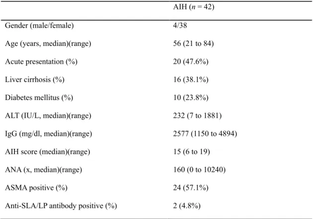
Table 1. Patient characteristics for AIH patients
AIH (n = 42)
Gender (male/female) 4/38
Age (years, median)(range) 56 (21 to 84)
Acute presentation (%) 20 (47.6%)
Liver cirrhosis (%) 16 (38.1%)
Diabetes mellitus (%) 10 (23.8%)
ALT (IU/L, median)(range) 232 (7 to 1881)
IgG (mg/dl, median)(range) 2577 (1150 to 4894)
AIH score (median)(range) 15 (6 to 19)
ANA (x, median)(range) 160 (0 to 10240)
ASMA positive (%) 24 (57.1%)
Anti-SLA/LP antibody positive (%) 2 (4.8%)
Table 2 Relationship between presence of anti-PCK2 antibody and clinical features in AIH Anti-PCK2 positive
(n=21)
Anti-PCK2 negative (n=21)
P value
Gender (male/female) 2/21 2/21 1
Age (years, median)(range) 56 (24 to 68) 53 (21 to 84) 0.58
Acute presentation 10 11 1
Liver cirrhosis 7 4 0.48
Diabetes mellitus 5 5 1
ALT (IU/L, median)(range) 165 (17 to 1881) 293 (7 to 1507) 0.92 IgG (mg/dl, median)(range) 2827 (1250 to 4610) 2570 (1150 to 4894) 0.56 AIH score (median)(range) 15 (9 to 18) 14 (6 to 19) 0.66 ANA (x, median)(range) 160 (0 to 10240) 640 (0 to 2560) 0.38 ASMA (x, median)(range) 20 (0 to 640) 20 (0 to 640) 0.64