Fukushima Medical University
福島県立医科大学 学術機関リポジトリ
This document is downloaded at: 2021-11-08T00:24:52Z
Title Safety and effectiveness of transcatheter closure of atrial septal defects: Initial results in Fukushima Prefecture
Author(s)
Kobayashi, Atsushi; Kunii, Hiroyuki; Yokokawa, Tetsuro;
Sakuma, Yuya; Oikawa, Masayoshi; Yoshihisa, Akiomi;
Yamaki, Takayoshi; Nakazato, Kazuhiko; Ishida, Takafumi;
Takeishi, Yasuchika
Citation Fukushima Journal of Medical Science. 64(3): 151-156
Issue Date 2018
URL http://ir.fmu.ac.jp/dspace/handle/123456789/711
Rights © 2018 The Fukushima Society of Medical Science
DOI 10.5387/fms.2018-13
Text Version publisher
Vol. 64, No. 3, 2018
[Original Article]
Safety and effectiveness of transcatheter closure of atrial septal defects : Initial results in Fukushima Prefecture Atsushi Kobayashi1),*, Hiroyuki Kunii
1),*, Tetsuro Yokokawa
1,2), Yuya Sakuma
1), Masayoshi Oikawa
1), Akiomi Yoshihisa
1,3), Takayoshi Yamaki
1), Kazuhiko Nakazato
1),
Takafumi Ishida
1)and Yasuchika Takeishi
1)1)
Department of Cardiovascular Medicine, Fukushima Medical University,
2)Department of Pulmonary Hypertension,
3)Department of Advanced Cardiac therapeutics
(Received June 28, 2018, accepted October 1, 2018)
Abstract
Background : Atrial septal defect is the third most common type of congenital heart disease. Sur- gical closure was the standard treatment for atrial septal defects before transcatheter closure was approved as minimally invasive treatment in Japan in 2006. In our hospital, this procedure has been performed since 2015.
Objective : To evaluate the safety and effectiveness of transcatheter closure of atrial septal defects in our hospital.
Methods : Thirty patients (mean age 57.0 ± 19.7 years, 11 males), who underwent transcatheter closure of atrial septal defect between September 2015 and February 2018 at the Fukushima Medical University Hospital, were enrolled. All procedures were performed under general anesthesia with angiographic and transesophageal echocardiographic guidance. Safety and effectiveness were eval- uated by the procedural results and complications.
Results : All 30 patients successfully underwent transcatheter closure of atrial septal defects and no patient developed complications. New York Heart Association functional class was improved, and the right ventricular area and right atrium area were decreased, postoperatively.
Conclusion : Transcatheter closure is a safe and effective treatment for atrial septal defects, and thus could be an alternative option to open heart surgery.
Key words : atrial septal defect, transcatheter closure
Introduction
Atrial septal defect is a common congenital heart disease and has two types : primum and se- cundum. Primum atrial septal defect is a variant of common atrioventricular canal defects. Secundum atrial septal defect is a defect that develops in the fossa ovalis due to the defective septum primum.
Closure of atrial septal defects either percutaneously or surgically is indicated in patients with a hemody- namically significant shunt that causes enlargement of the right heart
1).
Catheter intervention for structural heart dis- ease has spread in recent years worldwide. Trans- catheter closure of secundum atrial septal defects has been covered by public insurance in Japan since 2006. In Fukushima prefecture, transcatheter clo- sure of atrial septal defects started from 2015 at Fu- kushima Medical University Hospital. We investi- gated the safety and effectiveness of transcatheter closure of atrial septal defects in our institution.
Corresponding author : Tetsuro Yokokawa, M.D., Ph.D. E
-mail : [email protected] https://www.jstage.jst.go.jp/browse/fms http://www.fmu.ac.jp/home/lib/F
-igaku/
*Two authors contributed equally to this work.
151
152 A. Kobayashi et al.
Methods
We enrolled 30 patients who underwent trans- catheter closure of secundum atrial septal defects between September 2015 and February 2018. Dur- ing this period, three patients were referred to our hospital due to atrial septal defects ; however, they were not indicated for transcatheter closure because of small superior, inferior, or posterior rims by trans- esophageal echocardiography.
Baseline data including age, sex, body mass in- dex, New York Heart Association class, past history, plasma levels of brain natriuretic peptide, echocar- diographic findings, hemodynamic findings, and pro- cedural data were collected.
Procedure of transcatheter closure of atrial sep- tal defects is shown below. The implantation meth- ods are similar between both devices of Amplatzer
ⓇSeptal Occluder (Saint
-Jude Medical, Zaventem, Belgium) and Figulla
ⓇFlex II ASD Occluder (Oc- clutech GmbH, Jena, Germany). All procedures were performed under general anesthesia with angi- ographic and transesophageal echocardiographic guidance. Before the procedure, we determined the size of occluder device by transesophageal echo- cardiography but not by a sizing balloon. First, a guide wire was inserted to the atrial septal defect through the right femoral vein. The occluder was screwed to the tip of the delivery cable, immersed in saline and drawn into the loader. A long guiding sheath and dilator were advanced over the guidewire through the atrial septal defect to the left atri- um. After deployment of the left
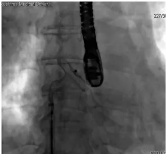
-sided disc in the left atrium, the system was pulled back. The oc- cluder was thereafter fully deployed by withdrawing the sheath to expand the right atrial disc. Then, a residual shunt was evaluated by transesophageal echocardiography. Figure 1 shows an angiographic picture during the occluder deployment. The pa- tients were discharged from the hospital 3 to 5 days after atrial septal occlusion. They received thieno- pyridine daily for a month and aspirin daily for 6 months. During the follow
-up, blood sampling for brain natriuretic peptide measurement and transtho- racic echocardiography were carried out at 1, 3, 6, and 12 months after atrial septal occlusion.
Data were analyzed using the Statistical Pack- age for Social Sciences version 25 (SPSS Inc., Chi- cago, IL, USA). Continuous data are expressed as mean ± SD, and skewed data are presented as me-
dian and interquartile range. Categorical variables are expressed as numbers and percentages. The data were analyzed using Wilcoxon signed rank test. A P value of < 0.05 was considered statisti- cally significant.
Results
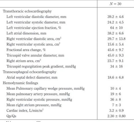
The baseline characteristics of the 30 patients are shown in Table 1. The echocardiographic find- ings at baseline demonstrated preserved left ven- tricular systolic function, enlargement of right ven- tricle and both atria as shown in Table 2. The atrial septal defect diameter measured by transesophageal
Fig. 1. An angiographic image in transcatheter clo- sure of atrial septal defect
The occluder was deployed by withdrawing the sheath to expand the right atrial disc.
Table 1. Baseline characteristics N = 30
Age, years 57.0 ± 19.7
Male 11 (37)
BMI, kg/m
222.7 ± 3.7
NYHA class, I/II/III/IV 17/12/1/0
Atrial fibrillation 8 (27)
Hypertension 5 (17)
Diabetes mellitus 5 (17)
Chronic kidney disease 12 (40)
Laboratory data
BNP, pg/mL* 39.9 (17.4
-123.6)
Values are mean ± SD, *median (IQR), or number (%).
BMI, body mass index ; NYHA, New York Heart Asso-
ciation ; BNP, brain natriuretic peptide ; IQR, interquar-
tile range
echocardiography was 18.6 ± 6.8 mm. The hemo- dynamic findings showed high mean pulmonary blood flow/systemic blood flow ratio of 2.30 ± 0.80.
Five out of eight patients with atrial fibrillation un- derwent pulmonary vein isolation for atrial fibrilla- tion before transcatheter closure of atrial septal de- fects.
All patients successfully underwent transcathe- ter closure. Amplazter
ⓇSeptal Occluder and Fig- ulla
ⓇFlex II ASD Occluder were used in 18 and 12 patients, respectively. No device
-related complica- tions were observed during the follow
-up period of 312 ± 485 days (4 to 807 days) until April 2018 (Ta- ble 3). A slight residual shunt was detected imme- Table 2. Echocardiographic and hemodynamic findings
N = 30 Transthoracic echocardiography
Left ventricular diastolic diameter, mm 39.2 ± 4.6 Left ventricular systolic diameter, mm 24.2 ± 4.5 Left ventricular ejection fraction, % 64 ± 10
Left atrial dimension, mm 38.2 ± 6.6
Right ventricular diastolic area, cm
229.7 ± 13.8 Right ventricular systolic area, cm
215.6 ± 5.4
Fractional area change, % 45.6 ± 9.7
Tricuspid valve annular diameter, mm 45.0 ± 9.3
Right atrium area, cm
223.7 ± 9.1
Tricuspid regurgitation peak gradient, mmHg 34 ± 16 Transesophageal echocardiography
Atrial septal defect diameter, mm 18.6 ± 6.8
Hemodynamic findings
Mean Pulmonary capillary wedge pressure, mmHg 10 ± 4
Mean pulmonary artery pressure, mmHg 19 ± 6
Right ventricular systolic pressure, mmHg 36 ± 8
Mean right atrium pressure, mmHg 7 ± 3
Cardiac index, L/min/m
23.2 ± 0.9
Qp/Qs 2.30 ± 0.80
Values are mean ± SD.
Qp/Qs, pulmonary blood flow/systemic blood flow ratio
Table 3. Procedural results
N = 30
Successful device deployment 30 (100)
Atrial septal defect occluder
Amplatzer
ⓇSeptal Occluder 18 (60)
Figulla
ⓇFlex II ASD Occluder 12 (40)
Device size, mm 22 ± 7
Complication
Death 0
Erosion 0
Device embolization and malposition 0
Cerebral embolism 0
Bleeding at puncture site 0
Blood transfusion 0
Infective endocarditis 0
Atrioventricular block 0
Supraventricular tachycardia 0
Values are mean ± SD, or number (%).
154 A. Kobayashi et al.
IV III
II I
0 0
4 26 1 month
(n = 30) 0 0
4 23 3 months
(n = 27) 0 0
2 22 6 months
(n = 24) 0 0
2 13 12 months
(n = 15) 0
1
12 17 Before
(n = 30)
1
3
9 17
4
23 20 13
2
2 2
Months after atrial septal occlusion
New York Heart Association