Development of new endotoxin measurement assay using bioluminescence method
9
0
0
全文
(2) Maria Nonoguchi, et al.. 182. Endotoxin Factor C. Activated factor C Factor B. Activated factor B Clotting enzyme. Proclotting enzyme. Coagulogen. Coagulin Gelation Turbifdity. Benzoil‒Leu‒Gly‒Arg‒Luciferin Luciferin relaese Luciferase+ATP. Right. Turbidimetric kinetic assay. Bioluminescence method. Fig. 1. The clotting enzyme, which is generated by the Limulus reaction, along with Fig. 1. The clotting enzyme, which is generated by the Limulus reaction, along with the endotoxin activates the the endotoxin the luminescent substrate; the released luciferin luminescent substrate;activates the released luciferin emits light in the presence of luciferase and ATP. emits light in the presence of luciferase and ATP.. II. Materials and methods. endotoxins with turbidimetric kinetic assay using leukocyte-rich plasma samples (LRP).. 1. Specimens. In our previous study, we have reported that. Between April 2018 and April 2019, patients. this new measurement method has higher. who were transported to the Iwate Medical. measurement accuracy than the conventional. University Advanced Critical Care Center and. one. 1, 2). were suspected of having a bacterial infection. .. On the other hand, a bioluminescence. were approached for enrollment in this study.. technique for measuring endotoxin using. Written informed consent was obtained from all. high-intensity luciferase has been developed. the patients or the patients’family members.. 3). by Kuroda et al. This technique was later 4). This study was a prospective study, The. improved by Onodera et al. In this method,. “International Consensus Definition of Sepsis. the clotting enzyme, which is generated by. and Septic Shock, 3rd Edition (Sepsis-3)”was. the Limulus reaction, along with the endotoxin. used to identify bacterial infection, sepsis, and. activates the luminescent substrate; the. septic shock. The Acute Physiology and Chronic. released luciferin emits light in the presence. Health Evaluation II (APACHE II) scores 5) and. of luciferase and ATP (Fig. 1). Recently, an. sequential organ failure assessment (SOFA). automated method was developed, and it is. scores. being used clinically for the determination of. 2. Leukocyte-rich plasma (LRP). endotoxin in dialysate. 3, 4). 6). were used as indices of severity.. . However, at present,. After collecting 2 mL of blood from each. no paper has examined endotoxins in human. patient, 0.1 mL of heparin was added to the. blood using this automated method. In this. whole blood sample and incubated at 37℃ for. study, we compared the diagnostic of the. 5-10 min. Next, 800 µL of the 37℃ - equilibrated. turbidimetric kinetic assay using LRP and the. blood and 400 µL of the 37 ℃ - equilibrated. automated bioluminescence method using the. 6% dextran T500 (Pharmacosmo, Holdaek,. same samples for detecting infectious diseases.. Denmark) were mixed and incubated at 37℃.
(3) Original: Endotoxin measurement by new bioluminescence method. 183. for 15 min. The pellet contained red blood. method, the turbidimetric value was also. cells aggregated by dextran, whereas the. expressed in EU, where 1 pg/ml = 7 mEU/ml.. supernatant layer was collected as the LRP.. 4. Endotoxin measurement by the automated. We have named this method the LRP37 7). bioluminescence method. method .. The automated bioluminescence system. Dextran T500 was autoclaved at 121 ℃ for. developed by DKK TOA Corporation (Tokyo,. 90 min before use to ensure that endotoxin. Japan) was used. The samples obtained by. levels were below the detection limits of the. LRP37 method was pretreated by dilution and. Limulus test (i.e., endotoxin-free).. heating method as the same method described. 3. Endotoxin measurement by turbidimetric. above, and the 0.2 ml samples were added. kinetic assay. into LAL tube. After confirming dissolution,. After 10-fold dilution of the test samples,. the BL tube containing the luminescent. which was obtained by the LRP37 method,. substrate was inserted into LAL tube and. with a plasma pretreatment solution (0.02%. the mixture was automatically stirred for 5. Triton X-100 solution, FUJIFILM Wako. second. Then, it was automatically heated. Pure Chemical Corporation, Osaka), they. to 37℃ for 19 min to release luciferin by the. were mixed well in a vortex mixer to ensure. Limulus reaction. ATP and luciferase in the. destroying leukocytes. Then, the samples. BL tube were then automatically added into. were heated at 70℃ for 10 min and cooled in. the mixture. The bioluminescence reaction. ice water for 5 min to prepare a pretreated. occurs immediately, and the amount of. specimen. Endotoxin single test Wako. luminescence was measured, and the amount. (FUJIFILM Wako Pure Chemical Corporation). of endotoxin was finally calculated using a. was dissolved in 0.2 ml of the pretreated. software that incorporated a calibration curve.. ®. Bioluminescence-type endotoxin analyzer. (FUJIFILM Wako Pure Chemical Corporation,. Luminitz-ET ® (DKK-TOA Corporation) was. Osaka) was used for measurement. The time. used for this assay.. taken for the amount of transmitted light to. 5. Statistical analysis. decrease by 8% was defined as the gelation. Comparisons between the two groups were. time, and endotoxin concentration was. made using Wilcoxon signed rank test. The. calculated using a calibration curve created. systematic bias between the two measurement. from the gelation time of standard LPS (E.coli. methods ware investigated using Bland-. 0111:B4, FUJIFILM Wako Pure Chemical. Altman analysis. The Spearman rank order. sample, and Toxinometer MT-5500. Corporation). 8, 9). .. correlation coefficient was employed for the. The maximum set-up time for this. analysis of correlations. We used at cut-off. instrument is 200 min, and the lowest. value obtained through receiver operating. endotoxin measurement limit in plasma. characteristic (ROC) analysis. A p-value < 0.05. is approximately 0.3-0.5 pg/ml. Moreover,. was considered statistically significant. Two-. because the endotoxin levels are expressed. group comparisons, correlations, and multiple. as endotoxin units (EUs) in the luminescence. liner regression analyses were performed.
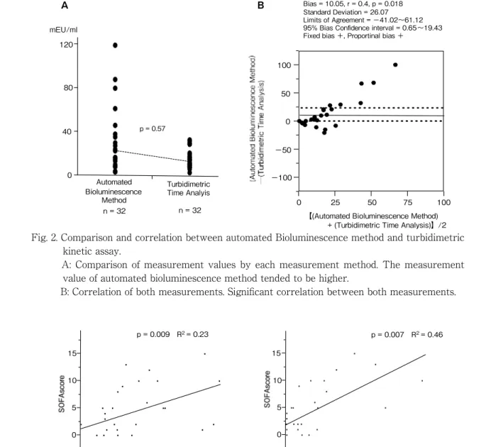
(4) Maria Nonoguchi, et al.. 184. A. B. Bias = 10.05, r = 0.4, p = 0.018 Standard Deviation = 26.07 Limits of Agreement = −41.02∼61.12 Diagnostic support tool 95% Bias Confidence interval = 0.65∼19.43 Fixed bias +, Proportinal bias +. mEU/ml 120. (points). 100 80. 50 0. p = 0.57. 40. −50 0. Automated Bioluminescence Method n = 32. −100. Turbidimetric Time Analyis. 0. n = 32. 25. 50. 75. 100. 【(Automated Bioluminescence Method) + (Turbidimetric Time Analysis)】/2. Fig. 2. Comparison and correlation between automated Bioluminescence method and turbidimetric kinetic assay. A: Comparison of measurement values by each measurement method. The measurement value of automated bioluminescence method tended to be higher. B: Correlation of both measurements. Significant correlation between both measurements.. p = 0.009 R2 = 0.23. p = 0.007 R2 = 0.46. 15. 15. 10. 10. 5. 5. 0. 0 0. 5. 10. 15. 20. 25. 30 mEU/ml. Turbidimetric Time Analysis. 0. 20. 40. 60. 80. 100. 120. mEU/ml. Automated Bioluminescence Method. Fig. 3. Evaluated SOFA score to determine severity of sepsis, and also assessed the correlation between these two values.. Fig. 3.JMP Evaluated SOFA score determineCary, severity of sepsis, and also assessed the III.correlation Resultsbetween these two values. using software (SAStoInstitute, NC,. USA). ROC analysis was performed using Dr.. Blood samples were obtained from 32. SPSS II software (SPSS, Chicago, IL, USA).. individuals; 24 infected patients and 8. 6. Ethics. healthy individuals. Results revealed that. Ethical approval for the study (Ethical. the endotoxin level was to be 10.61 ± 7.99. Committee No. H29-174) was provided by. mEU/ml using the turbidimetric kinetic. the Ethical Committee of Iwate Medical. assay, whereas the endotoxin levels were. University, Morioka, Japan (Chairperson: Prof.. found to be 18.8 ± 28.31 mEU/mL using the. Y Sato) on March 15, 2018.. automated bioluminescence method. The.
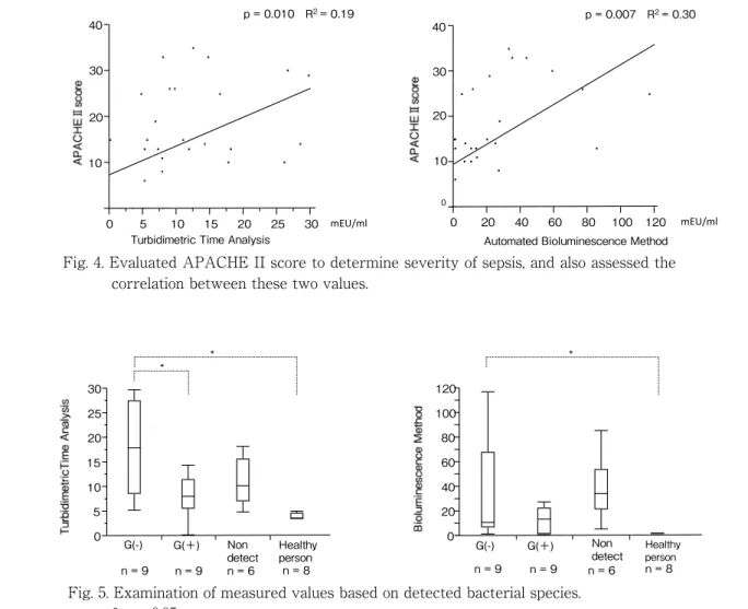
(5) Original: Endotoxin measurement by new bioluminescence method. p = 0.010 R2 = 0.19. 40. 185. p = 0.007 R2 = 0.30. 40. 30. 30. 20. 20. 10. 10. 0. 0. 5 10 15 20 25 Turbidimetric Time Analysis. 30. mEU/ml. 0. 20. 40. 60. 80. 100. 120. mEU/ml. Automated Bioluminescence Method. Fig. 4. Evaluated APACHE II score to determine severity of sepsis, and also assessed the correlation between these two values.. B * * Fig. 4. Evaluated APACHE II* score to determine severity of sepsis, and also assessed the correlation between these two values. 30. 120. 25. 100. 20. 80. 15. 60. 10. 40. 5. 20. 0. G(-). G(+). n=9. n=9. Non detect n=6. Healthy person n=8. 0. G(-). G(+). n=9. n=9. Non detect n=6. Healthy person. n=8. Fig. 5. Examination of measured values based on detected bacterial species. * p < 0.05. Fig. 5. Examination of measured values based on detected bacterial species.. automated bioluminescence method showed. diseases using both measurement methods. a slightly higher value and a larger standard. was compared. For turbidimetric kinetic assay,. error. The Bland-Altman analysis showed. sensitivity was 0.91, specificity was 0.89, and. a significant correlation between the two. AUC was 0.91 when the diagnostic cutoff value. measurement methods. In addition, systematic. was 5.22. For the automated bioluminescence. bias was also recognized, and it became clear. method, sensitivity was 0.95, specificity was 1.0,. that the difference between both measured. and AUC was 0.95 when the diagnostic cutoff. values tended to increase as the average of. value was 1.21 (data not shown). Additionally,. both measured values increased (Fig. 2). The. we evaluated SOFA (Fig. 3) and APACHE. measurement time was 138.01 ± 8.58 min for. II (Fig. 4) scores to determine the severity. the turbidimetric kinetic assay and 20.0 min. of sepsis, and also assessed the correlation. for the automated bioluminescence method;. between the severity and endotoxin values.. thus, the automated bioluminescence method. The correlation between turbidimetric kinetic. was significantly faster (p < 0.01) (data not. assay and SOFA score (p = 0.009, R2 = 0.23). shown). The diagnostic accuracy of infectious. or APACHE II score (p = 0.010, R2 = 0.19) and.
(6) Maria Nonoguchi, et al.. 186. that between the automated bioluminescence 2. method and SOFA (p =0.007, R = 0.46) or 2. in hemodialysis, has been applied to blood samples for the first time in this study.. APACHE II scores (p = 0.007, R = 0.30) were. Our results indicate the following: (1). observed, and the automated bioluminescence. there was a significant correlation between. method demonstrated a greater correlation.. the values measured by both methods,. The measured value of the bacteria type. even though the values measured by the. was compared (gram-negative bacteria;. automated bioluminescence method tended. 9, gram-positive bacteria; 9, Non-detect; 6,. to be slightly higher; (2) the measurement. Healthy individuals; 8). In both measurement. time was significantly shorter with the. methods, the measurement value was highest. automated bioluminescence method; (3). in cases of gram-negative bacteria detection.. measurement accuracy of infectious diseases. There was a significant difference between. was significantly high for both methods, but it. the cases of gram-negative bacteria detection. was higher for the automated bioluminescence. and healthy people. None of the healthy. method; (4) in both methods, the value of. individuals showed a value that exceeded the. gram-negative bacteria was significantly. cut-off value for infectious diseases (Fig. 5).. higher than that of healthy individuals; and (5) the values of both methods were significantly. IV. Discussion. correlated with patient severity.. Currently, the turbidimetric kinetic assay. Because both assays use the Limulus. method is used for measuring endotoxin levels. reaction as the measurement principle, the. in a plasma sample. Recently, we evaluated. correlation of the measurement values was in. the accuracy of infectious disease diagnosis. line with our prior expectations. On examining. using this method, and we found a sensitivity. the measurement values for each case, the. of 0.58 and a specificity of 0.97. 10). , which was. standard error of the measurement values. not sufficiently high. To solve this problem,. was larger in the automated bioluminescence. we added dextran to the whole blood sample. method, and it is our opinion that further. and separated the layer containing a large. studies are necessary to accurately capture. amount of white blood cells while heating. the kinetic characteristics of the luciferase. the sample to 37 ℃ before subjecting the. reaction. With respect to measurement time,. sample to turbidimetric kinetic assay. This. the automated bioluminescence method. 7). modification, named the LRP37 method ,. required 20 min in all cases. However, in the. significantly increased measurement accuracy.. turbidimetric kinetic assay, turbidity develops. In this study, we compared endotoxin levels. with difficulty at low endotoxin concentrations;. in samples obtained by the LRP37 method. therefore, the assay may take 2 hour or. using the turbidimetric kinetic assay and. more. Thus, the automated bioluminescence. the automated bioluminescence method.. method will be particularly useful for quickly. The automated bioluminescence method,. measuring low endotoxin concentrations.. which has recently been used in Japan as a. In this study, measurement sensitivity. method for measuring endotoxin in dialysate. was 0.95 when the specimen made by.
(7) Original: Endotoxin measurement by new bioluminescence method. 14). and neutrophils. 187. 15). LRP37 method was measured using the. monocytes. , and the. automated bioluminescence method. This. recovery of HLA-DR antigen by adsorption of. value was dramatically superior to the. human leukocyte antigen 16) act as mechanisms.. endotoxin measurement method currently. Therefore, we believe that PMX-DHP was. used in clinical practice. When comparing. effective even in cases that appeared to be. bacterial species detected in blood, ascites,. endotoxin-free. However, the turbidimetric. urine, sputum, and cerebrospinal fluid, both. kinetic assay using plasma with few blood cell. methods demonstrated high values in cases of. components may have low endotoxin detection. detection of gram-negative bacteria. However,. sensitivity. We must re-examine these cases. in cases where only gram-positive bacteria. using new measurements.. were detected and in cases where bacterial. There are no reports that show a correlation. infection was suspected but bacteria were. between disease severity and endotoxin. not detected, endotoxins increased slightly. In. levels, and the significant correlation. particular, in the automated bioluminescence. between the endotoxin level and SOFA or. method, the measurement value in the case. APACHE II score reported here suggests. of detection of gram-positive bacteria was. that endotoxin levels can serve as an effective. slightly higher, and did not differ significantly. marker of severity as well as a diagnostic. from the measurement value of gram-negative. marker for infectious diseases in the future.. bacteria. The reasons may be that both. Simultaneously, it may also provide sufficient. endotoxin assay methods demonstrated false. evidence for the treatment of sepsis using. positives, and the sensitivity of the bacterial. endotoxin adsorption therapy.. culture test may be insufficient, which may have led to gram-negative bacteria not being detected. However, for healthy subjects, both tests did not increase endotoxin levels. Considering these facts, there is the possibility that a small amount of endotoxin leaked from the intestinal tract into the blood and was detected in severe conditions. The automated bioluminescence method may have detected the leaked endotoxin more sensitively. The usefulness of direct hemoperfusion with polymyxin B immobilized fiber (PMXDHP), an endotoxin adsorption therapy, has also been reported in sepsis 11) and sepsis ARDS 12) caused by gram-positive infections without endotoxin production. It has been reported that the removal of anandamide with peripheral vasodilatory effect 13), the removal of activated. Acknowledgements The authors thank Shigeatsu Endo for valuable suggestion. We also thank Kayo Kishi for technical assistance. We would like to express the deepest appreciation to them. Conflict of interest: The authors have no conflict of interest to declare.. .
(8) 188. Maria Nonoguchi, et al.. References 1) Kan S, Takahashi G, Onodera C, et al.: Evaluation of an endotoxin-specific limulus amebocyte lysate assay using leukocyte-rich plasma for the diagnosis of gram-negative bacterial infection. J Infect Chemother 19, 299304, 2013. 2) Kan S, Takahashi G, Onodera C, et al.: Study on the Leukocyte-Rich Plasma as the Specimen for the Measurement of Endotoxin. Rinsyou Byouri 60, 1045-1052, 2012. 3) Noda K, Goto H, Murakami Y, et al.: Endotoxin assay by bioluminescence using mutant firefly luciferase. Anal Biochem 397, 152-155, 2010. 4) Onodera C, Takahashi G, Kan S, et al.: Experimental application of a synthetic luminescent substrate assay using endotoxinspecific limulus amebocyte lysate to human blood. J Infect. Chemother 18, 370-377, 2012. 5) Knaus WA, Draper EA, Wagner DP, et al.: APACHE Ⅱ : a severity of disease classification system. Crit Care Med 13, 818-829, 1985. 6) Vincent JL, Moreno R, Takala J, et al.: The SOFA (Sepsis-related organ failure assessment) score to describe organ dysfunction/failure. On behalf of the Working Group on Sepsis-Related Problems of the European Society of Intensive Care Medicine. Intensive Care Med 22, 707-710, 1996. 7) Takahashi G, Inada K, Sato K, et al.: A dextran-based warming method for preparing leukocyte-rich plasma and its clinical application for endotoxin assay. BioTechniques 68, doi: 10.2144/btn-2020-0005, 2020. 8) Oishi H, Takaoka A and Hatayama Y: Automated limulus amebocyte lyste (LAL) test for dcdotoxin assay using a new Toxinometer ET-201. J Parenter Sci Technol 39, 194-200, 1981.. 9) Kambayashi JM, Yokota M, Sakon M, et al.: A novel endotoxin-specific assay by turbidivetry with Lumulus amoebocyte lysate containing βglucan. J Biochem Biophys Methods 22, 93-100, 1991. 10) Takahashi G, Sato Nobuhiro, Kojika Masahiro, et al.: Study of endotoxin measurement by endotoxin scattering photometry and high sensitive endotoxin assay. Jpn J Crit Care Endotoxemia 12, 85-90, 2008. 11) Kawamata T, Imaizumi H, Yoshida M, et al.: Polymiyxin B-immobilized fiber improves hyperdynamic state in MRSA septic patients. Intensive Care Med 23, 130-131, 1997. 12) Tsushima K, Kudo K, Koizumi T, et al.: Direct hemoperfusion using a polymyxin B immobilized column improves acute respiratory distress syndrome. J Clin Apher 17, 97-102, 2002. 13) Wang Y, Liu Y, Sarker KP, et al.: Polymyxin B binds to anandamide and inhibits its cytotoxic effect. FEBS Lett 470, 151-155, 2000. 14) Tsujimoto H, Ono S, Hiraki S, et al.: Hemoperfusion with polymyxin B-immobilized fibers reduced the number of CD16+CD14+monocytes in patients with septic shock. J Endotoxin Res 10, 229-237, 2004. 15) Kumagai T, Takeyama N, Yabuki T, et al.: Apheresis of activated leudocytes with an immobilized polymyxin B filter in patients with septic shock. Shock 34, 461-466, 2010. 16) Ono S, Tsujimoto H, Matsumoto A, et al.: Modulation of human leukocyte antigen-DR on monocytes and CD16 on granulocytes in patients with septic shock using hemoperfusion with polymyxin-B-immobilized fiber. Am J Surg 188, 150-156, 2001..
(9) 岩手医誌 72 巻,5 号(令和 2 年 12 月)181-189 頁.. 生物発光法を用いた新しいエンドトキシン測定法の開発 野々口マリア,高橋 学, 菅 重典,秋丸理世,児玉善之, 横藤 壽,下山 賢,森野豪太, 稲田捷也,井上義博 岩手医科大学医学部,救急・災害・総合医学講座. (Received on January 17, 2020 & Accepted on February 14, 2020). 要旨 エンドトキシン測定法は少量の血球を含んだ血清を 用いて測定する比濁時間法が利用されるが,高い測定 精度を報告できていない.我々はこれまで多くのエン ドトキシンは白血球と結合あるいは白血球内に取り込 まれた状態で存在することに着目し,多白血球血漿を 用いた測定で測定値が高くなることを報告してきた. 今回は多白血球血漿を用いて比濁時間法と生物発光法 を用いたエンドトキシン測定について比較した. 測定は当院に搬送された感染が疑われる患者検体 24 検体と健常者 8 検体を使用し,多白血球血漿検体を. 作成して比濁時間法と生物発光法で行った. 測定時間は生物発光法において比濁時間法に比較し て有意に短時間で測定値を得た.エンドトキシン測定 値は,生物発光法において高値となった.また多白血 球血漿検体を用いることで重症度と測定値が相関し た. 生物発光法は測定時間と測定精度で感染症の診断 マーカーとして有意に優れているという結果であっ た.. 189.
(10)
図

関連したドキュメント
LAMP assay can be used as a rapid confirmatory test for HIV-1 group-M
Abstract Aims: The purpose of this study was to develop high-sensitivity analytical methods for the determination of lansoprazole and 5-hydroxy lansoprazole, glibenclamide and
To examine whether Flk1-Nano-lantern BAC Tg mice are useful for fluorescence imaging, we compared the fluorescent intensity of Venus in ECs of Flk1-Nano-lantern BAC Tg mice with
Histologic appearance varies markedly from area to area in the same case, varying from vascular granulation tissue heavily in filtrated with both plasma cells and lymphocytes to
In the present study, the mechanism of injury following acute ischemiareperfusion of the canine small intestine was clarified by evaluating changes in the plasma endotoxin levels in
Thalidomide (50 mg/kg) was administered 22 h and 2 h before endotoxin injection (1 mg/kg). Blood samples were collected at designated intervals to measure concentra- tions of NOx
Mainly, by using the extrapolation method, families of estimates can be derived which are valid for any nonsingular matrix and thus can be used for nonsymmetric problems. In
Instead an elementary random occurrence will be denoted by the variable (though unpredictable) element x of the (now Cartesian) sample space, and a general random variable will
